Maltseva Marina Arnoldovna
Neurologist, specialist in the field of extrapyramidal pathologies, doctor of the highest category
Shabunina Ekaterina Mikhailovna
Neurologist, category 2
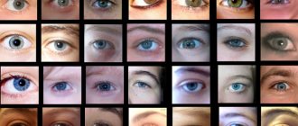
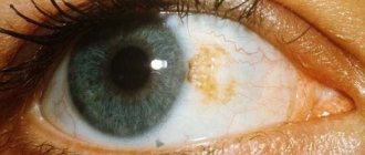
They say that the eyes are the mirror of our soul, but they are also a mirror of the processes occurring in our body. The condition of a person's eyes can tell a lot about his psychological and physical state. Rolled eyes, dilated pupils, changes in the color of the whites of the eyes - all this and much more can be a symptom of serious diseases or any problems in the human body.
Definition of disease
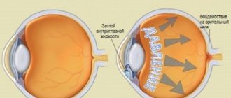
Nystagmus is involuntary oscillatory movements of the eyeballs. It usually occurs from birth or early childhood, although it can also be diagnosed in adults due to various eye diseases, brain diseases, injuries, and intoxications.
There are ocular and neurogenic (central) nystagmus. In the first case, visual impairment occurs at an early age or is the result of disturbances in the functioning of the visual apparatus. In the second case, the appearance of the disease may be associated with lesions of the vestibular component of the oculomotor system.
Depending on the nature of the movements, nystagmus is divided into three types:
- Pendulum-shaped. The eyes move from side to side.
- Jerky. The eyes move slowly in one direction and then quickly return.
- Mixed. It combines the signs of jerk-like and pendulum-like nystagmus.
Based on the direction of eye movement, four groups of nystagmus are distinguished:
- Vertical.
- Horizontal.
- Rotational.
- Diagonal.
Depending on the amplitude of oscillatory eye movements:
- Large-caliber (amplitude more than fifteen degrees).
- Medium-caliber (amplitude from fifteen to five degrees).
- Small-caliber (amplitude less than five degrees).
Other types of nystagmus:
- Physiological. This is jerky nystagmus, in which oscillatory eye movements occur due to changes in pressure in the vestibular nuclei.
- Latent. Vibrations occur when the eyes are closed or half-closed. This nystagmus occurs due to fatigue and increased intraocular pressure.
- Installation. Jerky movements appear at maximum diversion of the pupil to the side.
- Motor imbalance nystagmus. It combines symptoms of other types of disease. Its occurrence is associated with weakening of the tone of the oblique muscles of the eyes.
- Sensory derivation nystagmus. Develops due to impaired visual acuity. As a result of the disease, visual acuity decreases. The cause of its appearance is associated with central vision impairment at an early age.
- Return-incoming. The eyes diverge in different directions during oscillatory movements. Such nystagmus may be a sign of parasellar tumors.
Eye movements in this disease cannot be controlled. The amount of vibration may vary depending on the direction of gaze, head position, concentration or fatigue.
It happens that in people with this diagnosis, the head is in an unnatural position, in which the amount of vibration is minimal. The gaze cannot linger on a certain object, but passes by it. Because of this, the brain receives a fuzzy picture. This is why people with nystagmus have poor vision.
Rapid eye movement sleep behavior disorder
Rapid eye movement sleep behavior disorder (REMSD) is characterized by increased muscle tone during this period of sleep, vivid, often frightening dreams, accompanied by complex movements and phrases that correspond to the content of dreams. Patients with idiopathic RPDH are at high risk of developing synuclein-associated neurodegenerative diseases (Parkinson's disease, dementia with Lewy bodies, multiple system atrophy). The probability of getting sick within 10 years is 40–65%. Currently, RBDG is considered as a marker of the prodromal period of synucleinopathies.
Table. RBDG Screening Questionnaire (one point is awarded for each positive answer)
Introduction
Rapid eye movement sleep behavior disorder (REMSD) is a parasomnia characterized by vivid, frightening dreams accompanied by simple or complex motor phenomena during the rapid eye movement (REM) phase of sleep. This disorder was first described by the American scientist CH Schenck in 1986 [1]. They examined four men aged 67–72 years who reported unintentionally injuring themselves or their spouse as a result of aggressive behavior during sleep. After conducting polysomnography, CH Schenck found an increase in muscle tone and the number of rapid eye movements during REM sleep, as well as many episodes of psychomotor agitation, accompanied by complex gestures and phrases that corresponded to the content of dreams. In the 1980s a similar phenomenon was observed by V.L. Golubev (personal communication).
Under normal conditions, REM sleep usually accounts for 15–25% of a night's sleep, and it is believed that it is in this phase that a person dreams 80%. This period is characterized by low-amplitude electroencephalographic (EEG) activity, skeletal muscle atonia, rapid eye movements, heart rate and blood pressure variability [2]. With RBDG, the mechanism for generating muscle atonia is disrupted; motor activity during the dreaming process is not completely suppressed, resulting in motor “execution” of dreams [3].
Prevalence
In the general population, the incidence of RPDH is approximately 0.05%. This figure reflects the prevalence of a severe form of RPDH, in which injury occurs to the patient or the person who sleeps next to him. Patients with milder forms of the disease usually do not see a doctor. RPDH can manifest as early as 20 years, but most often occurs between the ages of 52 and 62 years, although there are reports of its diagnosis at both earlier (9 years) and later (84 years) ages. Men make up 87% of sick patients [4]. The frequency of episodes of the disease varies greatly - from several episodes per night to single episodes over the course of a month. Patients are usually unable to determine the cause of the episodes or the increase in their frequency.
Etiology
RBDG is usually divided into primary, or idiopathic, and secondary. Three large-scale studies have shown that idiopathic RPDH is observed in 25–43% of cases, while 48–75% of cases of RPDH are associated with taking various drugs that can provoke RPDH, most often antidepressants, or neurodegenerative diseases [4, 5 ].
Rapid eye movement sleep behavior disorder is specific to diseases belonging to the group of alpha-synucleinopathies (Parkinson's disease, multiple system atrophy, dementia with Lewy bodies) [6, 7]. On the one hand, patients with idiopathic RPDH develop neurodegenerative diseases in 40–60% of cases [8, 9]. Prospective analyzes show that, on average, 12 years after the onset of symptoms of idiopathic RPDH, approximately 50% of patients have symptoms of parkinsonism or cognitive impairment [8, 10, 11]. There are also data on two patients with idiopathic RPDH who, over a period of 20 years, did not develop other symptoms of a neurodegenerative disease, but at autopsy they were found to have Lewy bodies in the structures of the brain stem [3, 12]. On the other hand, according to various sources, in patients with Parkinson’s disease, the frequency of RBDG varies from 18 to 47%, and the rapid eye movement sleep phase without atonia occurs in more than 58% of cases [8, 13, 14]. The mentioned phase of rapid eye movement sleep without atonia is not a clinical phenomenon, but an electrophysiological finding characterized by the persistence of increased tone on the electromyogram (EMG) during rapid eye movement sleep. It is usually considered as an intermediate stage on the path of development of RBDG [8, 15]. However, there have been no long-term, prospective studies of the frequency of rapid eye movement sleep phase transition without atonia in REMDR [6].
Rapid eye movement sleep disorder may accompany other neurodegenerative diseases. There is evidence of cases of RBDG in progressive supranuclear palsy, Guam disease (amyotrophic lateral sclerosis - parkinsonism - dementia), spinocerebellar ataxia type 3, narcolepsy, limbic encephalitis with antibodies to voltage-gated potassium channels, Guillain-Barré syndrome, etc. [16].
Taking antidepressants, especially those belonging to the class of selective serotonin reuptake inhibitors, can cause RPBD [17]. Last year, the results of a study by Canadian scientists were published who observed patients with idiopathic RPBD who were taking antidepressants. Patients were examined annually until the first symptoms of parkinsonism or dementia appeared [18]. The study was designed for eight years. It was shown that patients with antidepressant-induced RPDH had a lower risk of developing neurodegenerative disease than patients with RPDH who were not taking antidepressants (22 vs. 59% at five years). The authors hypothesized that antidepressant use provokes the development of RPDH at an earlier stage of the neurodegenerative process, which explains the development of alpha-synucleinopathy in less than five years. Most likely, RPBDG in such patients is a marker of the onset of a neurodegenerative disease [18].
Pathogenesis
The mechanism for maintaining muscle atonia during the rapid eye movement sleep phase is associated with the nuclei of the pons and medulla oblongata. In the 1960s–70s. French researcher M. Jouvet first showed that cats with a damaged area of the brain near the locus coeruleus ( peri-locus ceruleus area
) in the rapid eye movement sleep phase, they raise their head, move, and try to catch the mouse [19]. Subsequent studies found that damage to the ventral portion of the sublaterodorsal nucleus in rats (which corresponds to the area near the locus coeruleus in cats) causes the disappearance of atonia during rapid eye movement sleep, which is accompanied by simple and complex movements [20].
The development of RBDG in humans is associated with damage to which specific structures remains unclear. Several cases of the development of RBDG in stroke and multiple sclerosis with lesions in the pons have been described [21–23]. There is also a known case of the development of RBDG in an acoustic neuroma, with complete restoration of the normal sleep phase with rapid eye movements after tumor resection [24].
In pathological studies of patients with RPDH, the lesion is predominantly observed in the area of the locus coeruleus, substantia nigra and raphe nuclei [3]. A study of the brains of patients with multiple system atrophy suffering from RPDH showed a decrease in the number of cholinergic neurons of the pedunculopontine nucleus, laterodorsal tegmental nucleus and neuromelanin-containing neurons of the locus coeruleus [25]. It was the lesion of the pedunculopontine nucleus and the laterodorsal tegmental nucleus, according to the researchers, that underlay RBDG in these patients. This, most likely, explains the effectiveness of the use of cholinesterase inhibitors in some patients with RPDH [26], as well as in a significant proportion of patients with progressive supranuclear palsy [27].
However, further studies have shown that cholinergic mechanisms are indirectly involved in the development of rapid eye movement sleep phase disorders [3]. Neither lesions of the pedunculopontine nucleus nor lesions of the laterodorsal tegmental nucleus underlie the pathogenesis of RPBD, as well as lesions of the locus coeruleus, which is inactive during the rapid eye movement sleep phase [3], and therefore cannot be the cause of the development of RPBD.
B. Boeve et al. [3] hypothesized that a structure located near the locus coeruleus, similar to the sublaterodorsal nucleus in rats, which has direct projections to spinal GABAergic inhibitory interneurons, is responsible for the decrease in muscle activity during rapid eye movement sleep. When it is damaged, the excitatory effect on the inhibitory interneurons of the spinal cord disappears, resulting in increased muscle activity. In addition, damage to the sublaterodorsal nucleus can lead to a decrease in excitatory influences on the giant cell nucleus of the reticular formation, which in turn reduces the inhibitory effect on spinal interneurons or directly on motoneurons of the anterior horns. It still remains unclear whether damage to the giant cell nucleus of the reticular formation can lead to the occurrence of RPDH [3].
Clinical picture
Clinical manifestations of RBDG are characterized by three main symptoms: vocalization, motor restlessness and the specific nature of dreams (most often frightening) [3, 10].
Vocalizations during sleep are also observed in many healthy people. This includes grumbling, talking, laughing, humming, etc. Such phenomena can occur in any phase of sleep and, as a rule, are not related to the content of dreams. With RBDG, the most characteristic sounds are loud screaming, howling, laughter and cursing. The voice and intonation of a sleeping person are sometimes very different from those inherent in him while awake.
Motor restlessness in the form of periodic twitching of the limbs also occurs in healthy people. However, with RPBDG the movements are more purposeful. RPBD is often described as the "performance" of a dream, suggesting that movements and vocalizations correspond to the content of the dream. But there is another point of view, according to which the content of dreams depends on the movements a person makes in his sleep. With the loss of muscle atonia during the rapid eye movement sleep phase, motor activity increases, depending on which the content of dreams can be formed. People often describe how external stimuli influence the content of their dreams. For example, a person who falls asleep in a car driving on a rough road may dream of an earthquake. In general, these two hypotheses are not mutually exclusive and can be implemented alternately [3].
Typically, the movement begins with a rhythmic twitch of the limb, then progresses to a strike, a turn (in an attempt to defend), running, jumping, etc. It is at these moments that traumatization occurs to the patient or the person sleeping next to him. Attempts to wake the patient during such an episode are woven into the fabric of the dream, and the sleeper’s reaction to them can lead to bruises, contusions, fractures and even subdural hematomas [6].
RPBD is not only a sleep pathology, but also a change in dreams [3]. The dreams that patients with RPBD see are realistic, colorful, plot-driven, and filled with emotion. Over time, the content of dreams becomes more and more frightening; patients often dream of manifestations of aggression and attack, they try to escape, defend themselves, push away danger, and it is at such moments that they can injure themselves or their bed partner [4, 10]. This phenomenon is associated with the activation of dopaminergic structures of the midbrain and forebrain. The dreams of RPDH sufferers are filled with nightmares where insects, animals or people threaten them or their loved ones. Patients almost always play the role of protector. Many patients can retell the contents of their dreams if they are awakened during the episode. And unlike ordinary dreams, which are stored in memory until noon at best, the content and even the smallest details of dreams with RBDG are remembered for a long time.
The timing of these episodes coincides with the most typical time of rapid eye movement sleep - the second half or even the last third of sleep time, unless the person has narcolepsy, obstructive sleep apnea, or the time of rapid eye movement sleep increases due to with long-term sleep deprivation [3, 6, 28].
Diagnostics
Diagnosis of this disorder according to the International Classification of Sleep Disorders (2005) is indicated if:
1) phases of sleep with rapid eye movements without atonia were detected, defined as a prolonged or periodic increase in tone, recorded on EMG leads from the chin, or an increase in phasic muscle activity on EMG leads from the legs;
2) at least one of the following signs has been recorded:
a) a history of behavior that has led or could lead to injury during sleep;
b) an episode of RPDH recorded using polysomnography;
3) there is no epileptiform activity according to EEG data;
4) these disorders cannot be explained by a neurological or somatic disease, mental disorder, or taking medications.
The “gold standard” for diagnosing RPDH, as recommended by the American Academy of Sleep Medicine, is polysomnography, which requires appropriate equipment and an experienced clinician to interpret the data. Another challenge is that patients with underlying neurological syndromes such as parkinsonism and cognitive impairment are often unable to undergo this study. In addition, in patients with dementia, the background EEG may be so altered that it becomes difficult, if not impossible, to detect rapid eye movement sleep. Quite often, with this form of pathology, even an experienced clinician using video recordings cannot say with certainty where the waking EEG is and where the sleep EEG is. Some patients may not have rapid eye movement sleep episodes at all on polysomnography.
Currently, tests are used to diagnose RPDH, the specificity of which has been confirmed in comparative studies in comparison with polysomnography.
A test for diagnosing sleep disorders, proposed by Mayo Clinic specialists (Mayo Sleep Questionnaire - MSQ), includes 16 questions regarding limb movements during sleep, restless legs syndrome, sleepwalking, obstructive sleep apnea, etc. In a pilot study [6], the test was performed twice – the first time the patient himself answered the questions, and the second time his partner or caregiver answered the questions. The study showed that the correlation with polysomnography data was higher for questionnaires completed by partners/caregivers, regardless of the presence of cognitive impairment in the patient. After which testing was recommended to be carried out not among patients, but among their partners or caregivers.
The Rapid Eye Movement Sleep Behavior Disorder Screening Questionnaire (RBDSQ) contains 10 questions that address the clinical characteristics of RBDSQ. The maximum number of points that could be obtained by passing the test is 13 (table). In one study, three groups of patients took the test: 54 patients with polysomnography-confirmed RPBD, 160 patients in the control group in whom this diagnosis was excluded based on polysomnography, and 133 healthy people who did not undergo polysomnography. In the group of patients with RPBDG, the average score was 9.5, in the control group – 4.6. This means that the use of this questionnaire in diagnosing RPDH has a sensitivity of 96% and a specificity of 56% (the test is positive if the result is more than 5 points) [29].
The search for the optimal diagnostic tool for identifying RBDG led to the creation of a test that contains only one question, formulated by R. Postuma et al. [30]: “Have you ever been told or noticed yourself that you make movements in your sleep that you dream about (for example, pushing, waving your arms in the air, moving your legs as if running)?” The question allowed us to determine the presence of RBDG with a sensitivity of 93.8% and specificity of 87.2%. The study involved 484 people, including 242 patients with RPBDH, confirmed by polysomnography, and 242 people without RPBDG, also according to polysomnography. The authors concluded that this question has high diagnostic value for screening patients for the presence of RBDG.
Treatment
For patients with RBDH who have a history of trauma associated with this disorder, it is recommended that the first step is to secure the sleeping area.
One of the most effective drugs for relieving episodes of RPDH is clonazepam. In small doses (0.25–0.5 mg), it eliminates episodes of psychomotor agitation, but maintains high muscle tone during the rapid eye movement sleep phase [4, 5, 10]. There is evidence of the effectiveness of triazolam, but there is no effect when taking other drugs in this group. The effectiveness of clonazepam may be due to a weak serotonergic effect that other benzodiazepines do not have. However, a large number of side effects and the symptomatic nature of the action of clonazepam significantly limit its use. A contraindication to its use in patients with RPDH is the presence of cognitive impairment and obstructive sleep apnea.
For the treatment of RPDH, melatonin is used in a dose of 3–12 mg as monotherapy or in combination with clonazepam. There are results of a double-blind, placebo-controlled study of the use of melatonin in patients with RPDH [31]. Those who took melatonin at a dose of 3-12 mg at night daily decreased the duration of the rapid eye movement sleep phase without atonia, and in half of them the clinical manifestations of RMDH disappeared by the end of the study. In addition, after stopping melatonin, symptoms did not return immediately and did not reach the severity they were before treatment. This allows us to conclude that melatonin has a modifying effect on the course of the disease [31]. However, long-term prospective studies have not been conducted to assess the durability of the effect obtained. It should be noted that melatonin preparations have a high safety profile. A group of such drugs includes Melaxen (Unipharm Inc., USA).
In the presence of parkinsonian symptoms, correction of antiparkinsonian therapy plays an important role in the treatment of RPDH. Considering that dopaminergic structures of the midbrain and forebrain are responsible for the emotional content of dreams [3], taking high doses of levodopa or dopamine receptor agonists may increase the realism of dreams and motor activity during an episode of RBD.
Cholinesterase inhibitors (donepezil) [26], antipsychotics (quetiapine) [3], and clonidine (due to its ability to suppress the rapid eye movement sleep phase) are now used quite successfully. Tricyclic antidepressants, as well as selective serotonin and norepinephrine reuptake inhibitors, can worsen the course of RPDH. This should be taken into account when prescribing antidepressants to this group of patients [14].
Conclusion
RPBDG is the earliest and most specific marker of neurodegenerative diseases belonging to the group of alpha-synucleinopathies (Parkinson's disease, multiple system atrophy, dementia with Lewy bodies). The presence of RBDG predicts the development of a neurodegenerative disease within 12 years with a 50% probability. This makes patients with this syndrome an ideal group for studying other possible predictors and markers of synucleinopathies, as well as the effectiveness of neuroprotective therapies.
Causes
Nystagmus is a rare condition. It occurs in one person out of several thousand. It usually occurs in newborns or young children.
The cause of its occurrence is associated with various neurological disorders, sometimes of a hereditary nature (albinism, Leber congenital amaurosis).
In adults, the cause of nystagmus can be:
- Loss of vision (due to cataracts or eye injury).
- Brain diseases (tumors, multiple sclerosis, stroke).
- Traumatic brain injuries.
- Exposure to toxic substances (alcohol, some medications).
- Malformations of the skull and brain (Arnold-Chiari anomalies).
- Hydrocephalus.
- Diseases of the vestibular apparatus (labyrinthitis, vestibular neuronitis, vestibular nerve neuroma).
Dilated pupils
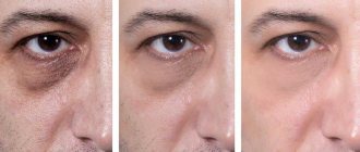
The second most common complaint is severely dilated pupils. This eye condition can also be a symptom of a wide variety of diseases.
Firstly, when using various drugs and a number of medications, as well as alcohol, patients may have severely dilated pupils. Dilation of the pupils can also be caused by various abnormalities of the thyroid gland. A head injury can also cause the pupils to become very dilated.
Dilated pupils may not be a symptom of serious injury or illness. This may be a simple reaction to light or a manifestation of a person’s individual characteristics. In addition, an increase in the size of the pupil occurs when a large amount of adrenaline is released into the blood.
Clinical Brain Institute Rating: 3/5 — 1 votes
Share article on social networks
Symptoms
It happens that people with nystagmus do not feel any signs of the disease. But sometimes the following complaints appear:
- Visible oscillatory movements of the eyes from side to side.
- Dizziness, loss of orientation in space.
- Nausea due to inability to fixate on an object with the gaze.
- Oscillopsia (feeling of continuous vibration of surrounding objects).
Nystagmus may be indicated by hearing impairment (most often in one ear), loss of coordination, decreased tone of body muscles, and double vision.
Treatment
Stages of diagnosing the disease:
- Initial examination by an ophthalmologist.
- Additional studies (visual acuity, condition of the oculomotor system and optical media of the eye, condition of the retina and fundus).
- MRI, electroencephalogram, echo-encephalography.
- Examination by a neurologist.
Classifications of nystagmus
Basic principles of treatment of nystagmus:
- Vision correction with glasses or contact lenses. Contact lenses are most often used because when the eye moves, the center of the lens moves with it, resulting in clearer vision.
- Taking Baclofen or Botox injections. But the effect is temporary.
- Surgery on the eye muscles will ensure a shift in the relative resting position of the eyes to the median position and will give a good cosmetic result.
- Treatment of the underlying disease that caused nystagmus.
Treatment of nystagmus is a long and very difficult matter. And it is not always possible to achieve the desired result.
It is necessary, first of all, to eliminate the cause that led to the development of nystagmus.
Causes of nystagmus
During treatment, it is necessary to stop taking medications that provoke nystagmus. These drugs include sleeping pills and potent drugs, such as benzodiazepines and barbiturates.
By medication
Drug treatment involves the use of various vasodilating drugs that nourish the tissues of the eye, especially the retina. Doctors can also recommend various vitamin and multivitamin complexes to completely strengthen the body.
It must be remembered that medications are only auxiliary in nature.
Surgically
Surgical treatment is performed to reduce oscillatory eye movements.
For jerky nystagmus, surgery is performed to move the “rest zone” to the midline position. To do this, stronger muscles are weakened and weaker ones are strengthened. Thanks to this, the position of the head is straightened, nystagmus is reduced, and visual acuity is increased. With the help of surgery, a person can not only get rid of uncontrolled eye movements, but also restore vision. Also, after surgical interventions, photophobia disappears.
Folk remedies
Treatment of nystagmus using traditional medicine should be combined with traditional methods of treatment.
The most famous recipes for folk remedies:
- Parsley juice. Drink freshly squeezed parsley juice before meals at least three times a day.
- Dill. Infuse dill seeds with hot water for fifty to sixty minutes in a dark place. Drink half a cup of infusion every hour throughout the day.
- Caraway. Pour boiling water over one tablespoon of cumin and leave for an hour. Take the infusion half a cup twice a day.
13.1.3 Types of involuntary movements of healthy eyes
The group consisting of some involuntary eye movements is sometimes called spontaneous physiological nystagmus. This group includes tremor, drift and saccades. These movements are inevitably present even when the subject strives to maintain continuous fixation of some stationary point stimulus with his gaze. It is thanks to them that those inhibitory processes that involuntarily arise in a long-term stimulated area of the retina are overcome. Some quantitative characteristics of involuntary eye micromovements can be judged from the data given by A. Levy-Shoen (1969), which are based on the results of studies by various authors (
).
The listed types of micromovements of healthy eyes are of interest to psychologists and physiologists, in particular in connection with solving problems of ergonomics of visual fatigue [Yarbus A.L., 1965; Volkov V.V., 1989; Somov E.E., 1992].
Involuntary eye movements that provide binocular vision include vergence (not only voluntary, but also involuntary) and fusional eye movements, as well as cycloduction. The vergence reflex has four components:
- tonic - the result of the innate ability, immediately after waking up from sleep, thanks to nervous impulses, to give the muscles of the eye a certain tone;
- proximal - the result of psychogenic awareness of the proximity or distance of the object of fixation;
- fusional - the manifestation of an optomotor reaction in response to the disparity of retinal images; as a result, without changes in the refraction of the eye, the images appear on the corresponding points of the retina (with bitemporal disparity, convergence is excited, with binasal disparity, divergence is excited);
- accommodative - the result of the normal influence of accommodation on convergence (the manifestation of a synkinetic reaction to closely located stimuli). Normally, by 1.0 dioptre, accommodation accounts for an increase in convergence of 4 prism dioptres. The ratio of accommodative convergence to accommodation is denoted AK/A. The result is expressed in prism diopters per unit of accommodation measured in diopters.
In this group, cycloduction stands out separately, which manifests itself in the fact that when the head is tilted to one or the other shoulder, both eyeballs experience a true torsion effect: they rotate compensatoryly like a steering wheel. In this case, each eye turns the upper end of the vertical meridian in the direction opposite to the side of the head tilt. The inclination towards the nose is called incycloduction, towards the temple - excycloduction. According to RS Jampel (1970), in this case, it is not so much the entire eyeball that rotates around the Y axis, but rather its anterior section (around a point on the outer limbus). In this way, the sensory constancy of the location of the vertical sutures of the retina relative to the horizon is maintained. When the head is tilted by 30°, the eye involuntarily compensatoryly deviates by 7-8° in the opposite direction [Kirillov Yu.A., 1976].
If we compare the amplitude of involuntary eye movements when fixating a stationary stimulus, then, as established by E.S. Avetisov (1977), in the presence of bifixation, the eyeballs are noticeably more stable. The magnitude of the bifixation field horizontally is 5-10 minutes, and vertically does not exceed 4 minutes. Keeping the object of fixation within a narrow fusional field is the result of involuntary static work of the oculomotor muscles, coordinated by the sensory apparatus of binocular vision.
- < Back
- Forward >
Prevention
Prevention of nystagmus includes the following recommendations:
- Timely diagnosis of the disease.
- A balanced diet and regular moderate physical activity.
- Compliance with occupational hygiene.
- Timely treatment of diseases that can cause nystagmus.
- Full healthy sleep.
- Quitting drinking alcohol and smoking.
Inexpensive drops for dry eyes
You can take a color blindness test here.
Carnival colored lenses https://eyesdocs.ru/linzy/karnavalnye/osobennosti-vidy-marki.html