Scleral lenses get their name because, when put on, they are placed on the sclera (the outer dense layer of the eye). They are easy to distinguish by their large diameter (when compared with standard optical models). Scleral lenses are used for two purposes - decorative and therapeutic. The products have a number of features and require strict adherence to the wearing regime.
Share
Tweet
Share
Cool
Send
What are scleral lenses?
Scleral lenses are those lenses that have a larger diameter than regular lenses. Their main feature is that they rest on the sclera of the eye, and not on the cornea, like standard contact options. In simple terms, scleral lenses cover the entire surface of the eye.
They appeared earlier than the already familiar corneal ones. And they were originally used to treat keratoconus. This is a pathology in which the cornea of the eye changes and takes on the shape of a cone. Scleral lenses, due to their structure, help straighten the cornea and, thus, correct vision.
Treatment options for keratoconus
Contact optics cannot cure keratoconus. Modern surgical methods, in particular the installation of so-called intrastromal rings and cross-linking, help correct vision in this disease, as well as stop its progression. In this way, you can stabilize the cornea, stop the process of its protrusion and improve vision. Another option is to install intraocular optical implants behind the iris.
To select the optimal treatment method, you should always consult an ophthalmologist.
History of appearance
Today, scleral vision correction is considered an innovative treatment method, although in fact these lenses have a long history. The first mention of them occurs in the 19th century. At that time, a device for correcting vision was made from glass using a cast of the eye. In the 20th century, scleral prescription lenses began to be made from polymethyl methacrylate. But such lenses, just like glass ones, did not allow oxygen to pass through, so they were more harmful to health than corrected vision. In this regard, the use of scleral lenses was suspended until the 70s. Then the first oxygen-permeable models were created. In 2008, a fundamentally new technology for manufacturing this type of lenses appeared, and in 2014 this technology was patented.
What is treated with such lenses?
Lenses for the entire eye (scleral lenses) are, first of all, a device for correcting vision. In addition, they are used for diseases and disorders such as:
- Corneal ectasia.
- Sensitivity to light (after injury).
- Microphthalmia.
- Dry eye syndrome.
- Aniridia (absence of the iris).
- Overgrowth of the cornea (Stevens-Johnson syndrome).
- Aberration of the eye.
- Chemical and thermal burns of the eyes.
- Problems after laser vision correction.
- Problems after operations.
In addition, scleral lenses are used in various ophthalmic experiments. Such studies are popular in psychology and in the study of the visual system.
Keratoconus cannot be treated with contact optics
Ophthalmologists do not consider contact optics to be the best way to correct vision in keratoconus. Most experts believe that it is much more effective to install intrastromal corneal segments. These are special implants that stabilize the cornea, increase visual acuity and are comfortable for the patient. Another option is to implant intraocular optics. It is placed behind the iris, so the implant does not come into contact with the cornea of the eye.
We remind you that the selection of an optical correction method and suitable lenses is the prerogative of an ophthalmologist.
MagazinLinz.ru team
Advantages and disadvantages
The main advantage of such lenses is the material from which they are made - polymacon. It is very elastic, yet strong, as well as safe and durable. The positive aspects also include:
- Elasticity - such lenses can be worn without removal for several days; they are harmless to the sclera and cornea of the eye.
- The presence of through pores - thanks to this, scleral lenses allow oxygen to pass through perfectly and thus, corneal edema can be avoided. Other lenses cannot boast of this property.
- Strength and rigidity of the material - the lenses are protected from mechanical damage and are very easy to care for. They can be worn for several years.
- Safety of the material for eye health.
- The shape and size of scleral lenses can be changed depending on a person's preferences without harm to health.
Today, such products are used not only for medicinal purposes. White scleral lenses are now very popular among young people. With their help you can add mystery and uniqueness to your image. Pupilless scleral lenses are also often used by young people to stand out from the rest and create a unique, unique look.
Among the disadvantages of such models, one can primarily highlight their cost. One pair of scleral lenses costs at least 3 thousand rubles, so not everyone can afford them. In addition, such lenses cannot be purchased at a pharmacy. They are made for each individual individually, taking into account the shape of the eyes and sclera.
Is it possible to wear lenses for keratoconus and what kind?
If you have keratoconus, you can wear contact lenses, but in each case the type of lenses is selected individually depending on the stage of the disease. Contact vision correction with lenses for keratoconus is more preferable than spectacle correction.
Important! The selection of lenses for keratoconus is carried out only by a doctor!
CLs used for keratoconus are divided into several types:
- soft;
- rigid gas-permeable;
- hybrid;
For grade 1 keratoconus, when astigmatism does not exceed 2.5 diopters, it is possible to use soft silicone hydrogel CLs. Silicone hydrogel lenses for keratoconus provide good contact with the cornea and the formation of a single optical system with the eye, which allows achieving good visual acuity.
If pathological changes in the corneal surface progress and the degree of astigmatism increases, soft CLs cease to help and cannot provide high visual acuity. Therefore, at this stage it is possible to prescribe rigid CLs that smooth out corneal irregularities.
Gas permeable hard contact lenses are divided into 2 large groups:
- corneal;
- scleral
Corneal lenses have a diameter of up to 10 mm. Even high degrees of astigmatism are well corrected.
Scleral lenses differ from corneal lenses in diameter, usually exceeding 13.5 mm. Scleral CLs rest on the sclera and thus do not have a damaging effect on the periphery of the cornea, but contact with the sclera can cause discomfort when worn.
Hard contact lenses are selected individually to the shape of the patient's cornea. When using hard CLs, you must strictly adhere to the wearing and storage regime to avoid the development of serious complications.
Hybrid lenses are CLs in which the central part is hard and the periphery is soft. This combination allows you to achieve good visual acuity due to the hard part, and more comfortable wearing and good fit due to the soft part. The disadvantage of these lenses is their tight fit at the edges, which leads to reduced wetting of the corneal surface with tears. Lack of hydration leads to dry eyes and the appearance of microcracks, which can trigger the progression of the disease.
Glasses for keratoconus
Spectacle vision correction for keratoconus is possible only in patients with stage 1 of the disease. The selection of glasses is carried out by a doctor or optometrist in a vision correction office. As the degree of astigmatism increases, wearing glasses becomes impossible.
Laser vision correction for keratoconus
If a patient is diagnosed with keratoconus, then laser vision correction is not performed, since there is no guarantee of results, and there is a high risk of developing secondary keratectasia. Therefore, before the laser vision correction procedure, it is necessary to undergo a thorough examination to avoid the development of complications.
If during the preoperative examination a suspicion of keratoconus is detected, it is recommended to postpone the operation and undergo an examination after about six months. This will allow you to compare the condition of the cornea over time and give an accurate conclusion.
Read an article by an ophthalmologist about methods of treating keratoconus.
Yulia Chernova, ophthalmologist, especially for Mirmam.pro
How to choose?
Before purchasing scleral lenses, you need to be prepared for the fact that selecting them is a long and difficult task. Cheap Chinese analogues can cause the development of many diseases (hypoxia and corneal edema, vascular ingrowth and many others). Therefore, it is better to purchase such a problematic item to order. However, lens manufacturing can take up to 4 months, so be patient.
The lens manufacturing process consists of the following steps:
- The parameters of your cornea are measured.
- An initial set of scleral lenses is created, which will then be adjusted to suit the structure of your eyes.
- Approbation stage. You will need to wear a trial pair of lenses under your doctor's supervision for several weeks.
- Final fitting stage. At this stage, the lenses are painted in the required color. If you have them for medicinal purposes, then they are left transparent.
One more point should be noted. Black scleral lenses come in different types:
- Diameter 20-24 mm - they completely cover the sclera and make the eye black.
- Diameter up to 18 mm - in this case the cornea turns out to be contrasting.
- Diameter 13-15 mm are semi-scleral lenses.
Never order scleral lenses online. Be sure to consult with an ophthalmologist to determine the parameters of your cornea and sclera. Remember that even as an accessory for actors, these lenses are made to order.
There are times when even properly fitted lenses can cause discomfort. To avoid this, it is important to learn how to put them on and take them off.
Other correction methods
There is a visual correction method called Piggyback. Its essence is that the patient first puts on a soft lens - for comfort, and on top of it - a hard one, to create the correct spherical surface of the front part of the eye.
As an alternative, you can use hybrid models that combine the advantages of different models. The edges of such products are made of soft polymer, and in the center they are ZhGP lenses. Note that both methods are not very common in our country, but they are actively used abroad.
How to put on scleral lenses?
Properly fitted lenses can be put on in about 20 seconds. But there are several points to consider.
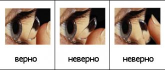
First, you need to learn to distinguish the front side from its “wrong side”. If you put the lens on incorrectly, it will not stay in place and will cause severe pain. A correctly inverted lens looks like a bowl with a convex base and upward-facing edges. An incorrectly turned accessory looks like a saucer with a flat bottom and smooth edges.
You should only put on your lenses after you have finished your makeup. If you do the opposite, then particles of cosmetics can get on the lens and cause irritation. You're guaranteed to have puffy, red eyes the next day.
Use only those cosmetics that have an eye on them. This ensures that the product will not damage the lenses.
Before putting it on, you need to put special drops in your eyes and wash your hands thoroughly. It is better to wipe them with a napkin rather than a towel, so that there are no lints left on your hands. If washing your hands is not possible, use a germicidal wipe and disinfectant.
Algorithm for putting on scleral lenses:
- Remove the lens from the container and place it face down on the pad of your index finger.
- The lower eyelid needs to be pulled back with the thumb of the hand in which you have the lens.
- Raise the upper eyelid with the index finger of the other hand.
- We apply the lens to the surface of the sclera so that the edges lie along the line of the drawn eyelid. If you did everything correctly, there will be no discomfort.
Cross stitching of the cornea
This procedure helps strengthen the structure of the cornea, which is weakened by keratoconus.
To strengthen the cornea, riboflavin (vitamin B2) and exposure to ultraviolet light in the UVA range are used. Riboflavin is instilled onto the cornea, and then the area is irradiated with UVA rays. Activated by ultraviolet rays, riboflavin binds to collagen fibers of the cornea. As a result of this process, the number of cross-links between the collagen fibers of the cornea increases, which helps strengthen the corneal tissue and stop the further development of keratoconus. This technique does not eliminate changes in the shape of the cornea that have already occurred in keratoconus (cone-shaped protrusion). Cross-linking the cornea stops further progression of keratoconus. This procedure can be used in conjunction with Intacs, which flatten the cone shape of the cornea itself.
The ability to strengthen the structure of the cornea for a long time and stop the progression of keratoconus is a huge advantage of this method. The technique is already used in some clinics in the country.
Cornea transplant surgery
Approximately 10-20% of patients with keratoconus require corneal transplant surgery. During surgery (called keratoplasty), the portion of the cornea affected by keratoconus is removed and replaced with a donor cornea. The success rate of obtaining good vision as a result of the operation is quite high - about 90%.
Usually, after surgery, vision is not restored immediately. In some cases, a few weeks are enough, in others it takes up to 1 year to obtain good vision.
It should also be borne in mind that the probability of survival of a transplanted cornea after 5 years after surgery is 74%, after 10 - 64%, after 20 years - 27% and only 2% after 30 years. Partial keratoplasty, in which only a small portion of the cornea is removed, helps reduce the risk of graft rejection. In general, keratoplasty performed by highly qualified surgeons gives good results.
But even if the operation is successful and there are no complications, many keratoplasty patients still require contact lenses, usually rigid gas permeable (RGP), to restore vision due to corneal irregularity and high corneal astigmatism.
Prescribing contact lenses or glasses after surgery requires that your vision stabilize, which usually takes several months. But in some cases this time can vary greatly.
In general, we can say that various methods can be used to correct vision in keratoconus. The most common method is hard contact lenses. Some experts believe that surgery, usually recommended for moderate to severe keratoconus, should be considered a last resort and can be avoided in many cases with the use of scleral lenses.
How to remove lenses correctly?
Removing lenses is even easier than putting them on. First, remember to wash your hands thoroughly. Then, with the index finger of your left hand, you need to pull down the lower eyelid and release the lens from below. With your other hand, you need to carefully grab the lens and pull it down. After removing it, place it in a container and close it. The container itself can be shaken to clean the lenses.
Some care features
To clean scleral lenses, use the same solution as for regular lenses. They must be stored in a special container.
To deep clean lenses, you need to pour the solution into the container and add a protein tablet. In 12 hours, this way you can completely clean any product from all contaminants.
Never rinse scleral lenses under running water. This can introduce bacteria and trigger the development of allergies. Change your lens solution regularly and do not use it after the expiration date.
Remember that scleral lenses are not just a party accessory. They are used to solve various health problems, so they need to be treated responsibly. They need to be made only to order to avoid discomfort, allergic reactions and other side effects.