Tonometry is used to measure intraocular pressure levels. The pressure in the eyeball can be normal, decreased with eye hypotension, increased with glaucoma or intraocular hypertension. The level of intraocular pressure is determined in several ways: by palpation (indicative method), using impression or applanation tonometers, and non-contact method.
What is ophthalmotonus?
Ophthalmotonus is a pathological symptom signaling a violation of visual function.
An increase in intraocular pressure occurs due to impaired outflow of fluid localized under the membrane. The tubules through which fluid flows out are blocked, which leads to an increase in volume and increased pressure. Increased intraocular pressure leads to compression of the vessels of the fundus, as a result, areas of the retina are injured, and photosensitive structural elements die. When the optic nerve is compressed, the cells become deficient in oxygen and nutrients, which leads to atrophy and complete visual dysfunction.
A decrease in the level of ophthalmotonus is also a formidable pathological sign. Due to low pressure, blood circulation in the visual organ is disrupted, resulting in tissue atrophy.
High blood pressure may indicate the development of cardiovascular diseases, while low blood pressure may indicate liver failure or the initial stage of retinal detachment.
Important! Normal IOP (intraocular pressure) values range from 10–25 mmHg. Art., depending on the measurement method.
Impression tonometry
This technique was proposed by Schiotz and is based on indentation of the cornea with a special rod with a constant cross-section. For this purpose, piles weighing 5.5, 7.5 and 10 grams are used. The obtained values are expressed in linear quantities. They directly depend on the mass of the load and the level of intraocular pressure. To convert these linear values into mmHg, you need to use nomograms.
The accuracy of impression tonometry is lower than that of the appallation type of study. Moreover, this technique helps to determine the level of pressure if the patient’s corneal surface is not smooth.
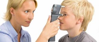
In modern ophthalmological practice, all the disadvantages of contact tonometry have been eliminated, thanks to the use of non-contact tonometers of various designs. These innovative devices implement all the achievements in the field of optics, mechanics and electronics. With non-contact tonometry, a portion of compressed air is sent to the center of the cornea from a certain distance, which has a certain volume and comes out under a certain pressure. As a result, the cornea becomes deformed and the interference pattern changes. These devices help measure intraocular pressure with high accuracy, without direct contact of the tonometer with the surface of the eye.
Indications for diagnostics
In ophthalmology, intraocular pressure is measured according to indications, that is, symptoms that patients complain about. These include:
How to get rid of eye pressure
- Visual dysfunction.
- Formation of a film before the eyes.
- Headache radiating to the temporal and ocular areas.
- Rapid eye fatigue.
- Twilight vision disorder.
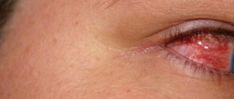
- Redness of the outer shell of the eye.
- Concentration on an object is accompanied by painful sensations.
- Pain when blinking eyelids.
- Dry mucous membrane.
- Changing the shape of the pupil.
- Narrowing of visual fields.
For persons over 40 years of age, this procedure is mandatory during a preventive examination and is carried out once a year, regardless of the complaints detected.
In patients diagnosed with glaucoma, examination is carried out once every trimester. If you have a genetic predisposition to glaucoma, for preventive purposes, experts recommend taking measurements once every 2 years, provided there are no pathological symptoms.
Patients suffering from hypertension also require constant monitoring of IOP levels, since a persistent increase in blood pressure leads to irreversible changes in the retinal vessels. Regular examination by an ophthalmologist allows you to assess the degree of damage to the optic nerves and prevent the development of complications.
Basic methods of determination
In ophthalmological diagnostics, tonometric and tonographic research methods make it possible to determine a rational method of treating glaucoma, as well as the dynamics and complications of the disease.
The measurement of ophthalmotonus differs in technique, including:
- Palpation (finger).
- Instrumental (contact, non-contact).
During the procedure, the ophthalmologist determines the degree of elasticity of the eyeball, based on identifying the level of deformation during external exposure.
Finger technique
Palpation is the simplest technique for determining the degree of ophthalmotonus. The technique is used in cases where there are contraindications to direct contact of the instrument with the eyeball or when there is a likelihood of obtaining inaccurate results as a result of injury to the cornea after eye surgery. There are two options for carrying out the technique:
- Direct palpation of the eyeball after anesthesia is used during the operation.
- Transpalepebral palpation is carried out through the eyelids by pressing the fingers of both hands above the upper ocular cartilage, and the degree of eye hardness is assessed.
Iphthalmotonus can be determined by scleral compliance. So, at a normal level of pressure, shocks are felt during squeezing, an increased level requires more pressure, and the shock becomes weaker.
The study determines the difference in pressure between both eyes. To interpret the results obtained, there is a system for assessing ophthalmotonus, where:
- T-pressure is normal.
- T+1 – some compaction compared to the norm.
- T+2 is a significant excess of the norm, but indentation of the fibrous membranes is observed.
- T+3 – excessive increase in density, inability to press in the eyeballs.
Measurement by this method is inaccurate and is not acceptable for clinical research. Due to the ease of implementation and the absence of the need for devices, the method can be used in emergency situations.
How to determine IOP at home?
Patients diagnosed with late-stage glaucoma should have their intraocular pressure measured throughout the day. For home measurements, special portable devices are used.
Determination of ophthalmotonus using a home ICare One meter
The device for home determination of IOP is called ICare. The measurements are based on the application of replaceable sensors to the cornea of the eye.
The advantages of this method are:
- High accuracy of results.
- Instant product of measurements.
- Absence of corneal reflex.
- No discomfort during the study.
- Eliminating the possibility of infection.
The device has a built-in memory, so the last ten measurements are always saved.
What is this?
The pressure itself is the force that acts on the walls of the organ from the inside. This reaction helps maintain the shape of the visual organ and regulate the constant amount of nutrients in it. The value of this force depends entirely on the following factors:
- Pupil width;
- Production and transportation of internal substance;
- Level of corneal tone;
- The sensitivity of the lining of blood vessels and the degree of their filling.
When a person is healthy, he has a clear interaction between the regulation of these elements. IOP values may fluctuate throughout the day; this process is considered normal. After waking up, vascular tone is higher. However, such fluctuations are insignificant, for this reason they are fundamentally unable to negatively affect the condition of the organ.
Important! The norm of this characteristic does not depend on gender or age. To prevent the development of the inflammatory process, it is important to periodically measure the indicator. We will describe below how IOP is usually measured.
Decoding the results
The normal level of ophthalmotonus has approximately the same values in both eyes. The norm at which the difference is acceptable should not exceed 3–4 mm Hg. Art. If the difference in the IOP value of both eyes is 4–5 mm Hg. Art. even with normal ophthalmotonus indicators, this is a sign of glaucoma.
IOP levels change with age, but age-related changes in average values are insignificant. Thus, at the age of 20, blood pressure slowly increases, and after 70 years, it decreases.
Based on the data obtained, the ophthalmologist can make a preliminary diagnosis, depending on which an individual treatment regimen is selected.
Measuring intraocular pressure is an important diagnostic criterion in identifying diseases of the visual apparatus. If you have eye diseases, it is necessary to constantly monitor the IOP value, which will help reduce the risk of complications. Today, there are many methods for determining the level of eye pressure, but ophthalmologists prefer non-contact techniques that do not require direct exposure to the organ. Improvement of methods and devices for measuring IOP simplifies the process of diagnosing eye pathologies.
Measuring intraocular pressure
Intraocular pressure is determined by the difference in the rate of increase and decrease of moisture in the chambers of the eye. The first ensures the secretion of moisture by the processes of the ciliary body, the second is regulated by the resistance in the outflow system - the trabecular meshwork in the corner of the anterior chamber3.
The only absolutely accurate method for measuring intraocular pressure (“true”) is manometric. To measure pressure, a manometer needle is inserted into the anterior chamber through the cornea, making direct measurements. This method, naturally, is not applicable in clinical practice.
In clinical practice, a wide variety of devices and instruments are used to measure intraocular pressure using the indirect method of determining IOP. With this method, the desired pressure value is obtained by measuring the response of the eye to a force applied to it. Thus, an experienced doctor can approximately estimate the level of intraocular pressure without instruments - by palpation, by the resistance of the eyeball when pressing on it with fingers.
The application of a certain force to the eye (flattening or indentation of the cornea) inevitably affects the hydrodynamics in the chambers of the eye. A certain amount of moisture is displaced from the chambers. The larger this volume, the more the resulting indicator differs from the “true” intraocular pressure (P0). The result obtained in this way is called “tonometric” pressure (Pt)5.
In Russia, Maklakov tonometry and non-contact tonometry are most often used. In addition, some medical institutions use ICare tonometers, Goldmann tonometers and, in some places, even Pascal tonometers.
Of these five methods, four can determine the “true” intraocular pressure - ICare tonometers, Goldmann tonometers, non-contact tonometer, and Pascal tonometer. Although these instruments also exert some pressure on the ocular membranes during measurement, their effect on ocular fluid dynamics is believed to be minimal. For example, when measuring, the Goldmann tonometer displaces moisture from the chambers of the eye in a volume of 0.5 μl. This results in an overestimation of the pressure figure by approximately 3%. That with average figures, IOP differs from the truth by less than 1 mm Hg. Art. This difference is considered to be insignificant, and therefore the intraocular pressure measured by such devices is called true.
True intraocular pressure is considered normal
ranging from 10 to 21 mmHg.
Tonometry using a non-contact tonometer is often mistakenly called pneumotonometry. However, these are completely different methods. Pneumotonometry is currently practically not used in Russia. Non-contact tonometry is used very actively. It is positioned as a way to determine true intraocular pressure. The method is based on flattening the cornea with air flow. It is believed that the more accurate the data from such tonometry are, the more measurements are taken (four measurements per study are considered sufficient to obtain an average figure on which one can already rely)4,5. The numbers produced by non-contact tonometers are comparable to the numbers obtained when measuring IOP with a Goldmann tonometer (9-21 mm Hg is considered normal)3.
Tonometry using ICare is also comparable to the results obtained by Goldmann. The convenience of this tonometer is its portability and the ability to be used for examining children from an early age without anesthesia4. In addition, ICare tonometers are convenient for self-monitoring of intraocular pressure by patients at home. But the high cost of such a tonometer - 3000 euros (according to representatives of Icare Finland Oy in Russia) - makes it, unfortunately, difficult to access for most patients.
Tonometry with weights was proposed by Maklakov in 18841. Maklakov’s tonometer entered clinical practice a little later. But this method occupies a strong position in the arsenal of Russian ophthalmologists. In Russia, Maklakov tonometry is the most common method for measuring intraocular pressure. It has been actively used and continues to be used in all CIS countries, as well as in China5. The method did not take root in Western Europe and the USA.
Unlike other tonometry methods we use, Maklakov tonometers displace a slightly larger volume of moisture from the chambers of the eye, thus more significantly overestimating the results of measuring intraocular pressure. This method gives us the so-called “tonometric pressure”.
Tonometric intraocular pressure is considered normal
ranging from 12 to 25 mmHg2.
It is important to know that it is not correct to compare intraocular pressure indicators obtained with a Maklakov tonometer with indicators obtained with ICare, Goldmann, Pascal tonometers or a non-contact tonometer. Data obtained using different tonometry methods are interpreted differently. Meanwhile, patients and even doctors often sin by comparing and equalizing pressure values obtained using a Maklakov tonometer and a non-contact tonometer. Such a comparison has no basis; moreover, it is potentially dangerous, because The upper limit of normal IOP for a non-contact tonometer is considered to be 21 mm Hg, and not 25 mm, as with Maklakov tonometry.
In addition, despite the fact that all of the listed methods, with the exception of Maklakov tonometry, show the “true” intraocular pressure, the numbers obtained when measured with different devices in most cases differ somewhat. Therefore, it is highly recommended that patients with glaucoma always measure intraocular pressure using the same method. Only in this case does comparison of measurement results make logical sense.
The “gold standard” of tonometry in the West is tonometry using a Goldmann tonometer. Although it is believed that the Pascal tonometer (dynamic contour tonometry) is less dependent on the condition of the eye membranes, and therefore more accurate. Tonometry according to Maklakov is recognized as quite accurate, minimally dependent on the researcher and a very reliable technique. Of the above methods, tonometry with a non-contact tonometer is the least reliable and is intended more for screening (quick superficial examination) rather than for the management of glaucoma patients4.
This article does not cover transpalpebral tonometers (tonometers that measure intraocular pressure through the eyelid). Despite the fact that they are very often used in Russian medical institutions, there are no studies showing sufficient comparability of measurement results with known tonometers4.
Bibliography
1) T.I.
Eroshevsky, A.A. Bochkareva, “Eye Diseases”, 1983 2) “National Guidelines for Glaucoma”, 2011 3) Josef Flammer, “Glaukoma, a guide for patients”, 2006 4) European Glaucoma Society “Terminology and Guidelines for Glaucoma, 3rd Edition”, 2008 5) Becker-Shaffer's Diagnosis and Therapy of the Glaucomas, 8e, 2009 Author
: Ophthalmologist A.E. Vurdaft, St. Petersburg, Russia.
Page updated date:
29.03.2019