Home » Medicine in Israel » Diagnostics in ophthalmology: modern methods and technologies
Change font: A
A
Vision problems can begin at any age; many of them can be easily corrected if therapy is started in a timely manner. Unfortunately, most ailments do not manifest themselves in the early stages, and identifying them without appropriate research can be quite difficult. Diagnostics in ophthalmology allows you to detect a problem at the initial stages and take the necessary set of measures to eliminate the pathology or prevent it from progressing further.
Indications for diagnostic examinations in ophthalmology
For a healthy person who has no vision problems, a scheduled visit to the ophthalmologist once a year is enough. But sometimes unpleasant symptoms arise that should not be ignored and should be diagnosed in ophthalmology as soon as possible. These include:
- itching and burning in the eyes;
- decreased visual acuity;
- redness of the sclera or eyelids;
- increased lacrimation;
- increased sensitivity to light;
- feeling of dryness, “sand” in the eyes.
In addition to these conditions, any eye injury or foreign object entering the eye that causes severe discomfort is grounds for a visit to an ophthalmology center for diagnosis.
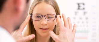
Autorefractometry
A diagnostic method in ophthalmology, which makes it possible to determine the level of myopia, farsightedness or the presence of astigmatism in a patient. The examination procedure is carried out using a special apparatus, which, using beams of infrared rays, travels to the retina of the eye and collects information about the state of visual abilities. Indications for use:
- selection of glasses or contact lenses;
- eye injury;
- before the operation;
- after suffering from keratitis.
Description of the method
When carrying out visometry, special objects are used, presented in tables - optotypes that have a certain ratio of width to base.
The most popular option (in Russia) is the table (Sivtsev-Golovin) of the Russian alphabet. This table consists of 12 rows of letters. The top row is the letters of the largest size and then in descending order.
One hundred percent vision assumes the ability to recognize letters from line 10 at a distance of 5 meters. This quality of visual acuity is determined by a parameter of 1 unit. If the patient sees only the 9th line, then such vision is indicated by a value of 0.9 and so on. The lower the visual acuity, the lower the main indicator. Some subjects are able to read lines 11 and 12 from a 5-meter distance. This visual acuity is defined as 150-200%. People who cannot even see the top lines have very low quality visual acuity. To carry out visometry in such patients, an approximation of 1-2-3 steps to the table is used. If the patient is unable to distinguish optotypes, visometry can be performed using light rays. Using light perception, examination is carried out in the most extreme cases, when the results of a standard examination indicate complete or partial loss of vision.
Computer perimetry
The computer perimeter is used in diagnosing abnormalities of the retina and optic nerve, helping to identify glaucoma, retrobulbar neuritis, and multiple sclerosis. This technique is successfully used for hypertension, hemorrhages and brain injuries, and suspected brain tumors. Contraindications to performing this examination are:
- alcohol or drug intoxication;
- emotional instability;
- mental retardation.
Biomicroscopy
One of the diagnostic methods in ophthalmology, which allows you to examine the condition of the inner layer of the cornea using an ultra-precise microscope. During the testing process, the specialist checks the structure of the cells and determines the changes that have begun. This method allows you to promptly pay attention to the early stages of pathological processes in the cornea and promptly stop them. Indications for biomicroscopic examination of the eye:
- eyelid injuries;
- inflammation of the iris;
- conjunctivitis;
- scleritis;
- glaucoma;
- eyeball injuries;
- neoplasms or cysts on the eyelids or conjunctiva.
Do I need to be examined if there are no vision problems?
Some visual pathologies in the early stages may be asymptomatic. For example, a disease such as glaucoma may not initially manifest itself, but if appropriate measures are not taken in time, glaucoma leads to irreversible loss of vision. The same applies to retinal pathology. Certain disturbances in its functioning can only be identified during a detailed examination of the fundus of the eye - and without the intervention of a specialist, there is a risk of serious deterioration in visual functions.
Many modern people spend long hours at the computer, forgetting to take at least minimal breaks. At the same time, the visual system may undergo changes that are not immediately noticeable, similar to ordinary fatigue, and without urgent treatment can lead to serious problems.
If we talk about children, then we cannot do without the professional attention of an ophthalmologist - there are often cases when an objective, competent diagnosis of possible deviations in the development of the child’s visual system and timely treatment help prevent the development of dangerous ailments.
Adults are recommended to visit a specialized clinic and undergo an eye examination at least once a year, children - at the age of 3-12 months, 3, 5, 7 years and then annually.
For pregnant women, ophthalmological examinations are required with a thorough examination of the condition of the fundus at 6, 10 - 14 and 32 - 36 weeks of pregnancy.
Diagnostic examinations of the visual system are mandatory before microsurgical interventions for the patient. This allows you to identify possible contraindications, determine the individual parameters of the operation as accurately as possible and predict its result.
advantages of diagnostics at the excimer clinic
- In our clinic, consultations are conducted only by highly qualified specialists who have extensive experience in conducting all types of modern diagnostic procedures.
- Modern equipment available in the arsenal of doctors at the Excimer clinic allows you to analyze the state of the visual system with the highest accuracy, which, if any abnormalities in the functioning of the eyes are detected, is extremely important for making the correct diagnosis and choosing an effective treatment method.
- All studies are carried out in a short time.
Popular articles about vision
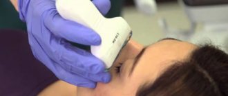
Ultrasound of the eye
Ultrasound diagnostics in ophthalmology provides an opportunity to study the structure of the eye, optic nerve and extraocular muscles, and check the level of blood circulation in them. Used to determine many diseases of the vision and nervous system:
- cataract;
- identification of a foreign object inside the eyeball;
- transformation of the length of the muscles of the eyeball;
- determination of orbital parameters;
- the beginning of the inflammatory process;
- glaucoma;
- cataract.
Retinal tomography
Diagnostics of the optic nerve head using a high-precision laser scanner. The device performs three-dimensional analysis and identifies minimal deviations from the norm. This technique allows not only to identify the disease at the initial stage, but also to monitor changes during the treatment process.
Varieties of this technology:
- laser scanning topography;
- confocal laser scanning topography;
- laser scanning ophthalmoscopy-polarimetry;
- electro-optical fundus modulation.
There are a great variety of diagnostic methods and technologies in ophthalmology, which of them is more appropriate in each specific case is decided by a qualified doctor, who also interprets the results of the study and prescribes the correct therapy.
Objective methods of visometry
Objective visometry techniques are used in cases where examination of children using standard methods is impossible.
Typically, the use of this type of research is used in cases where there is a suspicion of aggravation and simulation.
The following objective methods exist:
- Optokinetic nystagmus method.
- Detailed study of visual evoked potentials.
- Method of formed selective vision.
To analyze visual acuity using the optokinetic nystagmus method, special objects with a periodic structure are used. For example, a geometric lattice or a chessboard can be represented as such an object. Such an object in motion is presented to the patient being examined. The specialist monitors the patient's reaction to a moving object and determines changes in eye movement. As a rule, when recognizing the movement of an object, the patient's eyes involuntarily begin to move. The minimum size of the object is taken as the basis for determining the quality of visual acuity.
Using the study of visual evoked potentials, visual acuity is determined without taking into account eye movements. However, it is worth noting that some patient attention is required when using this technique. This method is based on recording electrical potentials in the occipital region as a response to visual stimuli. The patient is shown an object in the form of a fireclay field, the cells of which change places and become smaller. The dynamics of these changes occur with a given frequency. The minimum fixation size is considered to be the one that causes synchronous oscillations (on the EEG) with changes in the area of the translated object.
.The method of selective vision is based on the fact that the child prefers to look not at homogeneous objects, but at structured ones. The preferred gaze test is carried out as follows. Two objects are fixed in front of the patient being examined. The first object is a uniformly colored gray object, the second is represented as an object with vertical stripes of different colors. If the patient sees objects, then more often he chooses a bright striped object to fixate his eye. To assess visual acuity using such a test, not only eye movements are taken into account, but attention is paid to the rotation of the neck and head.
The inability to perform this test may indicate a violation of the motor function of the eyes, but a violation of the primary sensory system may often be suspected (not always justified).
Visual acuity
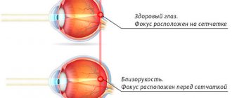
Visual acuity is one of the most important characteristics indicating the overall quality of visual function of the eyes and the health of this area in general. This indicator indicates the ability of the eyes to distinguish objects, separate them and determine any details in visible areas. Normally, the eyes should distinguish two points distant from each other, the resolution angle of which is 1 minute (1/60 degrees). For example, for a distance of 5 meters this figure will be 1.45 millimeters. Visual acuity is considered a qualitative indicator of the visual function of the eyes, which can be represented in numbers. The norm is 100% vision. But, unfortunately, today not everyone is able to maintain such indicators for a long time. To effectively monitor the quality of visual acuity, various research techniques are used. Among others, the above-described visometry technique is considered the simplest and most highly effective.
Dimension table examples
As mentioned above, special tables are used to measure visual acuity using visometry.
Below are examples of the most common table options for conducting isometric research:
- Sivtseva-Golovina. The Russian alphabet is presented as optotypes in such a table. This is the most popular table used to conduct this research in Russia.
- Orlova's table. The optotypes on this table are black and white figures - a mushroom, a star, a car, an airplane, etc. This table is used for examining young children (from 3 to 7 years).
- Landolt table. The optotypes on this table are represented by half rings with breaks in various places (right, left, bottom, top). Landolt elements can be included in many tables for visometry. It is believed that these optotypes allow for a more accurate assessment of visual acuity, since they exclude the recognition of objects and do not make it possible to memorize the sequence of object locations.
- Snellen chart. The optotypes in this table are depicted in the Latin alphabet. This table is also considered a very common variation of aids for checking visual acuity in adult patients.
There are other versions of tables for vizmetry. In each specific case, the most suitable table is selected, with the help of which it will be convenient and effective to assess the patient’s visual acuity.