Open-angle glaucoma is a progressive, chronic eye disease accompanied by increased intraocular pressure, which over time leads to damage to the optic nerve. Among its clinical symptoms, it is necessary to highlight decreased visual acuity and deterioration of accommodation. The disease is often accompanied by painful sensations. To diagnose the disease, tonometry, gonioscopy, OCT, perimetry and ophthalmoscopy are performed. Complex treatment methods include: drug therapy, laser methods, surgical operations (sinusotrabeculectomy, sclerectomy).
Pathogenesis of primary glaucoma
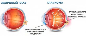
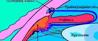
The pathogenetic path of glaucoma development has two main directions: hydrodynamic and metabolic. In the first case, we are talking about a violation of the outflow of intraocular fluid, which leads to an increase in the level of intraocular pressure. Hypertension causes a decrease in the level of perfusion pressure in the arteries of the eyeball and impaired microcirculation. With a prolonged increase in pressure inside the eye above 21-22 mm Hg. (26 mm Hg, if we are talking about tonometric pressure and not true) damage to two weak structures of the eye develops (the cribriform plate of the sclera and the trabecular diaphragm in the filtration system).
Production and reabsorption of intraocular fluid
The cribriform scleral plate contains several portions, the posterior of which is the toughest and thickest. The holes in the plates form tubules through which nerve fibers pass.
When the layers of the cribriform plate are displaced in the event of its deflection, which usually occurs against the background of increased intraocular pressure, the nerve fibers and vessels that enter these tubules are damaged. Peripheral nerve fibers are most susceptible to mechanical damage, because it is in this zone that the layers of the cribriform plate are displaced to a greater extent relative to each other.
That is, with intraocular hypertension, nerve cells are not directly affected, but through impaired microcirculation. With the development of open-angle glaucoma, a partial blockade of the scleral sinus primarily occurs, as the trabecula is displaced under the influence of high intraocular pressure.
With high pressure inside the eye, the filtration of fluid along the uveoscleral outflow tract, which is the most ancient in terms of ontogenetic development, also changes.
The metabolic mechanisms that take part in the formation of glaucoma include primary and secondary disorders. Primary ones arise even before the increase in intraocular pressure; in addition, they act even when intraocular hypertension is eliminated. Secondary disorders are directly related to the mechanical effect of intraocular hypertension on the hemodynamics of the eyeball.
The causes of metabolic disorders are hemodynamic disturbances, which lead to hypoxia and tissue ischemia. Also, pseudoexfoliative dystrophy in the anterior chamber of the eye, impaired glycosaminoglycan metabolism, or activation of lipid peroxidation can lead to the development of glaucoma.
The age-related decrease in the work of the ciliary muscle also has a negative effect, because its vessels also supply blood to the avascular trabecular diaphragm. In this case, the metabolism of the drainage system of the eyeball suffers.
Due to the division of the pathogenesis of glaucoma into two main mechanisms, there are two main approaches to the treatment of this disease. In the first case, the efforts of doctors are aimed at reducing the level of intraocular pressure, and in the second case, correction of metabolic and hemocirculatory disorders is carried out.
Diagnostics
As a rule, in the early stages, the open-angle form is diagnosed accidentally, during standard medical examinations by an ophthalmologist. The main symptom that raises suspicion at this stage is increased intraocular pressure.
Basic diagnostic tests:
- Optical coherence tomography (OCT) is an informative examination that allows you to study the structure of tissues for changes and lesions. In terms of the level of intervention, it is similar to a regular ultrasound, and in terms of information content, it is similar to a biopsy.
- Perimetry examination - determining the size of the visual field.
- Tonometry – measurement of IOP.
- Ophthalmoscopy – examination of the fundus of the eye, examination of the vascular network, shape and color of the optic nerve.
- Pachymetry – measurement of thickness and examination of the structure of the cornea.
Drug treatment (drops)
For the optic nerve, the safe level of intraocular pressure in patients with glaucoma is usually underestimated. In this regard, you need to focus on indicators that are 3-5 mmHg below the upper limit of the normal value; To achieve this, doctors try to influence the hydrodynamics of the eyeball in a dosed and controlled manner.
Another principle used in the treatment of glaucoma is that no drugs should inhibit the mechanisms that maintain fluid balance (mechanisms regulating the flow of aqueous humor, metabolic processes, blood microcirculation) for a long time.
Quite often, to eliminate intraocular hypertension, miotics (pilocarpine) are used to narrow the pupillary opening. You can also prescribe alpha agonists (brimonidine, apraclonidine, clonidine), prostaglandin derivatives (unoprostone, latanprost), adrenaline preparations (dipivalyl epinephrine, epinephrine bitartrate), beta blockers (timolol, betaxolol), carbonic anhydrase inhibitors (dorzolamide) for these purposes.
The main advantage of miotics is their direct intervention in the pathogenesis of glaucoma. They lead to a narrowing of the pupil, while the root of the iris moves away from the angle of the anterior chamber of the eye, and aqueous humor penetrates more easily to the drainage system. Also, when these drugs are prescribed, the trabecular diaphragm stretches, which increases its permeability, and the lumen of Schlemm's canal expands.
Beta blockers are most often prescribed for glaucoma. They can be non-selective (timolol), that is, they affect all types of receptors, or selective (betaxolol), which mainly affect the second type of receptors.
To inhibit the synthesis of aqueous humor, alpha-agonists (clonidine, brimoidine) are used. The effectiveness of this group of drugs is comparable to the effectiveness of timolol.
Drugs that inhibit carbonic anhydrase are considered a fairly new group. The severity of their hypotensive effect is practically no different from the effectiveness of beta blockers and acetazolamine.
Adrenaline stands apart in the group of drugs for reducing intraocular pressure. It can only temporarily reduce the synthesis of aqueous humor, as well as improve its outflow by narrowing the pupillary opening. Most often in ophthalmology, epinephrine dipivalate (0.1%) is used, which has a very high level of bioavailability (17 times higher than adrenaline). This helps reduce the likelihood of side effects due to the reduced concentration of the solution used.
New drugs for the treatment of glaucoma are prostaglandin F-2-alpha analogues (unoprostone, latanoprost). The uniqueness of drugs in this group is that they activate the uveoscleral outflow of aqueous humor without causing side effects. The severity of the hypotensive effect of these drugs is quite high.
Beta blockers are currently the drug of choice for glaucoma. However, even in the case of complete normalization of intraocular tone, it is recommended to combine them with cholinomimetics (1-2 times a day in minimal concentrations). This combination improves the outflow of intraocular fluid without leading to complete spasm of the ciliary muscle. Since there is an addictive phenomenon when using these drugs, you should replace the medications once a year for a 2-3 month period.
To normalize metabolic and hemodynamic disorders, both drug therapy and physical therapy, surgical treatment affecting blood vessels and the optic nerve can be used. Antiplatelet agents, vasodilators, angioprotectors, immunomodulators, antioxidants, vitamin complexes and some other groups that can improve metabolic processes are used as medications.
Symptoms of the disease
Early signs of the open-angle form of pathology are practically invisible. This can simply be expressed by discomfort in the eyes, fatigue, tension in the eyeball, pain, redness of the eyes, decreased night vision, rainbow halos appear when looking at bright light.
At the next stage, a clear clinical picture begins, which is expressed by a distinct narrowing of the visual fields. This is already the beginning of degenerative processes in the retina and the death of optic nerve cells. The field of view changes from the periphery to the center, in a circle, in the form of a tunnel that narrows.
At the last stage, the tunnel will block all light, this is an irreversible stage of complete blindness.
Laser treatment of glaucoma
Laser surgeries are often used to correct the level of intraocular pressure. They vary depending on the type of glaucomatous process.
In the closed-angle form of the disease, laser iridectomy is used, leading to normalization of communication between the posterior and anterior ocular chambers. If this operation is performed in time, a patient with angle-closure glaucoma can be completely cured.
In open-angle forms of the disease, laser trabeculoplasty is performed. This technique is traditional and helps to establish the outflow of aqueous humor through the structures of the drainage system.
When laser cauterization of the tissues of the anterior chamber of the eye occurs, wrinkling of the structures, stretching of the trabecular apparatus occurs, the scleral sinus opens, and the intraocular fluid flows out more easily.
There is also a relatively new operation of hydrodynamic activation of fluid outflow. It is carried out using a pulsed laser, which has a perforating effect (YAG laser). This type of laser intervention leads to an increase in the size of the intertrabecular spaces, helps to remove pigment accumulations and exfoliation from the depths of the trabeculae. It also causes partial thinning of the trabeculae. Typically, such an operation is prescribed in cases where traditional treatment with an argon laser has not been successful.
Laser treatment can also be used to eliminate postoperative complications. In the case of a functional block of the angle of the anterior chamber of the eye, goniospasis can be performed, in case of non-through coloboma of the iris - reperforation, in the case of excessive external filtration - plasty of the filtration pad, in case of bleeding into the cavity of the anterior chamber - coagulation of the hyphema.
Prevention of open-angle glaucoma
There is no special prevention; standard prevention of any eye diseases will do; the main emphasis is on early diagnosis; preventing open-angle glaucoma is much easier than treating it.
Remember:
- In any case, neglected pathology will lead you to blindness.
- Loss of your field of vision at the time of contacting an ophthalmologist cannot be restored.
- The treatment that is possible for the open-angle form of the pathology is effective relative to the stage at which it is prescribed.
- Age after 45 years is a risk zone.
- Do not neglect preventive examinations at least once a year and be healthy!
Share this useful article with your friends on social networks. Leave comments, where you can share your experience and help other readers. All the best.
Antihypertensive surgical treatment
With drug therapy, it is often possible to achieve stabilization of the process in glaucoma, but these methods should not be overestimated. The most effective treatment remains surgical treatment, which helps create new pathways for the outflow of aqueous humor from the eye cavity.
There are several types of surgical treatment, which differ in the point of application and the specific mechanism of implementation:
1. Surgical removal of the angular or pupillary block, as a result of which goniosynechia and mesodermal tissue are removed from the angle of the anterior chamber. The operations of goniotomy, filtering iridocycloretraction and diathermogoniopuncture “ab interno” are suitable for this purpose. 2. Performing external or internal fistulization of Schlemm’s canal. To do this, valve trabeculotopy, trabeculectomy, sinusotomy or deep non-penetrating sclerotomy are performed. 3. Surgical stimulation of the uveoscleral outflow tract includes sinusotomy with cyclodialysis and goniocyclodilation. 4. Surgical intervention on the ciliary body, which leads to a decrease in the production of aqueous humor. The operation is called cyclocryoapplication or cyclodiathermy.
Due to less reliable results and lower effectiveness, operations that stimulate natural fluid outflow pathways, rather than creating bypasses, are used less frequently. Such operations include sinusotomy and non-penetrating sclerectomy. Cyclodestructive operations are also rarely used, during which the doctor destroys part of the ciliary body. This helps reduce the rate of aqueous humor production.
The low efficiency of filtering operations is associated, first of all, with fibrosis of new fluid outflow pathways. This postoperative complication is recorded in approximately a third (20-30%) of patients in the long-term period. In addition, the ineffectiveness of fistulizing operations is noted in refractory glaucoma. This group includes juvenile, aphakic, neovascular glaucoma, and, in addition, all types of glaucoma (primary and secondary) that require repeated surgical treatment.
Methods for increasing the effectiveness of surgical treatment
In modern ophthalmic surgery, two new approaches to the treatment of glaucoma have been created, which have helped to increase the effectiveness of fistulizing operations.
In the first case, a small area of the episclera and sclera in the surgical area is excised. By reducing the number of cells in this area, the body’s fibroblastic reaction to the wound surface is also reduced.
The second approach is more radical and involves the use of antimetabolites during and after surgery. The most effective and promising in this case is the administration of cytostatics (fluorouracil and mitomycin). The experiment found that these substances suppress the activity of fibrocellular and collagen structures that can synthesize substances after antiglaumatosis operations.
Due to the use of cytostatics after surgical treatment of glaucoma, a rather pronounced avascular filtration cushion is formed.
As a result, the hypotensive effect becomes more pronounced, especially with previous unsuccessful operations, as well as in young patients.
Antimetabolites significantly reduce the risk of fibrous tissue developing in the filtering pathways created during surgery. Intraoperative administration of mitamicin was more effective than administration of 5-fluorouracil. However, the latter drug can also be used in the postoperative period, while mitamicin is not suitable for this. The high effectiveness of prescribing antimetabolites for non-penetrating sinusotomy has also been shown. These encouraging results will allow us to expand the indications for such surgical interventions and use them even in advanced glaucoma.
It should be noted that antimetabolites can negatively affect the cornea, causing toxic effects. In this regard, caution must be exercised when prescribing them.
How to prevent the development of OAG?
OAG is a dangerous and very common pathology that requires special attention from everyone. Preventive measures to minimize the risk of its development are as follows:
- Correct optical correction of myopia;
- Reading and performing visual work in a sufficient level of illumination;
- Wearing dark glasses in the summer to protect your eyes from UV;
- Use of protective equipment when working with toxic substances;
- Including foods rich in vitamin A in the diet;
- Regular preventive examinations with an ophthalmologist.
The Sfera ophthalmology clinic invites you to undergo diagnostics and treatment of OAG. We employ leading domestic ophthalmologists and have everything necessary to stop the development of this dangerous disease.
Prospects for the development of glaucoma treatment methods
Scientists and doctors continue to work on developing new drugs for the treatment of glaucoma that help reduce intraocular pressure levels. However, this is not the most effective way to develop treatment methods, since already now, with a combination of drugs, the production of intraocular fluid is reduced by 50-55%, and when using one drug - by 30-55%. If you reduce the synthesis of aqueous humor even more, then the likelihood of developing irreversible consequences increases, including cataracts, disruption of the trabecular filter, and endothelial keratopathy. In this regard, pathogenetic therapy of glaucoma should be aimed at normalizing the outflow of fluid, and not at reducing its production. This can be achieved by spasm of the ciliary muscle, but this route has also been exhausted. The main efforts should be directed to the development of methods (drug therapy, physiotherapy) that will improve the nutrition of the ciliary muscle and restore metabolic processes in it. This will help reduce the concentration of glycosaminoglycans and improve the outflow of fluid through the endothelium of Schlemm's canal, and also activate the uveoscleral outflow of moisture.
In addition, new laser filtering operations should be developed, which are currently being studied at the experimental level.
It is also necessary to improve and develop treatment methods (physiotherapeutic, medicinal, surgical) to restore the optic nerve fibers damaged as a result of the glaucomatous process.
Clinical manifestations of OAG
The symptoms of the disease depend on its form. You can see it in the table below:
| Form | Clinical manifestations |
| Simple primary open angle glaucoma | It spreads to both eyes and does not manifest itself in any way at the initial stages. As it develops, the patient notes:
|
| Pseudoexfoliative |
|
| Pigmented |
|
| Normal pressure | Diagnosed at the age of thirty-five. It affects both organs of vision and is manifested by normal intraocular pressure and an open anterior chamber angle. |