Terminal glaucoma
In the terminal stage, object vision is lost, that is, only light perception is present. Sometimes the patient completely loses the ability to see.
Other types of open-angle glaucoma are pseudofoliation, pigmentary and hypersecretory. In pigmentary glaucoma, pigment deposition completely covers the trabecular zone and disrupts the outflow of aqueous humor. As a result, the level of intraocular pressure increases. The pseudofoliation form of glaucoma is accompanied by deposition of the substance on the iris, cornea, and ciliary body in the area of the anterior chamber angle of the eye. Often this form develops along with cataracts. With hypersecretory glaucoma, there is an increase in the production of intraocular fluid. The outflow of aqueous humor is preserved.
The types of angle-closure glaucoma are malignant and angular.
First aid
It is quite rare for an acute attack of angle-closure glaucoma to begin suddenly. Its onset is usually preceded by the following signs:
- blurred vision;
- the appearance of rainbow circles around the light source;
- short-term loss of vision.
These manifestations can occur during emotional excitement, after a long stay in the dark, and they often disappear on their own.
At the first sign of an acute attack of glaucoma, immediately call an ambulance. But even before her arrival, in order to avoid complete loss of vision, it is necessary to reduce intraocular pressure by instilling 1-2% pilocarpine according to the following scheme: in the first hour, one drop every 15 minutes, then one drop every half hour. A compress of mustard plasters can be applied to the calf muscles.
Taking diuretics and saline laxatives will not interfere. If necessary, antiemetics are used.
If drug methods of stopping an attack are ineffective, emergency surgical intervention is necessary. The patient should be transported in a lying position.
Hidden glaucoma
During preventive examinations with measurement of intraocular pressure levels, latent glaucoma can be detected in patients 40-45 years old. In this case, it is necessary to prescribe optimal drug therapy and recommend a diet and a general sanitary and hygienic regime. This will preserve visual functions.
Normally, the level of intraocular pressure should not change by more than 5 mmHg. If the patient has glaucoma, the amplitude of these fluctuations increases. The leading sign of the glaucomatous process is an increase in pressure of more than 27 mm Hg, as well as an amplitude of ophthalmotonus fluctuations of more than 5 mm Hg. In the case of transition from initial glaucoma to developed, another sign of glaucoma is usually added: a change in visual functions in the form of a narrowing of the visual field and a decrease in its acuity.
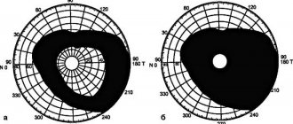
An important sign of glaucoma (narrowing of the visual field) usually begins on the nasal side.
How to check your field of vision yourself?
The outer boundary of the visual field of the right eye can be approximately determined as follows.
A person sits and looks at some point located directly at eye level. Covers the left eye with the left hand, extends the right hand forward, and aligns its thumb with the point that the right eye fixes. Then the outstretched right arm, without bending, moves towards the temple. The right eye continues to fixate the point located straight ahead. The lateral vision of the right eye monitors the position of the thumb of the right hand.
Next, the person notices the moment the finger disappears from the lateral field of vision. If the right hand at this moment is located at a right angle (90°) to the direction of gaze, then the outer border of the visual field is normal. If this angle is smaller, then the outer boundary of the field of view is narrowed.
The inner (nasal) border of the visual field can most often be disrupted with glaucoma.
To check this boundary of the visual field of the right eye, the starting position is the same. The outstretched right arm, without bending, is moved towards the nose. With lateral vision, they monitor the position of the thumb of the right hand and note the moment it disappears from the field of view.
Then the movement of the hand is stopped and the right eye is moved towards the nose. If at the same time the thumb of the right hand “disappears” behind the bridge of the nose and if its tip is visible, then the inner border of the field of vision is normal. If the finger emerging from behind the bridge of the nose is clearly visible and at a great distance from it, this means that the inner border of the field of vision is narrowed.
The left eye is closed with the index finger of the right hand through the eyelid. Four fingers of the palm of the left hand, folded together, are brought to the bridge of the nose. With the right eye in a straight position, with lateral vision they try to see the fingers on the bridge of the nose.
Dynamics of glaucoma
The dynamics of visual functions should be determined by systematic and long-term (at least six months) observation. In this case, dynamics can be of several types:
- Stabilized dynamics are characterized by the absence of changes in the visual field.
- The unstabilized dynamics of the developed stage is characterized by a narrowing of the field of view along individual radii by 5-10 degrees or more.
- Unstabilized dynamics of advanced glaucoma is accompanied by a narrowing of the visual field by 2-3 degrees.
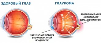
An important sign of the glaucomatous process is excavation of the optic nerve head. It occurs as a result of protrusion and expansion of the cribriform plate under the influence of intraocular hypertension. More often, this symptom appears in the later stages of glaucoma and is accompanied by atrophy of nerve fibers, as well as glial tissue.
When performing ophthalmoscopy, you can see that the retinal vessels have a sharp bend at the point of transition over the edge of the nerve disc. Sometimes they completely disappear beyond the edge of the excavation. It is necessary to distinguish pronounced physiological excavation from disc excavation due to glaucoma.
Another sign of glaucoma is swelling of the retina, which can be detected based on the size of the blind spot, which increases in size.
Classification of glaucoma - types and stages
Glaucoma is usually called a chronic eye disease, the course of which is accompanied by a triad of certain symptoms:
- Constant or periodic increase in IOP;
- Characteristic changes in the visual field;
- Marginal excavation of the optic nerve.
The most popular classification signs of this disease are the following.
By origin of glaucoma: primary and secondary
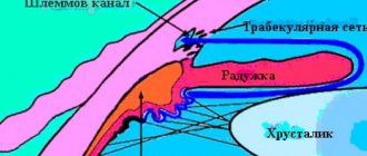
Primary glaucoma is characterized by strictly intraocular localization of pathological processes. They arise in the angle of the anterior chamber of the eye (AOC), its drainage system, or in the head of the optic nerve. Pathological processes precede the appearance of clinical symptoms, representing the first stage in the development of the disease.
In secondary glaucoma, both intracapsular and extraocular disorders can be its cause. This disease may be a side, optional consequence of other pathologies.
According to the mechanism of increasing IOP: open-angle and closed-angle
In open-angle glaucoma, progression of the pathological triad is noted with the presence of an open glaucoma.
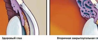
Angle-closure glaucoma is characterized by an internal block of the ocular drainage system, namely the overlap of the APC by the root of the iris, as the main pathogenetic link.
By IOP level: hypertensive and normotensive
With hypertensive, Pt is moderately increased (26 - 32 mmHg) or high (starting from 33 mmHg), and P0 is respectively (22-28 mmHg) and (starting from 29 mmHg).
With normotensive conditions, Pt values reach 25 mm Hg, and P0, respectively, reach 21 mm Hg.
According to the course of the disease: stabilized and unstabilized
With stabilized glaucoma, during a 6-month observation period of patients, deterioration in the state of the visual field, as well as the optic nerve head, was not detected.
Unstabilized glaucoma is characterized by deterioration in the visual field, as well as the optic nerve head, which are recorded during repeated examinations. Assessment of the dynamics of the glaucomatous process also includes the level of IOP with its correspondence to the “target pressure” in general.
According to the degree of changes in the optic nerve: initial, developed, advanced, terminal
The division of the glaucomatous continuous process into four stages is quite arbitrary. In the diagnosis, the stages are designated using Roman numerals (I-IV), where I is the initial stage and IV is the terminal stage (taking into account the state of the visual field, as well as the optic nerve head).
At the initial stage I, the boundaries of the visual field are normal, but minor changes (scotomas) in the paracentral parts of the visual field are detected. There is an extended excavation of the optic disc, not reaching its edge.
At the developed stage II - lesions of the visual field of the paracentral sections are pronounced and combined with its narrowing by more than 10 degrees in the superonasal and/or inferior nasal segments, extended excavation of the optic disc, in some sections it reaches its edge (it is of a marginal nature).
At the advanced stage III, the boundaries of the visual field are narrowed concentrically and in one segment or more, they are less than 15 degrees from the point of fixation. A marginal extended subtotal excavation of the optic disc is observed, reaching its edge.
At the terminal IV stage - complete loss of vision or preserved light perception with incorrect projection. Occasionally, the visual field is preserved by a small island in the temporal sector. Total excavation of the optic disc is observed.
By age of patients: congenital, infantile, juvenile, adult glaucoma
In congenital glaucoma, the defects are caused by the pathology of the development of the auricular system of the eye or the drainage system. It manifests itself during the first three years of a child’s life, due to recessive heredity (sporadic cases are possible). The pathogenesis of the disease is due to dysgenesis of the anterior chamber angle and increased IOP. Clinical symptoms include: photophobia, lacrimation, enlargement of the eye, blepharospasm, corneal edema with an increase in its size, excavation and atrophy of the optic disc.
Infantile glaucoma develops in children 3-10 years old, due to heredity and pathogenesis identical to simple congenital glaucoma, increased intraocular pressure is noted, without changes in the size of the cornea and eye, excavation of the optic disc increases as the disease progresses.
Juvenile glaucoma can occur between 11 and 35 years of age, caused by hereditary disorders in chromosome 1 and TIGR. The main role in the pathogenesis of the disease belongs to trabeculopathy and goniodysgenesis. There is an increase in IOP, changes in the optic disc and visual functions - according to the glaucomatous type.
The onset of glaucoma in adults is over 35 years of age. It is a chronic pathological process, with the pathological triad described above, without other eye diseases and congenital pathologies.
Currently, the classification of glaucoma is especially widely used, taking into account the form and stage of the disease, the state of IOP, as well as the dynamics of visual functions.
To shorten records, you can use alphabetic and numerical designations in the medical history, without indicating that the glaucoma is primary.
For example, the full diagnosis of the disease: “Primary angle-closure glaucoma, developed, unstabilized with moderately increased IOP,” in abbreviation will look like: “Angle-closure glaucoma, unstabilized IIB.” Or: “Angle-closure glaucoma IIB”, if there is little data on the dynamics of visual functions.
The full diagnosis record: “Primary open-angle glaucoma, not stabilized, advanced with normal IOP,” in an abbreviated version, looks like: “Unstabilized open-angle glaucoma IIIA.”
In recent years, the existing classification has been expanded by numerous varieties of forms of primary glaucoma, as well as an approximate assessment of the localization of the main resistance to the outflow of intraocular fluid from the eye.
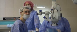
Our doctors who will preserve your vision with glaucoma:
An ophthalmologist deals with the diagnosis of the disease and postoperative care of patients.
Laser surgeon, main focus of work is modern laser methods for treating glaucoma.
In the medical department, everyone can undergo examination using the most modern diagnostic equipment, and based on the results, receive advice from a highly qualified specialist. We are open seven days a week and work daily from 9 a.m. to 9 p.m. Our specialists will help identify the cause of vision loss and provide competent treatment for identified pathologies.
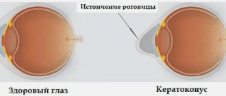
Various types of rings and segments are implanted into the MGC for keratoconus. You can find out the cost of various implants and their installation, and make an appointment at the Moscow Eye Clinic by calling a multi-channel phone (daily from 9:00 to 21:00, free for mobile phones and regions of the Russian Federation) or using the online registration form.
SIMILAR ARTICLES: Swelling of the eye glaucoma Medical examination of patients for glaucoma Differential diagnosis of secondary glaucoma Glaucoma of the optic nerve Cataract or glaucoma Surgery for glaucoma Fedorov Clinic
Clinical course
Depending on the clinical course of glaucoma, various stages of the disease are distinguished:
- Compensated glaucoma is accompanied by a normal level of intraocular pressure and stabilization of visual functions. This can be achieved with proper treatment and adherence to the regimen.
- Subcompensated glaucoma is accompanied by a slight increase in intraocular pressure. At the same time, its regulation is disrupted. There are signs of subcompensation, including pathological transformations of the elastocurve.
- Uncompensated glaucoma leads to severe dysregulation of ophthalmotonus. However, these violations do not reach an extreme degree.
- Decompensated glaucoma is an acute attack of the disease. At the same time, there is no regulation of ophthalmotonus and other important functions of the eye.
Due to the fact that glaucoma is the cause of blindness in a large number of the population in developed countries, this disease has important social significance. In 80% of cases, an open-angle form of the disease is diagnosed. Another feature is the inconspicuous onset, and therefore glaucoma is diagnosed already in a developed or advanced stage, when there are gross impairments of visual function. At these stages, it is quite difficult and sometimes impossible to achieve stabilization of the clinical course.
Prognosis and prevention
Glaucoma should not be left untreated as it will eventually lead to vision loss. Despite the mild form of the disease, asymptomatic in the initial stages, the visual cells die. If any manifestations of the disease are detected, you should immediately consult a doctor, since progression is inevitable, as is a constant increase in pressure inside the visual organs. The first stage without proper treatment develops into the second, the second into the third, and the third into loss of vision, the thermal stage.
With the help of modern equipment, it is impossible to forget about glaucoma forever, but it is quite possible to stop its development and preserve your vision for many years.
Consequences
Delayed treatment for glaucoma can lead to the following irreversible consequences:
- the appearance of scomots (a dark spot covers part of the object that the patient is trying to examine);
- tubular vision (peripheral vision is seriously impaired, color perception is reduced);
- neovascularization of the cornea (new vessels grow into the corneal tissue, which leads to deterioration of vision, in some cases to complete blindness);
- chronic swelling of the corneal tissue (characterized by decreased corneal transparency, inflammation of the conjunctiva, photophobia);
- cataract (the lens of the eye becomes partially or completely cloudy, stops transmitting light, vision deteriorates, even to the point of loss);
- blindness (the optic nerve atrophies, leading to loss of vision).
Self-medication for glaucoma is unacceptable, since it can only accelerate the development of the disease.