Dystrophic (associated with tissue nutrition disorders), degenerative changes in the cornea of the eye in some cases lead to severe deformation of this outer transparent protective layer. The cornea stretches forward, acquiring a peculiar pointed, conical shape. Accordingly, the refractive characteristics roughly change - the refraction of light with this shape excludes clear distinction of objects.
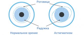
This condition is called “keratoconus” and is a serious ophthalmological pathology that requires timely, qualified intervention. The clarity of vision quickly decreases, various optical aberrations arise (distortion of the shape of visible objects, a halo effect around light sources, “rainbow” blurring of contours, doubling or multiple duplication of objects), the ability to binocular vision is lost; in some cases, pain develops and the cornea becomes cloudy. A thorough examination is necessary, including precise topological measurements of the geometry of keratoconus, biomicroscopy, tomography, ophthalmometry, etc. This condition does not develop in reverse, and to eliminate it, they resort to one or another, depending on the specific clinical picture, ophthalmic surgery (cross-linking, implantation of corneal rings, keratoplasty).
Keratoconus, fortunately, is a relatively rare disease: in the total volume of ophthalmological pathology, its share ranges from 0.01% to 0.6% of cases. It should be emphasized, however, that these estimates are approximate and ambiguous, since the incidence rate is influenced by a number of factors - for example, geographic or racial (there is statistical evidence that Mongoloids get sick more often). There is also no consensus on whether the risk of developing keratoconus depends on gender. Medical and statistical studies are hampered, in particular, by the difficulties of differential diagnosis in the early stages - for example, between the initial development of keratoconus and complex astigmatism.
It has been reliably established that the “start” of keratoconus deformity in most cases occurs at puberty, during the period of rapid puberty. Almost always (95% of cases) both eyes are affected. As for the nature of the development of the disease (possible options are slow growth, spasmodic course with periods of temporary improvement, spontaneous suspension, rapid progression of pathology, acute keratoconus), it is purely individual and differs even in the right and left eyes of the same patient.
Stage I (first degree)
- Myopia and a weak degree of myopic astigmatism are detected (possibly with a plus or zero sphere);
- K-index <= 48 D;
- no biomicroscopic signs;
- A “protrusion spot” or a characteristic pattern on computer topography is revealed - bow-tie;
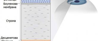
- Pachymetry readings are normal or slightly less than 500-550 microns.
Possible complications of operations
Despite the fact that techniques and equipment for surgery for keratoconus are being improved every year, complications may arise during their implementation, as with any medical procedure.
Their severity and frequency directly depend on the complexity and volume of the operation: for example, with collagen cross-linking, there are practically no negative consequences of the operation, which cannot be said with the most complex and extensive operation - penetrating keratoplasty (corneal transplant). In the latter case, rejection or clouding of the transplant, failure of the sutures, etc. are possible.
For a complete list of complications that may occur during or after surgery, please visit the specific surgery page.
Stage IV (grade 4)
- A very strong decrease in visual acuity, sometimes to the point of counting fingers in front of the face;
- Inability to measure refraction and perform computer topography;
- K-index > 55 D;
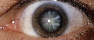
- Biomicroscopic signs: significant deformation of the cornea - a cone is clearly visible, thinning at the apex, opacities, local edema may be detected, fractures of the cornea, scars on it, Fleischner's ring, Vogt's striae may be detected;
- Pachymetry readings are less than 370 µm.
Keratoconus
Taking into account the nature of the course of keratoconus (speed of progression, tendency to relapse), treatment can be differentiated: non-surgical or surgical.
Conservative treatment of keratoconus consists of vision correction using semi-rigid lenses (hard in the center, soft in the periphery), which seem to press the cone of the cornea. In the initial stages, especially with a non-progressive, stable course of keratoconus, spectacle correction can also be effective. Courses of vitamin therapy, tissue therapy, immunomodulators and antioxidants are prescribed; eye drops (taurine), subconjunctival and parabulbar injections of ATP, methylethylpyridinol. For keratoconus, physiotherapy (magnetic therapy, phonophoresis with tocopherol, and other procedures) is effective.
With the development of acute keratoconus, emergency care is required: instillation of mydriatics (mesaton, midriacil, etc.) into the eye, application of a pressure bandage to the eye to prevent corneal perforation.
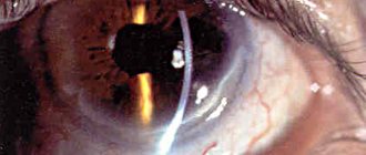
A relatively new, well-proven method of conservative treatment of keratoconus is corneal cross-linking, which consists of removing the surface epithelium from the cornea, instilling a riboflavin solution onto it and subsequent irradiation with UV rays. This procedure allows you to strengthen the cornea, increase its resistance to deformation, stop the development or achieve regression of keratoconus. After corneal cross-linking, conventional spectacle and contact correction with soft lenses becomes possible.
At the initial stage of keratoconus, with sufficient thickness of the cornea, it is possible to perform an excimer laser procedure (PRK + FTC), which allows to correct astigmatism, increase visual acuity, strengthen the anterior layers of the cornea and slow down the progression of keratectasia.
In some cases, in order to reduce corneal deformation, thermokeratoplasty is used - the application of precise applications to the periphery of the cornea with a coagulator, which makes it possible to achieve flattening of the cornea.
Keratoconus surgery uses the method of implanting corneal rings. Stromal (corneal) rings change the surface of the cornea, normalize refraction and stabilize the cornea.
The classic operation for keratoconus is penetrating or lamellar keratoplasty, which involves removing your own cornea and implanting a donor graft in its place. Keratoplasty is accompanied by almost 100% engraftment of the graft and allows you to correct visual acuity to 0.9-1.0 in approximately 90% of cases. Penetrating keratoplasty can be attempted even in end-stage keratoconus.
Prevention and prognosis
This disease often leads to the development of complications. However, if the disease is detected in a timely manner, the chances of a quick and successful cure are really high. The later keratoconus is detected, the more difficult the treatment becomes.
Due to the fact that the exact causes of the development of the disease have not yet been determined, clear preventive algorithms aimed at preventing such a disease have not been developed. The most effective means of prevention is considered to be regular visits to an ophthalmologist and careful attention to your body. At the moment, only timely diagnosis gives a significant chance of defeating the disease. It is also recommended to pay special attention to other ophthalmological diseases. Patients suffering from eye problems should visit a specialist more often than others.
People whose relatives have had problems with this disease should also regularly set aside time for preventive visits to the ophthalmologist.
Conservative treatment of keratoconus
It is useless to treat progressive chronic pathology. But at different times, doctors offered different methods. For example, in 1984, Professor Zinaida Davydovna Titarenko proposed the treatment of keratoconus using ultrasound therapy. But this only allowed for temporary relief of symptoms.
In 1998, Candidate of Medical Sciences Elena Nikolaevna Gorskova substantiated the theory of using immunomodulators, which should lead to an improvement in the biochemical parameters of tear fluid and stop the development of keratoconus. A positive effect was noted, but after some time the patients still required surgery.
Lenses for keratoconus are used only in the first two stages of pathology development. But they do not have a therapeutic effect, but only serve to improve vision. The disease will still continue to develop, and sooner or later surgery will be required.
One type of conservative therapy, which is sometimes still classified as minimally invasive surgery, is cross-linking. It consists of removing the top layer of the epithelium, saturating the cornea with riboflavin and exposure to ultraviolet radiation. All this causes a series of biochemical reactions that help strengthen collagen and thicken the cornea. The operation lasts no more than an hour, after which you need to wear therapeutic lenses for several days. The effect lasts 8-10 years, then keratoconus needs to be treated again.
Causes
Keratoconus is considered a relatively rare disease. A positive diagnosis is made in 0.1-0.6% of all cases of corneal deformation. The etiology of keratoconus remains a field of debate. The exact cause of the pathology has not yet been established, but several hypotheses have been put forward regarding the nature of the appearance of cone-shaped deformation of the cornea:
- hereditary (genetic);
- metabolic (metabolic disorder);
- endocrine (hormonal imbalances during puberty);
- immune (pathological reactions of the immune system).
The most likely causes of keratoconus are hereditary and metabolic. They are often combined when talking about a complex hereditary-metabolic pathology leading to the formation of the disease. This means that a predisposition to keratoconus is transmitted from parents to children and is activated during moments of restructuring of metabolic processes in the body. This hypothesis explains the characteristic age of onset of the disease and allows us to conclude that keratoconus is associated with changes in metabolism and hormonal levels.
The hereditary-metabolic theory is supported by the identified correlation with other autoimmune hereditary diseases, such as:
- eczema;
- hay fever (seasonal allergic rhinoconjunctivitis);
- atopic dermatitis;
- bronchial asthma.
A cone-shaped curvature may be a consequence of keratitis of traumatic or viral etiology. The development of keratoconus has also been recorded against the background of endocrine system disorders, for example, with Addison's disease. Patients with Down or Marfan syndrome may suffer from congenital curvature of the cornea.
Some of the unfavorable factors that contribute to the occurrence of keratoconus include:
- hyperactive exposure to ultraviolet radiation;
- the use of hormonal drugs, often glucocorticosteroids;
- radiation exposure;
- contaminated air.
Keratoconus can be a consequence (complication) of vision correction using excimer laser (LASIK) - iatrogenic keratoectasia.
Main signs of the disease
Knowledge of the main symptomatic signs of the disease can be of great help for the timely detection of an ophthalmological disorder. Any person should be attentive to their body. After all, any signals about the onset of a disease can and should be recognized. This will help prevent the occurrence of dangerous complications and increase the chances of successful treatment.
Keratoconus may present with the following symptoms:
- Gradual or sudden decrease in vision, first in one eye, later in the second eye.
- “Double image” syndrome - the patient is shown one object, but he sees many identical objects scattered chaotically.
- Distortion of the image, merging of characters, blurred contours.
- Severe photophobia.
- Painful sensations, rapid eye fatigue, “sand in the eyes” syndrome.
- Diffused light effect. For example, when looking at a bright light, a person suffering from keratoconus sees a bright or blurry aura around the point.
Indirectly, the presence of such a disease may be indicated by the failure of the effect of vision correction when wearing glasses or contact lenses.
If you discover the above or other signs of ophthalmological pathology, you must definitely visit a specialist. It is impossible to diagnose keratoconus on your own. Self-medication in cases of eye diseases does not bring positive results and poses serious risks.
Diagnostics
The first signs of keratoconus are detected during a standard ophthalmological vision test. Refractometry (a method for determining the refraction of light in the lens) reveals signs of astigmatism and myopia. Also held:
- keratopachymetry (determining the thickness of the cornea);
- photokeratometry (determining the radius of asymmetry of the anterior surface layer of the cornea);
- biomicroscopy of the eye (examination of different environments of the eye using a slit lamp);
- computer corneal topography;
- endothelial microscopy;
- optical coherence tomography.
Causes of keratoconus
The causes of this disease have not been precisely established to date. It is believed that risk factors for its development are hereditary factors, structural features of the cornea, and the adverse effects of certain environmental factors (increased background radiation, increased intensity of ultraviolet radiation from the sun).
In addition, experts believe that the causes of the development of keratoconus may be connective tissue pathology and endocrine diseases.
Symptoms
A patient with this disease who consults a doctor for the first time will complain of:
- deterioration of visual acuity;
- halos around light sources;
- rapid eye fatigue;
- increased sensitivity, irritation;
- duality in the eyes;
- three-dimensional letters when reading;
- itching and burning;
- Initially, visual acuity deteriorates in the evening, and then in daylight.
In acute keratoconus, the patient will complain of severe vision loss and severe eye pain. This type of disease lasts about three weeks, after which vision slowly improves. Acute keratoconus may be accompanied by corneal rupture and scarring.