Excludes: congenital cataract (Q12.0)
If it is necessary to identify the cause, use an additional external cause code (class XX).
Cataracts in chronic iridocyclitis
Secondary cataracts in eye diseases
Glaucomatous flecks (subcapsular)
If it is necessary to identify the drug that caused the lesion, use an additional external cause code (class XX).
ICD-10 text search
Search by ICD-10 code
Search by alphabet
- A
- B
- IN
- G
- D
- E
- AND
- Z
- AND
- TO
- L
- M
- N
- ABOUT
- P
- R
- WITH
- T
- U
- F
- X
- C
- H
- Sh
- SCH
- E
- YU
- I
ICD-10 classes
- I Certain infectious and parasitic diseases (A00-B99)
In Russia, the International Classification of Diseases
10th revision (
ICD-10
) was adopted as a single normative document for recording morbidity, reasons for the population’s visits to medical institutions of all departments, and causes of death.
ICD-10
introduced into healthcare practice throughout the Russian Federation in 1999 by order of the Russian Ministry of Health dated May 27, 1997. No. 170
The release of a new revision (ICD-11) is planned by WHO in 2021, 2018, 2022.
| Pharm. groups | Active substance | Trade names |
| Other Metabolics | Taurine* | Taurine |
| Taufon |
Official website of the RLS ® company. Home Encyclopedia of medicines and pharmaceutical assortment of goods of the Russian Internet. The directory of medicines Rlsnet.ru provides users with access to instructions, prices and descriptions of medicines, dietary supplements, medical products, medical devices and other goods. The pharmacological reference book includes information on the composition and form of release, pharmacological action, indications for use, contraindications, side effects, drug interactions, method of use of drugs, pharmaceutical companies. The medicinal reference book contains prices for medicines and pharmaceutical market products in Moscow and other cities of Russia.
Transfer, copying, distribution of information without the permission of RLS-Patent LLC is prohibited.
When quoting information materials published on the pages of the website www.rlsnet.ru, a link to the source of information is required.
Lots more interesting things
© REGISTER OF MEDICINES OF RUSSIA ® RLS ®, 2000-2020.
All rights reserved.
Commercial use of materials is not permitted.
The information is intended for medical professionals.
ICD-10 code H26
You are here: Home > ICD-10 > H00-H59 > H25-H28
ICD-10 code H26 for Other cataracts
Excludes: congenital cataract (Q12.0)
ICD-10
ICD-10-CM 10th Revision 2016
ICD-10-GM ICD-10 in Germany
ICD-10 ICD-10 in Russian
ICD-10 International Statistical Classification of Diseases and Related Health Problems 10th Revision
ICD-10
ICD-10 is the 10th revision of the International Statistical Classification of Diseases and Related Health Problems (ICD), a medical classification list by the World Health Organization (WHO).
It contains codes for diseases, signs and symptoms, abnormal findings, complaints, social circumstances, and external causes of injury or diseases.
What is a traumatic cataract?
Cataract is a disease characterized by clouding of the lens (natural lens) of the eye. The disease is one of the most common ophthalmological pathologies, so the problem is constantly being studied, the causes and methods of treating cataracts are determined. One of the causes of the pathology is a wound or contusion of the organ of vision, resulting in the development of traumatic cataracts of the eye.
More than 70% of patients risk going blind in one or both eyes due to the rapid development of the pathology. Therefore, if you suspect eye problems, you should immediately contact an ophthalmologist.
Key Facts
Cataracts of the eye - what kind of disease is it? Sooner or later, every person comes to this question. And this is not surprising. Cataract, like glaucoma, is an age-related disease; its appearance and development is caused by age-related changes that occur in the body and affect all organs and systems, including the visual one. Cataract is not contagious, it is not an infectious disease, it is not transmitted by contact or airborne droplets. In simple terms, cataracts are clouding of the lens.
Ophthalmologists consider lens clouding to be a natural process, so cataracts cannot be fully considered a disease. It is rather a condition of the eye apparatus, which, however, requires specialized medical care and observation by an ophthalmologist. The only method that can lead to a complete cure is replacing the lens of the eye for cataracts.
The risk group for cataracts includes all people over 60 years of age. With each passing year, the likelihood of illness increases, especially with diseases such as diabetes and hypertension. In addition to cataracts, these diseases lead to deterioration of the retina and decreased visual acuity, and are sometimes accompanied by glaucoma. Often, in older patients, glaucoma and cataracts are diagnosed at the same time, and this increases the likelihood of vision loss.
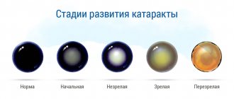
Cataract develops gradually; a number of stages are conventionally distinguished, characterized by a certain set of symptoms and signs of changes in the tissues of the lens. Doctors call the development of the disease “maturation of cataracts.” The duration of the process depends on the cause, age and other circumstances, and can last from several months to 10-15 years.
In the initial stage of cataract, the manifestations are mild, vision practically does not deteriorate.
In the stage of immature cataracts, there are areas of clouding of the lens, which affects visual acuity.
Mature cataracts are characterized by significant visual impairment and complete clouding of the lens.
At the last stage of overripe cataracts, deformation of the lens and loss of vision occurs.
Disease code according to ICD-10
ICD-10 is a regulatory document, the full name of which is the International Classification of Diseases, revised and supplemented for the tenth time.
The purpose of this classification is to provide everyone with access to information about diseases, their symptoms, treatment methods, and possible consequences.
As for traumatic cataracts, according to ICD-10 this disease is assigned code H26.1. Information about the disease is located in grade 7, which contains eye diseases, in block H25-H28, which corresponds to diseases of the lens.
As with other pathologies, the ICD provides information about treatment methods for people with traumatic cataracts. According to information from this document, there are drugs that can stop the development of the disease and allow you to live without surgery for many years without loss of vision.
Cataract: surgery, photo, treatment and prevention of the disease
Cataract is a violation of the transparency (partial or absolute) of the lens. Relatively often observed in people over 45 years of age. Characterized by a decrease in visual acuity from the periphery to the center.
ICD-10 code: Lens diseases (H25-H28)
What types of cataracts are there?
Cataracts are classified by location:
Beginning
The malaise develops unnoticed by the patient. The patient complains of blurred vision, double vision of burning objects (moon, car headlights), and spots. When examining the eye, with a dilated pupil, spoke-shaped gray opacities are noticeable against its black background.
The tops of the opacities are directed towards the center, and the bases - towards the periphery. Peripheral lens opacities do not impair visual acuity. Because of this, this period is asymptomatic, and the patient does not observe any deterioration in vision.
This phase lasts from a month to several years.
Immature (swelling)
At this stage, patients complain of an unexpected decrease in visual acuity. When examining the patient's eye, it is noticeable that the lens is enlarged, thickened and located in the area of the pupil, and has a gray-white color with a pearlescent tint. Nevertheless, in some places the lens itself still retains transparency, which is why, in side lighting, the presence of a moon-shaped shadow falling from the iris onto the cloudy layers of the lens is acceptable.
This stage can last for a long time, and then move on to the next one.
Mature
At this stage, complete diffuse clouding of the lens occurs. The patient does not see objects at all and is only able to recognize the direction of light sources, since light perception remains. At this stage, surgical interventions are performed. However, if the lens is not removed, the cataract progresses from this stage to the next.
Overripe
The dense cortex of the lens slowly liquefies and turns into a milky mass, within which the base of the lens floats.
The lens shrinks, the anterior chamber of the eye deepens, and iris trembling occurs. The patient's vision is 0%.
These processes are irreversible. At this stage, complications may arise: increased intraocular pressure, rupture of the lens capsule.
Causes
The causes of this disease can be: diabetes mellitus, high myopia, occupational diseases, radiation therapy.
Diagnostics
Diagnosis of cataracts consists of two main methods:
- Determination of visual acuity.
- Ophthalmoscopy in direct and lateral projections.
Diagnosis of cataracts using ophthalmoscopy
Treatment
In case of cataracts, it is necessary to promptly refer the patient for surgical removal of the lens and replacement with an artificial lens. However, treatment is different at each stage.
At the beginning and immature stage, conservative therapy is carried out (eye drops: Quinax, Catachrom). They prescribe 1-2 drops 2 times a day for a long time.
Eye drops taken for initial symptoms of cataracts
In the mature stage of cataract, surgery is actually performed to remove the lens and install an artificial lens in its place. The operation is carried out at the given stage, since in the overripe stage there is a high risk of complications.
Treatment of cataracts using eye lens surgery
At an overripe stage of cataract, open cataract extraction or deep enucleation of the eye is performed.
Surgery for cataracts
Forecast
The prognosis after timely lens replacement operations is extremely favorable for the patient. The patient noted improvement in vision from the first day. Visual acuity is restored within 15 days. Further, if necessary, for better visibility, you may choose glasses.
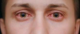
However, during this time, in order to eliminate possible complications, the patient must protect his eyes from possible injuries, avoid infectious diseases and sports. In case of redness of the mucous membrane of the eye, an emergency visit to an ophthalmologist.
Complications
It is rare for a patient to experience an acute attack of glaucoma, which is accompanied by pain in the eye and head, nausea and sometimes vomiting, even to the point of loss of consciousness.
In addition, general complications are likely:
- hypotension;
- hypertensive crisis;
- cerebrovascular disorder;
- acute urinary retention;
- mental disorders;
- other emergency conditions.
If they occur, you should immediately seek help from an ophthalmologist.
Prevention
To prevent cataracts, you need to take a course of vitamins A, E, B, P 2 times a year. Also avoid eye injuries, wear sunglasses both on sunny and foggy days, and follow a protein diet.
Cataracts are a relatively common occurrence in people over 45 years of age. And with modern medicine, lens replacement operations are performed by ophthalmologists in 15 minutes, and generally do not have any complications. The patient can go home 2 hours after the operation.
Although this disease is dangerous, it has extremely favorable results. As a result of the operation, patients can regain 80% of their vision. Which is quite good, considering that before the operation the vision was 0%.
SEE ALSO: Cataract lens What cataract or glaucoma cannot be treated What tests need to be taken before cataract surgery Timing for cataract What is glaucoma and cataract what is worse
Types of post-traumatic cataracts
Depending on the type of injury received, after which cataracts develop, there are several types of cataracts.
Types of post-traumatic cataract depending on its cause:
- contusion - the disease appears as a result of blunt trauma to the eye;
- wound - if a penetrating injury to the eye occurs, this can also lead to rapidly progressing eye pathology;
- chemical - occurs as a result of toxic substances getting into the eyes or the body as a whole;
- industrial - eye injury in the workplace - a common occurrence for welders and people working in hot shops;
- radiation - can occur after a high dose of radiation exposure.
Symptoms of the disease
In order not to miss time and get timely medical help, it is important to know the main signs of traumatic cataracts.
- decreased quality of vision, especially in the dark;
- Non-existent dots and stripes appear before your eyes;
- photophobia, especially concerned about intolerance to bright light;
- problems with performing actions that require special concentration (reading, embroidery);
- lack of perception of certain colors;
- double vision, blurred vision;
- the color of the pupil changes from black to gray, sometimes almost white.
With any of these signs, the patient should have no doubt about how to act in case of traumatic cataract - the only correct decision would be to consult an ophthalmologist.
Signs of complicated cataracts
Signs of complicated cataracts include:
- The affected pupil acquires a milky color. This external manifestation is explained by the accumulation of infiltrate in this area, reminiscent of pumice.
- Dull colors of the surrounding world.
- Fear of light.
- "Drops" on the eyes.
- Halos around illuminated objects.
- Vision cannot be corrected by known methods.
- Blurred vision. This means that infiltrate accumulates in the cornea. As the disease progresses, the amount of infiltration increases, vision becomes increasingly poor and, without treatment, may disappear altogether.
- Bifurcation of visible objects does not always appear, as it is formed due to destruction of the lens and retinal separation under the influence of calcified salts.
- Other visual impairments. Each person has their own individual symptoms of the disease. Some already at the initial stage lose the ability to read, as printed text becomes unclear, some complain of decreased visual acuity in low-light conditions indoors.
Symptoms of diabetes, myopia, glaucoma
In diabetes mellitus, the disease progresses rapidly. Within a few months, the patient may develop complete blindness. In this case, a patient with diabetic cataracts consults doctors with the following symptoms: “spots” in the eyes, a feeling of a veil before the eyes, reduced visual acuity in the dark.
READ MORE: Secondary cataract laser treatment
If a patient has myopia, the symptoms of cataracts in its early stages may be confused with their usual condition. Typically, patients with myopia seek help from specialists when the clouding of the lens has already become visible.
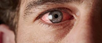
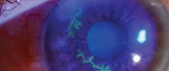
With glaucoma, the manifestations of cataracts are more noticeable, but the development of the latter is characterized by increased values of intraocular pressure and the accumulation of opacities, mainly in the immediate vicinity of the posterior capsule.
Photo 1. An eye with cataract disease due to advanced glaucoma. The iris is cloudy, bluish in color.
Therapy for a complicated disease differs from the methods used to treat the usual type of cataract.
Clinical picture
• General symptoms •• Painless progressive decrease in visual acuity •• Fog before the eyes, distortion of the shape of objects •• Ophthalmological examination reveals clouding of the lens of varying severity and localization.
Establishing diagnosis
After seeking medical help, the primary task of doctors is to confirm or refute the alleged diagnosis.
Methods for diagnosing traumatic cataracts:
- interviewing the patient - the doctor must understand what incident preceded the onset of the disease;
- studying the anamnesis - to make sure that the cataract is of an acquired traumatic nature, the doctor must exclude other possible causes of the pathology;
- ophthalmoscopy - examination of the fundus using a slit lamp;
- ultrasound scanning;
- inspection in transmitted light;
- visometry - measurement of visual acuity;
- biomicroscopic method - study of the structure of the eyes;
- perimetry - study of the visual field;
- tonometry - measurement of IOP (intraocular pressure);
- phosphene - recognition of the electrical sensitivity of the retina.
After making an accurate diagnosis, treatment will be prescribed, which almost always involves surgery.
Initial senile cataract
ICD-10 Category: H25.0
Content
Definition and general information [edit]
Senile (senile) cataracts are the most common form of the disease, having a significant share among cataracts of various origins.
Lens opacities can begin from the cortex (cortical cataract), from the nucleus (nuclear cataract), or subcapsularly (subcapsular cataract). Each of these species differs from each other not only clinically, but also in morphological and biochemical structure.
Etiology and pathogenesis[edit]
Clinical manifestations[edit]
For cortical cataracts
the first signs of opacification appear in the cortex of the lens at the equator. The central part remains transparent for a long time, so visual acuity does not change. Patients' complaints boil down to the appearance of "floaters", spots, diplopia or polyopia, when the patient sees several objects with one eye, especially luminous ones. The condition of initial cataract lasts differently for different people: for some this period is calculated in decades, for others the process progresses, and after 1-3 years the stage of immature or swelling cataract begins. In this case, the clouding covers almost the entire cortex of the lens, and therefore patients complain of a sharp decrease in vision. Swelling of the lens can lead to a narrowing of the auricle, which impedes the outflow of intraocular fluid and sometimes threatens the occurrence of ocular hypertension.
Subcapsular cataract
characterized by early and intense vision loss. For this type of cataract, hydration is specific in the form of subcapsular vacuoles, which are located mainly under the anterior capsule, sometimes occupying the entire capsular zone. In cases of subcapsular cataract localization under the posterior capsule, differential diagnosis between posterior senile subcapsular and complicated cataract is necessary. As cataracts progress, opacities spread towards the equatorial part of the lens. At this stage, the subcapsular cataract becomes cup-shaped. It is distinguished from complicated cataract by the fact that opacities and vacuoles (in the initial stage) are located in 1-2 subcapsular layers and are sharply delimited from the transparent cortex lying in front of them. With this type of cataract, visual acuity begins to decrease early.
Initial senile cataract: Diagnosis[edit]
To detect cataracts, the patient must be examined with a wide pupil using transmitted light. At the same time, against the background of the pink glow of the pupil, the opacities in the lens appear black, since some of the reflected rays are absorbed by the clouded fibers of the lens.
During a biomicroscopic examination using a slit lamp, the localization of foci of opacities, their prevalence and the degree of disintegration of the lens fibers are clarified.
When examining a patient with cataracts, the following is necessary:
• find out the etiology of the process;
• exclude other diseases of the organ of vision that may give a similar picture of the disease;
• diagnose the degree of maturity of cataracts;
• develop treatment tactics and determine the prognosis of visual functions in the early stages and in the long-term period of observation.
Blurred vision followed by decreased vision are typical symptoms of lens opacity. However, this symptomatology is also characteristic of other diseases: pathology of the retina, vitreous body.
Instrumental research methods
When establishing a primary diagnosis, all patients must:
- determination of visual acuity;
- other research methods (electrophysiological methods for studying the retina and optic nerve, ultrasound, endothelial microscopy, ultrasound biomicroscopy, tonography, FA, determination of retinal visual acuity, immunological methods, etc.) are used in accordance with the specific therapeutic and diagnostic situation.
Visual acuity
examined without correction, as well as using trial spherical and cylindrical lenses, thus determining the degree of clinical refraction and astigmatism in a subjective way. With cataracts, due to clouding of the optical system of the eye, visual acuity decreases, including light perception. In indications for surgical intervention that restores vision to a patient, the presence of correct projection of light is of great importance as a sign of preserved function of the visual-nervous system.
Peripheral vision
complements the central one with the ability to navigate in space and its functional activities at dusk and at night. In case of cataracts, in conditions of opaque media and the impossibility of ophthalmoscopy, perimetry helps to establish the localization of the painful focus in the eye, pathways, and nerve centers. In the absence of pathology of the visual analyzer due to cataract, the boundaries of the visual fields are normal.
Determination of objective refraction
and optical power of the cornea allows you to evaluate the refraction of the eye and the ability to calculate the optical power of the IOL.
Tonometry
indicates normal hydrodynamics or its change due to lens pathology.
Transmitted light examination or ophthalmoscopy allows you to determine the transparency of the refractive media of the eye, and examination of the fundus (if ophthalmoscopy is possible) allows you to assess the condition of the optic nerve, vascular system, central and peripheral parts of the retina.
Biomicroscopy
- an objective method that allows you to diagnose pathological changes and scars of the cornea, determine the depth of the anterior chamber and its contents, the size, size and shape of the pupil, as well as the degree of its reaction to light and mydriatics, the degree of iris dystrophy and the presence of exfoliations. Particular attention is paid to the nature of changes in the lens: the state of the anterior and posterior capsules of the lens, the size, degree of intensity and localization of opacities.
Using ultrasound biometrics
determine the length of the eye, the depth of the anterior chamber and the thickness of the natural lens. The data obtained are used not only to objectively characterize the parameters of the eyeball, but also to calculate the optical power of the IOL.
The condition of the membranes of the eye, the macular area, the presence of posterior vitreous detachment is assessed using ultrasound B-scanning
. The degree of vitreal destruction is determined based on the detection of single or multiple intravitreal inclusions and determination of their acoustic density.
In order to objectively characterize the condition of the retina and optic nerve, and predict visual functions, if there is a suspicion of concomitant pathology of the optic nerve analyzer, laser retinometry
and a comprehensive
electrophysiological study
(registration of sensitivity and lability of the visual analyzer, registration of ERG, electrooculogram and visual evoked potentials of the cerebral cortex). The obtained data are compared with the norm developed for various degrees of lens opacity in relation to all studied parameters.
Examination of the posterior corneal epithelium ( endothelial microscopy
) is carried out if there is a suspicion of a significant decrease in the number of cells of the posterior corneal epithelium.
A comprehensive ophthalmological examination for cataracts shows that, in addition to the lens, other structures of the eyeball may be involved in the pathological process: cornea, ligamentous apparatus, ST, vascular system, retina and optic nerve. Therefore, the volume and nature of cataract surgery depend not only on the initial state of the lens, but also on the pathology of other eye structures accompanying cataract, as well as the whole organism.
General examination
patients are aimed at detecting possible foci of infection, primarily in organs and tissues located near the eye. Before surgery, it is necessary to sanitize foci of inflammation of any location. Particular attention is paid to the sanitation of the oral cavity, nasopharynx and paranasal sinuses.
Blood and urine tests, electrocardiography and x-ray examination of the lungs help detect diseases that require emergency or planned treatment.
Differential diagnosis[edit]
Initial senile cataract: Treatment[edit]
Treatment of cataracts in the initial stage of the disease is carried out conservatively to prevent rapid clouding of the entire substance of the lens.
Rational ways to solve the problem of cataracts are not only the improvement of ophthalmic surgery, but also the development of conservative methods of treating this disease. For this purpose, instillations of drugs that improve metabolic processes are prescribed. These drugs contain cysteine, ascorbic acid, glutamine and other ingredients. At the same time, the very slow development of clouding in the lens in senile cataracts, which often lasts for years and even decades, makes it extremely difficult to truly assess the effectiveness of their conservative treatment. And yet, delaying the development of cataracts by 10 years using conservative methods can reduce the number of expensive extractions by 50%.
The most widely used drugs are:
• Oftan Katahrom - improves oxidative and energetic processes in the lens. The drug is used mainly for posterior subcapsular and cup-shaped lens opacities.
• Azapentacene (quinax) - inhibits the formation of quinone compounds and acts on the proteins of the lens during opacities in its various layers. Prescribed for senile cataracts.
• Taurine (taufone) is an amino acid containing sulfur. Helps improve energy processes in the lens and other tissues of the eye. Stimulates reparative processes. Used for senile, diabetic, myopic, radiation and other cataracts.
• Riboflavin (vitamin B2) - used for various types of cataracts.
• Vita-Iodurol - contains a number of active substances (calcium chloride, magnesium chloride, nicotinic acid, adenosine). Used for senile, myopic and contusion cataracts.
Drug treatment for cataracts is justified only for incipient opacities in the lens and is completely unfounded for already developed cataracts of various etiologies.
The indication for surgical treatment of cataracts is a decrease in visual acuity, leading to limited ability to work and discomfort in everyday life. The degree of cataract maturity does not matter when determining the indications for its removal.
A summary assessment of the clinical and functional study of patients with cataracts allows us to resolve certain strategic issues in the planned surgical treatment at the preoperative level.
• Detect risk factors for surgical and postoperative complications and carry out preventive measures in the preoperative period:
-carry out drug correction with angioprotectors in patients with severe hemodynamic and microcirculation disorders;
-conduct specific and immunocorrective therapy in patients with complicated cataracts in the presence of impaired immune status with mandatory monitoring after treatment;
- in case of hydrodynamic disturbances in patients with complicated cataracts (glaucoma, trauma, uveitis), not compensated by medication when using a minimal miotic regimen, carry out preliminary surgical treatment with mandatory subsequent monitoring of hydrodynamics and IOP;
- in the presence of dystrophic changes in the periphery of the retina or DR (if ophthalmoscopy is possible), conduct preliminary laser treatment for patients with complicated cataracts against the background of high myopia, diabetes, or trauma.
• Justify the choice of cataract removal method, the advisability of additional surgical interventions in order to prevent complications and create favorable conditions for surgery:
-if optimal conditions are available or possible, plan energy technologies for cataract extraction (phacoemulsification and/or laser extraction);
- for overripe cataracts, as well as low initial density and qualitative changes in endothelial cells, pseudoexfoliation syndrome and lens subluxation, use a combination of viscoelastics;
- to prevent rupture of the posterior capsule of the lens and prolapse of the vitreous during surgery, as well as to prevent postoperative complications in patients with pseudoexfoliation syndrome, degree I-II lens subluxation, plan implantation of an intracapsular ring during surgery.
• Predict the functional results of the operation and, based on a summary assessment of clinical, functional, immunological and electrophysiological research methods, determine indications and contraindications for intraocular correction of aphakia.
RELATED ARTICLES: ICD age-related cataract Cataract diseases of the optic nerve Prevention of the postoperative period of cataract What is recurrent cataract and what to do
Drug treatment
Treatment of traumatic cataracts using eye drops and drugs taken orally cannot guarantee complete relief from the pathology. Such treatment is permissible only in the early stages of the disease or is used during the selection of the optimal method of surgical intervention, as well as in the case of the patient’s categorical refusal to undergo surgery.
Some medications used to slow the progression of cataracts include the following:
- "Quinax";
- "Oftan-Katakhrom";
- "Taufon";
- "Vicein";
- "Vita-Iodurol".
ethnoscience
There are also traditional ways to stop the rapid development of traumatic cataracts.
Recipes against cataracts:
- Infusing potato sprouts with vodka. Prepare the composition at the rate of 5-6 tablespoons of sprouts per 0.5 liter of vodka. Leave in a dark place for 2 weeks. Use the product three times a day, 1 spoon for 3 months.
- A mixture of walnuts and sunflower oil. The crushed kernels are poured with oil in a ratio of 1:10. Let it brew for 5-7 days. Instill 2 drops into the affected eye 3 times a day.
- An infusion of calendula flowers (15 g per 0.5 liter of boiling water) can be consumed orally or washed out the eyes.
- Blueberry juice, diluted with water 1:2, is instilled into the eyes once a day, the duration of therapy is at least a month.
- Honey diluted with water (1:3) brings positive results when dropped drop by drop into the eyes for 30 days.
Both aloe juice and tincture of peony leaves are used to combat cataracts; positive dynamics are often noted when consuming certain foods (for example, buckwheat). But it must be remembered that the main type of treatment for traumatic cataracts is surgery, and long-term self-medication with folk remedies makes the prognosis of this disease unfavorable.
Surgery
In modern medicine, phacoemulsification is used - a type of surgical intervention in which the affected lens is removed and an artificial lens is placed in its place, fully performing its functions.
The advantages of this type of operation:
- minimal trauma;
- seamless implementation (the micro-incision closes independently);
- carried out in 1 day;
- carried out with a minimum of anesthesia (local anesthesia is used).
Modern technologies make it possible not only to get rid of traumatic cataracts, but also to eliminate glaucoma at the same time. After replacing the damaged lens, it is permissible to undergo laser vision correction to completely restore the quality of life.
Surgery is the only method that allows you to completely get rid of traumatic cataracts, in contrast to drug treatment, which only inhibits its development.
What to do?
Before prescribing removal of a complicated cataract, your doctor may first recommend milder methods of correcting the condition. Glasses are prescribed or lenses are selected in such a way as to ensure good vision. The doctor will tell you how often you need to come for follow-up examinations - this will depend on the stage of the disease and the degree and speed of its progress. If vision deteriorates sharply and severely, eye surgery is indicated. The specific type of intervention is determined by assessing the characteristics of the case. Some patients require surgery that affects only the lens and cornea; sometimes more extensive interventions are needed. If the root cause of the pathological condition is not eliminated, the ability to see will not be completely restored.
Many patients are indicated for lens removal surgery. Instead, a lens made of artificial material is installed. After the intervention, a recovery period is required. Rehabilitation requires careful adherence to medical instructions. If the patient neglects these tips, there is a high probability of complications and failure of the entire treatment as a whole.
Recovery after surgery
Many patients refuse cataract surgery because they fear the disease will recur. This is a false opinion - an artificial implant is installed once, there are no problems with it.
In addition, the very next day after the operation the patient can return to a full life (reading, sewing, watching TV, working on the computer).
The only recommendation for people with removed cataracts is regular examinations by an ophthalmologist to exclude the occurrence of retinal pathologies.