Macular edema was first described by ophthalmologist S.R. Irwin in 1953 as a complication after cataract surgery. The macula is the central part of the retina. Its swelling is also referred to as cystoid macular edema and Irving-Gass syndrome.
The macula area has a diameter of only 5 mm, but it is responsible for central vision. Any organic and functional disorders of the macula significantly affect visual perception, since they cause the loss of part of the visual field directly in front of the eyes.
Causes of macular edema
How does macular edema occur?
The cause of the problem is increased permeability of the vascular walls. As a result, fluid from the bloodstream enters the intercellular space. The retinal tissue in the macular area increases in volume, which interferes with the full functioning of the visual receptors.
Fundus picture with macula area indicated
Chronic diseases of other organs and systems
A common cause of macular edema is diabetes mellitus. Elevated glucose levels contribute to damage to the vascular wall (angiopathy develops). This creates favorable conditions for fluid to leak from the bloodstream into the retinal tissue. In addition, in diabetes, newly formed vessels grow into the retina, the walls of which are initially defective and permeable.
Diabetic macular edema, as a complication of diabetes, often develops when there is insufficient control over the increase in blood glucose levels and the disease is in the stage of decompensation.
Intraocular inflammation
- Uveitis – different types of inflammation of the uvea of the eye
- Cytomegalovirus retinitis is an inflammatory process in the retina caused by a viral pathogen.
- Scleritis is an inflammation of the outer layer of the eye.
Vascular problems
- Retinal vein thrombosis
- Large aneurysm (limited expansion) of the central retinal artery
- Vasculitis (genetically determined inflammatory processes in the walls of blood vessels)
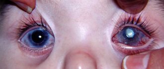
Eye surgeries
Macular edema can occur both after complex and extensive manipulations and after low-traumatic surgical interventions:
- Cataract removal with artificial lens installation
- Laser coagulation and cryocoagulation of the retina
- Laser capsulotomy
- Through corneal plastic surgery (keratoplasty)
- Scleroplasty
- Surgeries to improve fluid outflow in glaucoma
Postoperative complications causing macular edema most often resolve spontaneously and without consequences.
Eyeball injuries
Against the background of eye contusion, microcirculation disturbances in the retina may occur, leading to the development of edema. After penetrating wounds of the eye, edema can develop both against the background of injury and as a complication of surgical treatment.
Side effects of some medications
This condition is also known as toxic maculopathy. For example, macular edema can be caused by drugs based on prostaglandins (some drops for glaucoma), the vitamin niacin (better known as niacin), certain drugs for diabetes (based on rosiglitazone) and immunosuppressants (with the active ingredient fingolimod). Therefore, remember the medications you take in order to answer your doctor’s questions in more detail and quickly determine the cause of the problem.
Other intraocular pathologies
- Hereditary (retinitis pigmentosa)
- Acquired (epiretinal membrane, the presence of strands between the vitreous body and the macula, which can provoke edema and subsequent retinal detachment)
- Age-related macular degeneration (degeneration) of the retina
- Central serous chorioretinopathy (CSC)
Macula with retinitis pigmentosa
Exposure to radiation
Macular edema can be a complication of radiation therapy for cancer.
When and why does it occur?
It is important to understand that macular edema is not an independent disease; it is always a consequence and symptom of a more general pathological process or condition. The most likely and common of these reasons include:
- abnormal development of the clinical picture after cataract removal;
- retinal damage in diabetes mellitus;
- thrombosis of the blood vessels supplying the retina (usually the central vein);
- chronic inflammatory processes (for example, uevitis) and other diseases of the vascular system of the eye;
- systemic autoimmune diseases (especially collagenosis);
- aphakic glaucoma;
- oncopathology;
- focal retinal detachment;
- hereditary pathology of the retina (retinitis pigmentosa);
- intoxication;
- vascular diseases.
Any swelling, by definition, means that the tissue is saturated with excess fluid. If we are talking about such a vulnerable and highly specialized tissue as the substance of the retina (and the foveal zone of the retina is very thin and almost entirely consists of light receptors), then the consequences of edema can be - and often turn out to be - very serious. However, to date, the mechanisms of development of macular edema have not been fully studied and require further research. Each of the two main competing hypotheses, inflammatory and hypoxic, explains this pathological phenomenon only partially.
Diabetic macular edema (DME) is a particular problem in these cases, a distinction is made between diffuse (spreads to the foveal zone, leading to a pronounced thickening of the retinal layer) and focal (the central part of the macula is not affected, the thickening does not exceed two diameters of the optic nerve head).
In the case of central retinal vein thrombosis (CRV), so-called post-thrombotic macular edema (PTME) occurs, the cause of which is a violation of the blood-retinal barrier due to increased activity of vascular endothelial growth factor (VEGF).
Macular edema. Symptoms
- A cloudy spot that makes it difficult to see some details of the image before your eyes
- There are areas of distortion and blurred lines in the field of view
- The image in front of the affected eye may take on a pinkish tint.
- Decreased visual acuity at distance and near
- There is a cyclical pattern in the decrease in visual acuity - often the condition is worse in the morning
- Increased photosensitivity
Symptoms of DME
Diabetic macular edema is manifested by the following symptoms:
- Blurred image in the central field of view;
- Image distortion (straight lines look curved, wavy);
- Increased sensitivity to light.
Patients focus on the appearance of a pink tint to the image. They note a decrease in visual acuity at certain times of the day, often in the morning. The perception of color can change throughout the day.
Fig. 2 Vision of a sick person with macular edema
Diagnostics
The specialist makes a diagnosis after assessing the total information obtained from interviewing the patient and conducting all necessary examinations.
Interview and visual acuity testing (visometry)
The doctor may think about macular edema if you have:
- Characteristic combination of complaints
- Data on concomitant diseases that could become the basis for the development of edema (diabetes mellitus, etc.)
- Decreased visual acuity that cannot be corrected with glasses.
Visual field check
A feature of macular edema is the deterioration of central vision while maintaining normal peripheral vision. There are various techniques that a doctor can use to identify central vision problems. The most informative method is computer perimetry. With its help, areas of decreased clarity of central vision are identified, which are called central scotomas. The characteristic location of such a scotoma may indicate damage to the macula area.
Fundus examination
The condition of the macula is visually assessed using ophthalmoscopy and examination with a fundus lens. The first method allows you to get a general idea of the condition of the retina, the second - using a special lens and high magnification at the slit lamp - is suitable for a more detailed examination. Before the examination, the doctor will drop drops that dilate the pupil to achieve a better view of the macula.
FA (fluorescein angiography)
A technique that, using a special dye, identifies the area where fluid leaves the bloodstream due to increased permeability of the vessel wall. FA reveals the location of accumulation of this fluid in the retinal tissue, that is, it allows you to see the edema, its size and boundaries.
OCT or optical coherence tomography.
This method allows you to “scan” the retina, determining the thickness of all its layers, including in the macular area. OCT and FA provide the most information for diagnosing macular edema.
This is what an OCT image looks like
Macular edema on OCT
Causes
The main root causes provoking a pathological condition in the center of the retina are:
- retinopathy;
- thrombosis of the central retina and its branches;
- due to surgery;
- uveitis;
- retinal detachment;
- injuries to the visual apparatus;
- formations inside the eye cavity;
- glaucoma;
- intoxication with toxic substances;
- violation of venous outflow.
Not only ophthalmological problems lead to macular pathology, but also pathological conditions:
- infectious pathologies;
- arterial hypertension;
- tuberculosis infection;
- disturbances in kidney function;
- toxoplasmosis;
- diseases of the circulatory system;
- immunodeficiency;
- atherosclerosis;
- allergic manifestations;
- brain pathologies.
Macular edema. Treatment
The main goal is to stabilize visual function by eliminating increased vascular permeability. The treatment plan will depend on the cause of the swelling and its severity.
Medicines
Dosage forms that can be used are eye drops, tablets, products for intravenous and intramuscular injections. Anti-inflammatory medications, diuretics (diuretics), and medications that improve microcirculation are used. If macular edema is caused by the progression of a chronic disease, treatment is prescribed to improve control of the disease or stop further deterioration. The drug that itself caused the edema is discontinued or replaced with another.
Intravitreal injections
If in a particular case a more powerful therapeutic effect is required, they resort to delivering the medicinal substance as close as possible to the macula. To do this, the drug is injected directly into the eyeball. This procedure requires sterile conditions and good practical training of the doctor, and is therefore performed by an ophthalmic surgeon in the operating room under local anesthesia.
Corticosteroids. These are drugs with a powerful anti-inflammatory effect that can relieve tissue swelling.
Antiangiogenic factors. Designed to prevent the appearance of new defective vessels in the affected area. Often, with diabetes or retinal vein thrombosis, favorable conditions arise for the appearance of such vessels. Defects in the structure of their walls lead to increased transmission of fluid into the tissue. The result is swelling of the macula and retina.
Laser
Laser coagulation of the retina is performed to reduce swelling in the macula.
The procedure can be repeated to achieve better control over the process of fluid accumulation
If macular edema is present in both eyes, coagulation is usually performed in one eye, and after a few weeks in the other.
Surgery
In cases where edema is difficult to treat, and also to prevent complications of this condition, vitrectomy can be used. It involves the removal of the vitreous humor from the cavity of the eyeball.
Treatment of macular edema until it completely disappears takes several months (from 2 to 15). The only thing the patient can do to speed up the process is to follow all the recommendations of the treating doctor. With uncomplicated macular edema, vision is usually completely restored. But with long-term edema, irreversible structural damage may occur in the area of the macula, which will affect visual acuity. Therefore, if you suspect macular edema, do not delay visiting your doctor.
Making an appointment Today: 13 registered
Treatment
Treatment of diabetic macular edema is often carried out by ophthalmologists together with endocrinologists. Patients are prescribed a special diet and medications that normalize blood glucose levels.
With conservative (drug) therapy, patients are prescribed anti-inflammatory drugs in the form of injections, tablets, drops.
Laser coagulation
The most effective method of getting rid of most retinal diseases (dystrophy, rupture, detachment) is laser coagulation, this also applies to DME. However, this method of treatment for macular edema requires careful and balanced use, since laser beams can damage the macula. Therefore, coagulants (if there is such a need) are applied along the edge of the central zone of the retina to prevent further spread of edema.
Intravitreal drug administration (IVD)
Depending on the cause of macular edema, intravitreal (intraocular) injections of various drugs may be prescribed. This delivery method is the most effective in the treatment of retinal diseases.
The first type of drugs is anti-VEGF (vascular endothelial growth factor blockers). These include ranibizumab (Lucentis), aflibercept (Eylea), and bevacizumab (Avastin). The latter is not approved for use in ophthalmology in Russia, but some clinics use “Off Label”, i.e. not according to the indications specified in the instructions. Anti-VEGF reduces swelling and prevents its recurrence.
Drugs that contain synthetic analogues of glucocorticosteroids (hormones synthesized by the adrenal cortex) are highly effective in treating DME caused by retinal vein thrombosis. To get maximum results, they are injected directly into the vitreous body. Despite the high effectiveness of corticosteroid injections compared to other pharmacological forms (drops, tablets), the duration of the therapeutic effect in many cases is short due to the fact that the main active substance is absorbed immediately.
This is not observed when the innovative drug Ozurdex is implanted into the vitreous body, which is made in the form of a depot form (implant) and releases dexamethasone in doses over several months.
Our clinic has all the capabilities to treat diabetic macular edema even in the most complex cases. We offer fast and highly accurate diagnostics and the opportunity to begin treatment (including intravitreal injections) with retinal specialists on the same day of your visit!
THROMBOSIS OF THE CENTRAL RETINAL VEIN
Thrombosis of the central vein is associated with pathological tortuosity of the venous network of the eye, which occurs with hypertension, atherosclerosis, and diabetes. In young patients, thrombosis of the central vein is caused by infectious diseases of a general (influenza, sepsis, etc.) and local nature (carious teeth, sinusitis), as well as diseases with blood clotting disorders. Central vein thrombosis is characterized by the following manifestations:
- swelling and hemorrhages along the veins;
- swelling of the nerve disc is observed;
- the arteries are narrowed and immersed in the edematous retina.
Useful video
In case of swelling in the macular area, the main thing is timely consultation with an ophthalmologist and diagnostics. There are no specific preventive measures for the pathology. The only way to prevent the development of swelling is the treatment of provoking diseases of the visual apparatus.
Surgery is not always necessary to relieve swelling. If indicated, the elimination of edema occurs with the help of drug treatment. It is possible to completely restore vision after a period of 2 months to 1 year.
?
What is dangerous about macular edema and how to treat it?
The main factor influencing the decrease in visual acuity is diabetic macular edema.
The quality of central vision depends on the condition of the macula, so its impairment is the first symptom of swelling in this area. In addition to unclearness and blurriness, visual curvature of straight lines, the presence of a pinkish tint to the image, and increased photosensitivity may be observed. Deterioration of vision is often cyclical and is usually noted by patients in the morning.
Fact. The relationship between diabetes mellitus and macular edema can be represented by the 1/3 rule: in 1/3 of patients with diabetes mellitus, diabetic retinopathy develops (damage to the retinal vessels caused by diabetes mellitus), which in 1/3 of cases causes macular edema. And the latter, in turn, causes permanent vision loss in 1/3 of patients.
In order for macular edema to have a favorable prognosis, it is better to consult a doctor as early as possible, when changes in the retina are still reversible and there is a chance to preserve vision. At the Odrex Medical House you will be provided with medical care at the highest level, with maximum responsibility in establishing a diagnosis and determining treatment tactics.
Retinal disease
The retina is the most important structure of the eye, providing us with good vision. Compared to other eye diseases, retinal diseases are the most difficult to treat. Therefore, annual examinations by an ophthalmologist are very important, including examination of the fundus with pupil dilation, because Prevention is the best treatment!
All retinal diseases can be divided into three groups:
Dystrophic:
- retinal disinsertion;
- retinal dystrophy;
- retinal tear;
- macular degeneration and macular degeneration.
Vascular:
- Retinal angiopathy;
- diabetic retinopathy;
- hypertensive retinopathy;
- retinal hemorrhage.
Inflammatory:
- retinitis.
The most common manifestation of retinal diseases is macular edema. Macular edema can occur with diabetes mellitus, thrombosis and a number of other diseases.
Another symptom of retinal diseases is the presence of a fixed dark spot in front of the eye.
If at least one of the above symptoms appears, you should immediately consult an ophthalmologist. The earlier treatment is started, the greater the chance of restoring original vision.