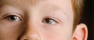
Nystagmus is an ophthalmological disease in which the eyeballs make involuntary oscillatory movements of high frequency.
Nystagmus is a serious pathology characterized by involuntary oscillatory movements (up to several hundred times per minute) of the eyes. Clinical symptoms describe rapid eye oscillations in horizontal, vertical, sometimes oblique directions or in circular, semicircular directions. There is a violation of the accommodative ability, which, in turn, is manifested by dysfunction of the visual organs.
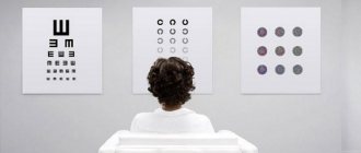
Diagnosis uses objective examinations, visometry, microperimetry, refractometry, electronystagmography, and CT scan of the brain. Conservative therapy is used for treatment, based on the use of antiepileptic and anticonvulsant drugs. Surgery to correct the position of the eyeballs is much less common.
General information
Nystagmus is a fairly common nosology in the practice of modern ophthalmology. According to world statistics, the congenital form of the pathology is diagnosed in 20-40% of visually impaired children. Often, it is possible to establish the etiology of involuntary eye movements.
The idiopathic type is observed with a frequency of 1:3000. The disease in the form of horizontal nystagmus is more common, while oblique and rotational nystagmus are quite rare. In the general statistics of eye diseases, horizontal nystagmus occupies 18%. Geographically, there is no epidemiology.
Why does the disease occur?
There are several dozen reasons for the development of nystagmus. And all of them are associated with one or another eye disease. Very often, such pathological movement of the eyeballs is provoked by congenital or acquired visual impairment. This may be clouding of the lens, atrophy of the optic nerve, the presence of albinism and retinal dystrophy.
But this pathology also occurs when the brain is damaged, for example, during injuries, when a concussion or bruise of the brain occurs. Another cause of the disease is inflammatory processes in the brain, which affect the pituitary gland, cerebellum and medulla oblongata. This phenomenon is often observed in drug addicts who use a substance such as mephedrone. It can also appear with significant alcohol consumption, but when sobering up occurs, everything goes away by itself and does not require any treatment.
There are three types of the disease - horizontal, vertical and rotational. All these three types can also have their own types - pendulum-shaped, jerk-shaped, mixed.
Causes of nystagmus
The congenital disease occurs against the background of various neurological disorders. This manifestation is evidenced by the appearance of symptoms against the background of congenital albinism or Leber amaurosis.
The acquired form has a number of underlying causes. Depending on the cause, the symptoms of nystagmus may differ. The main causes of the acquired form:
- Brain pathologies. Nystagmus in people in adulthood may indicate the presence of a malignant neoplasm or multiple sclerosis. The sudden appearance of such symptoms may indicate a stroke.
- Traumatic brain injury. Uncontrolled eye movements often result from damage to the optic nerves, as well as the cerebral cortex in the occipital lobe.
- Intoxication. This disease can occur as a result of the toxic effects of alcoholic beverages, an overdose of anticonvulsants or sleeping pills.
- Damage to the vestibular apparatus. Damage to the central or peripheral parts of the vestibular analyzer becomes the root cause of the manifestation of acquired symptoms. Most often, development is provoked by damage to the inner ear, and to be precise, the semicircular canals.
- Decreased visual acuity. Nystagmus can actively progress due to a significant decrease in visual acuity in the case of advanced cataracts, traumatic damage to the visual organs, or in the case of complete blindness.
Reasons for the development of nystagmus
Anomalies of intrauterine development, trauma during childbirth, disorders - including genetic ones - of visual acuity, transparency of the media of the eyeball, changes in the fundus, pathological processes in the optic nerve, retinal dystrophy, albinism can cause congenital nystagmus.
The causes of acquired nystagmus include:
- brain diseases (oncological formations, strokes, multiple sclerosis);
- malfunctions of the vestibular apparatus;
- poisoning, intoxication (medicinal, narcotic, alcohol);
- head injuries (with damage to the occipital region, optic nerves);
- refractive errors (myopia, farsightedness);
- strabismus;
- diseases with impaired media transparency (mature cataract, etc.).
Pathogenesis
Involuntary eye movements are based on decompensation of the tone of the membranous part of the labyrinth of the inner ear. In a normal state, nerve impulses are generated synchronously on both sides and transmitted at the same speed, which ensures the position of the eyes in a state of rest, and when moving, the organs can move synchronously. An increase in tone in the labyrinth of the inner ear on one side leads to desynchronization and the development of nystagmus.
In the case of damage to the peripheral and central parts of the vestibular analyzer, the emergence or change of clinical manifestations may be noted when the position changes. This is due to the secondary involvement of the semicircular tubules in the process of pathology.
The molecular mechanism of progression of congenital idiopathic nystagmus is currently not fully understood. Doctors suggest that the FRMD7 gene, or rather its mutation, inherited in an X-linked manner, is to blame. On the other hand, in clinical practice, cases of autosomal recessive and autosomal dominant inheritance are often observed.
Classification
Acquired and congenital nystagmus is diagnosed depending on the moment the first symptoms appear. The congenital form includes latent and manifest-latent variants. The acquired type is classified according to etiology into vestibular and neurogenic. From a medical point of view, the following stand out:
- Pendulum-shaped . It is characterized by phases of eye oscillations identical in speed and magnitude.
- Jerky . Expressed in rhythmic eye movements. At the same time, the eyeball moves slowly in one direction and quickly in the other. During the fast phase of movement, the subtype is determined. For example, if in the phase of rapid movement the eyes fluctuate to the left, this means a left-sided form, while in the phase of rapid movement, this means a right-sided form.
- Mixed . This form of the disease is a combination of inducing and jerking forms.
- Associate . In this case, the eyes move synchronously with the same amplitude in a jerk-like or pendulum-like manner.
- Dissociated . Eye movements are not synchronized in direction and amplitude with each other.
Types of disease
Horizontal nystagmus has its own symptoms, among which eye movements from right to left or left to right come first. This is the most common type of illness. However, the patient does not present any other complaints. The main cause of horizontal nystagmus is genetic pathology.
In second place in terms of frequency of detection is the vertical form, in which the eyeballs move from top to bottom or bottom to top. And finally, the rotational type of nystagmus is less common than others.
In this case, the scope of oscillatory movements and their frequency can be very different. Based on the magnitude of these fluctuations, nystagmus can again be divided into three types. This is large-caliber when the amplitude of movements exceeds 15 degrees, medium-caliber with an amplitude of movement up to 15 degrees and low-caliber with an amplitude up to 5 degrees.
Symptoms of nystagmus
In most cases known in practice, the first symptoms of the disease appear in early childhood or, directly from the moment of birth. The acquired form develops after exposure to an etiological factor. Patients complain of repeated involuntary eye movements. The patient is unable to focus on an object. The ability to adapt to changing conditions (changes in light intensity) is severely impaired.
The patient is unable to stop the nystagmus, but the magnitude of the oscillations can be reduced by changing the direction of gaze, focus on an object, or position of the head. The severity of symptoms can only be reduced if the patient takes a position with the lowest frequency of movements. The choice of position is determined specifically by the rest zone, in which the amplitude of movements decreases and accommodative ability improves.
Symptoms are more noticeable under conditions of stress, uneven tension or fatigue. The duration of symptoms is influenced by the nature of movements. With the pendulum variant, the duration of nystagmus is many times longer than in the case of the jerky variant of the disease. A change in manifestations is provoked directly by the appearance of objects in the field of view, a change in size or brightness. A significant role is given to the moment associated with visual concentration. The form of the disease is diagnosed by eye movement.
Clinical picture
As a rule, hereditary eye damage or the detection of ocular nystagmus in early childhood is persistent, which does not change with age.
In addition, with congenital pathology of the oculomotor system, there is a complete absence of changes in the refractive media (opacity of the cornea or lens), as well as in the photosensitive apparatus (retinal pigment degeneration, albinism, optic nerve atrophy, etc.).
Most often with the disease, oscillatory movements of the eyeballs are observed, the movement of which is rotatory or mixed in nature. It has been noted that nystagmus can intensify when the body is tilted back and forth or to the sides.
With a jerky form of nystagmus, the amplitude of oscillations decreases with head turns towards the fast phase. Often, visual acuity is significantly reduced and does not exceed 0.3 diopters.
The patient complains of being forced to hold his head in a certain position, the appearance of eyelid trembling, photophobia and blurred vision, which significantly reduces the patient’s adaptation and adaptability to living conditions.
Causes of eye redness, treatment methods, how to quickly relieve redness and whether you need to consult a specialist.
Existing methods for treating strabismus in children can be found in this publication:
Complications
A common complication of this disease is secondary alternating converging strabismus, which often develops in patients with the dissociated form. The characteristics of strabismus are also determined by the course of the disease. The pathology may be accompanied by amblyopia and mixed astigmatism. The acquired disease is complicated by a number of serious vestibular disorders, such as dizziness, problems with coordination, and headache. Due to the extreme need to constantly hold the head in one position, pinching of the cervical vertebrae may occur.
Diagnostics
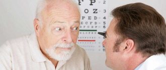
An objective examination of the patient is sufficient to make a diagnosis. Upon examination, it is possible to detect involuntary eye movements. The direction of nystagmus is determined by focusing the patient on the tip of a ballpoint pen. The ophthalmologist moves the instrument up, down, right and left. To study the etiology of the disease itself and further treatment strategies, the following are used:
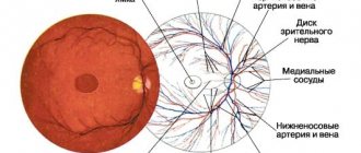
- Microperimetry . Allows you to accurately determine the point of fixation on the inner shell of the eye, study the sensitivity of the retina and record the parameters of optical nystagmus. This method allows monitoring the patient’s condition to determine the effectiveness of the measures taken.
- Electronystagmography (ENG) . Registers the biopotential that occurs between the retina and cornea. In people with involuntary eye movement, the electrical axis is greatly displaced, which is accompanied by a significant increase in the difference in corneoretinal biopotential, often up to 100-300 μV.
- Visometry . The decrease in visual acuity observed in patients is due to functional disorders of the visual apparatus.
- Refractometry . This diagnostic procedure is carried out to determine the type of refraction. Patients with nystagmus often experience disturbances in accommodation and myopia. Diagnosis of hypermetropia is carried out much less frequently.
- CT scan of the brain. Computed tomography is extremely necessary to identify various pathological neoplasms, as well as signs of changes in the structure of the brain.
Nystagmus surgery
Treatment of nystagmus
Oculomotor disorder, like nystagmus, is a very complex disease that has recently been treated in Russia.
And quite successfully. The only way to help is to perform an operation on the extraocular muscles, this is where treatment begins: the operation allows you to block movements during nystagmus with a straight gaze position. In Russia, surgical treatment of nystagmus is carried out by Igor Erikovich Aznauryan, Doctor of Medical Sciences, Professor, Academician of the Academy of Medical Sciences of the Russian Federation, head of the Yasny Vzor clinic.
It was he who developed and introduced into practice the tactics of high-tech radio wave surgery with the principles of mathematical modeling for oculomotor disorders.
Diagnosis of nystagmus
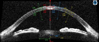
The accuracy of the operation is ensured not only by the mathematical calculation of the operation itself. This calculation is preceded by an accurate diagnosis. Now our ophthalmologist-surgeons have in their arsenal a device that is very necessary for planning the tactics of nystagmus surgery - the Gazelab nystagmograph, the only device in Russia and the CIS countries for studying patients with nystagmus.
Thanks to the device, we can determine the frequency and amplitude of nystagmus and the position of the eyes at which the nystagmus becomes less.
Make an appointment Call back
Surgery for nystagmus
In 78% of cases, we can completely compensate for nystagmus with direct gaze and a significant improvement in visual functions and a sustainable treatment result. In this case, “knifeless” technologies are used to minimize trauma to the eye tissue while preserving blood vessels and nerve elements.
We have developed a highly effective surgical technique for various types of nystagmus, thanks to which we can completely block or significantly reduce the frequency of oscillatory eye movements with direct gaze and significantly improve visual functions. These results are our pride, this is a huge step forward! Today, this is the only way to effectively help patients with nystagmus. The operations for nystagmus performed by our eye surgeons are unique. The high-tech radio wave surgery technique we have developed in combination with mathematical modeling allows us to:
- do not use traditional scissors and scalpels during the operation, which are used in the usual surgical techniques common today;
- to ensure the highest accuracy of the operation through the use of a mathematical modeling system for the operation, Doctor of Medical Sciences, Professor, Academician of the Academy of Medical Sciences of the Russian Federation, Head of the Yasny Vzor Clinic I.E. Aznauryan, STRABO;
- reduce the rehabilitation period by 5-6 times, the patient is discharged from the hospital the next day after the operation.
Surgeries for nystagmus are absolutely safe and very effective, their effectiveness has been proven over time. We use 4 types of operations depending on the type of nystagmus. Surgery for nystagmus is one of the stages of complex treatment of nystagmus. But not the only one.
To achieve a sustainable result and restore visual functions it is necessary:
- the use of an integrated approach that combines surgery and therapeutic treatment of nystagmus to restore visual function and binocular stereoscopic vision;
- treatment started in a timely manner - from the moment of diagnosis.
High degrees of astigmatism, hyperopia (farsightedness) and myopia in patients with nystagmus can be corrected using excimer laser correction, completely avoiding optical correction, which is often either impossible in patients with ocular torticollis, as in the case of correction with glasses, or is potentially inconvenient or dangerous , as is the case with contact lenses.
Until now, the presence of abnormal nystagmus has been a contraindication to refractive surgery due to the difficulty in centering the eye during the procedure.
However, at the laser correction center LaserCorr in Moscow, the possibility of performing surgery on patients with nystagmus in medicated sleep, during which any oscillatory movements stop, has been developed and implemented. Thanks to the eye-tracker technology in the laser and the customization function (the ability to perform the operation precisely and individually according to the parameters of the patient’s cornea), laser correction is possible under medicated sleep conditions. In this case, there is no need for the patient to subjectively monitor the fixation point in the laser, as is required in conventional laser correction.
In 78% of children, with timely treatment and the entire complex of surgical, optical, physiotherapeutic, and medicinal treatment, it is possible to improve visual functions to the level required for full social adaptation and attendance at a regular secondary school.
Treatment of nystagmus
The treatment strategy directly depends on the severity of symptoms and specifically the forms of nystagmus. To treat nystagmus the following is used:
- Conservative therapy . It is used when clinical manifestations progress against the background of central vestibulopathy. The most commonly used are neurotropic drugs from the anticonvulsant group, as well as antiepileptic drugs.
- Surgical intervention . Surgical intervention is used to form eye positions relative to the resting point, by restoring the physiological position. To do this, the structural features of the extraocular muscles are influenced.
Symptomatic treatment is mainly aimed at spectacle or contact vision correction. Experts recommend the use of contact lenses, since when the eye moves, the lens moves synchronously with the eye.
In individual cases, Botox injections are performed into the orbital cavity to limit small and frequent eye movements.
Treatment of the disease
The main treatment is aimed at improving the patient's vision. That is why glasses should be selected with special care for this disease. It is also necessary to use color filters of the density that best helps improve vision.
You should definitely use exercises to improve accommodation. To do this, you can use a device such as “Illusion” and even computer exercises, for example, “Zebra” or “Spider”.
Drug treatment includes eye vitamins and vasodilators, which help better nourish the retina. It is especially useful to take such preparations, which include blueberries, lutein, omega-3 fatty acids, beta-carotene, selenium, vitamin E,
Surgery is performed only in cases of severe disease, when this is the person’s only hope for improving vision. The operation is performed according to strict indications and most often in childhood.
Prognosis and prevention
With proper treatment and compliance with doctor's orders, the prognosis for the restoration of visual functions is very favorable. Corrective therapy of the underlying disease ensures elimination of the clinical manifestations of the pathology. But specific, preventative prevention has not currently been developed. Nonspecific preventive measures include timely diagnosis and treatment of brain pathologies, treatment of the vestibular system and, specifically, the organs of vision.
When detecting involuntary eye movements in a patient who has taken anticonvulsants or sleeping pills, it is necessary to adjust the dosage of the drugs.