Indications for pachymetry
The most common cases in ophthalmology when corneal thickness is a critical indicator, and its measurement is a necessary procedure:
- any painful condition of the eyelids, incl. inflammatory processes, swelling, developmental abnormalities, oncopathology, traumatic injuries;
- pathology of the conjunctiva (infections, allergic reactions, combined infectious and allergic inflammations, cystic formations, etc.);
- abnormal development and structure, inflammatory and degenerative processes in the cornea and scleral tissue;
- any pathological changes and reactions in the iris, its abnormal development;
- ophthalmotrauma, including the presence of a foreign body to be removed;
- cataract;
- glaucoma;
- pathological changes in the intraocular media caused by systemic endocrine disorders (primarily diabetes mellitus);
- preoperative examination and monitoring during postoperative rehabilitation;
- assessment of the dynamics of the condition of the affected structures during therapy.
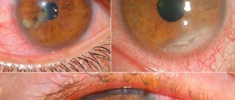
- keratoconus in various stages (as well as keratoglobus - a close, but much rarer variant of corneal deformity);
- corneal edema;
- glaucoma;
- dystrophic changes in the corneal tissue (eg, Fuchs endothelial dystrophy);
- the need for reliable postoperative monitoring after corneal transplantation;
- preparation for excimer laser vision correction.
Indications
This diagnostic procedure is carried out if the patient is suspected of having the following pathologies:
- corneal edema;
- keratoconus;
- keratoglobus;
- glaucoma;
- dystrophy.
Pachymetry is also indicated in the postoperative period after keratoplasty, during examination before laser correction of refractive errors.
Contraindications
In some cases, pachymetry cannot be performed, as there is a risk of complications. This research method is contraindicated in the following cases:
- for general intoxication;
- corneal damage;
- acute inflammatory diseases accompanied by the separation of pus - in these cases, ultrasound pachymetry is contraindicated.
This procedure should also not be performed if the patient has mental disorders, when acute psychosis or psychomotor agitation occurs, since in this case the patient can harm himself and others.
Contraindications
Contraindications to pachymetry are relative in nature and relate mainly to the ultrasound version of the study (optical pachymetry, being a non-contact method, has no contraindications). Ultrasound pachymetry is contraindicated or undesirable in infectious and inflammatory diseases, especially with a purulent component, and in cases of physical damage to the cornea with a violation of its integrity.
In addition, no planned ophthalmological procedures are performed on persons under the influence of drugs or alcohol, or in the presence of signs of mental inadequacy.
Determination of corneal thickness at the Ocodent clinic
Carrying out this diagnostic procedure in our medical center has a number of advantages. All research is carried out by highly qualified specialists who have all the necessary certificates and skills. In order to always be aware of modern methods of diagnosis and treatment of eye diseases, the staff of our clinic regularly undergoes retraining.
Diagnostics are carried out using modern high-precision equipment, using tools that meet all international quality standards. You can find out what services the Ocodent clinic offers and what their costs are on our website:
Norm
The standard thickness of the cornea in the frontal part of the eye is determined by the interval 0.49 - 0.54 mm, and in the limbus area - 0.7 - 0.9 mm. According to statistics, women have a slightly higher average corneal thickness than men. It should also be noted that the thickness of the corneal layer is not a constant indicator, it can vary throughout the day; Only with a significant deviation from the normative ranges is thickening/thinning considered as a pathology and requires, at a minimum, a thorough examination.
Types of pachymetry
There are three ways to conduct the survey. They differ in execution technique and information content.
For diagnosis use:
- Optical method. A slit lamp is used for examination. It directs a beam of light into the patient’s visual analyzer, the length and width of which is adjusted by the ophthalmologist. This method allows you to accurately diagnose the thickness of the cornea.
- Ultrasonic method. It is carried out using ultrasonic devices. They not only allow you to study the thickness, but also the structure of the eyeball.
- Computer technique. This type of pachymetry is performed using a tomograph. The device illuminates the structures of the visual analyzer and produces an image.
How is it carried out?
The slit lamp, one of the standard and traditional ophthalmic light sources, is equipped with a special attachment, inside of which there are two parallel plates; one is fixed, the other can rotate. The patient sits down on the device, which has special supports for the forehead and chin. A beam of light from the slit lamp is directed into the eye being examined (the plates in the measuring head are located perpendicular to the main optical axis). By slightly turning the movable plate with a precision mechanism, the doctor measures the thickness of the cornea by shifting the beam; a rotation of one degree corresponds to 1 mm of layer thickness.
The ultrasound version of pachymetry is a more accurate method - its error does not exceed 10 microns. The procedure is performed under local drip anesthesia (to eliminate possible discomfort when the sensor comes into contact with the surface of the cornea, although the pressure on the eyeball will be minimal in any case). All necessary measurements are displayed on the screen in real time, and additional indicators are calculated. After ultrasound pachymetry, prophylactic instillations are usually prescribed to avoid infection.
Types of research
Pachymetry can be performed in two ways:
- optical – non-contact method, which is performed using an optical coherence tomograph or slit lamp. This method is called coherent pachymetry;
- Ultrasound – contact measurement of the thickness of the cornea of the eye, which is carried out using an ultrasound machine.
The optical method makes it possible to measure the thickness of the cornea without touching it. A nozzle is put on the slit lamp, which measures the thickness of this membrane of the eye in different parts of it.
The ultrasonic method is considered more accurate, as it makes it possible to measure thickness up to 10 microns. Before the procedure begins, the patient must lie on a couch and be anesthetized using eye drops. After a few minutes, when the anesthetic effect reaches its maximum, the doctor uses a special device to touch the surface of the eye, applying minimal pressure to the cornea. The device automatically determines the thickness of the anterior wall of the eye and displays detailed research results on the monitor. After the procedure, an antibacterial drug is instilled into the patient’s eyes to prevent the development of inflammatory diseases.
Glaucoma research
It has been established that the likelihood of developing glaucoma directly depends on the size of the central part of the cornea.
More often, this disease occurs in patients with a corneal thickness of less than 550 microns. Therefore, it is believed that pachymetry and manometry data are sufficient to begin treatment of this disease - the combination of reduced membrane thickness with increased intraocular pressure may be the basis for prescribing special therapy.
Research in surgical practice
Pachymetry data play an important role in the preoperative preparation of patients in refractive surgery, as well as in assessing the results of surgical intervention.
For example, if the thickness of the cornea is insufficient, laser keratomileusis (LASIK) is contraindicated for patients. Such patients are prescribed PRK. At the same time, the results of determining the thickness of the cornea are used to select a corneal flap for plastic surgery.
Pachymetry video
Zolotorevsky Kirill Andreevich tells how the examination is carried out and what is the norm:
Author's rating
Author of the article
Alexandrova O.M.
Articles written
2029
about the author
Was the article helpful?
Rate the material on a five-point scale!
( 2 ratings, average: 3.00 out of 5)
If you have any questions or want to share your opinion or experience, write a comment below.
Interpretation of pachymetry results
The digital norm for corneal thickness is a relative indicator. The approximate range is from 0.410 to 0.625 millimeters. On average - 515 microns. The numbers may be higher at the edges of the shell. The numerical value of the thickness also depends on the anatomical features of the eye and the gender of the person. Therefore, decoding thickness measurements is the field of activity of a qualified specialist. You shouldn't do this yourself. Only a doctor is able to make a correct assessment and draw the necessary conclusions.
How is keratopachymetry performed?
The patient sits in a chair in front of the device, and the doctor instills anesthetic drops into his eyes, as a result of which the patient does not experience anxiety and discomfort that may occur when the sensors come into contact with the eye shell. After the effect of the anesthetic has begun, a special sensor is installed on the cornea, which automatically determines its thickness in different parts. The device displays the obtained data, measured in micrometers (µm), as digital indicators on a chart. The duration of the procedure is about 10 minutes for both eyes.
At the Constellation clinic, this examination is carried out using a French Compact Touch device, which has built-in software for conducting research and calculations using various methods. In addition, the device has high image quality on the screen, which makes the measurement complete and as accurate as possible.
Methods for measuring the thickness of the corneal structure
- The ultrasound method involves exposing the cornea to waves, reading the result with a device, and then displaying the shell thickness data on the screen. An ultrasound device sensor is applied to the cornea, it comes into contact with the corneal structure, and pressure is tried to be kept to a minimum. This technique allows you to determine the thickness of the cornea by calculation, taking into account the speed of movement of waves, the time of their passage through the corneal layers and recording their reflection. Before the procedure, an anesthetic is instilled, and after - antibacterial drops. The procedure is performed within a few minutes with the patient lying down.
- Optical method. The slit lamp is equipped with a glass nozzle consisting of two parallel plates: a static lower one and a rotating upper one. The light beam is directed to a specific point in the eye. The patient and the doctor sit on opposite sides of the biometer. Measuring the level of deflection of the movable plate on a graduated scale gives the thickness of the cornea in its different areas. One degree of rotation of the plate corresponds to one millimeter of thickness. There is no need to instill painkillers and disinfectants into the patient, since there is no eye contact with the device.
Non-contact research methods exclude the possibility of eye infection, but contact methods provide more reliable results. Therefore, in cases where increased accuracy is required, contact pachymetry is used. If the integrity of the cornea is compromised or a purulent process occurs, the eye is examined using a non-contact method.
Features of pachymetry
- Ease of research. No preparation is required for pachymetry, and the diagnosis itself takes no more than 20 minutes.
- Painless research. Regardless of whether it is performed contact or non-contact, you can be sure that you will not experience serious discomfort.
- Safety of the event. Pachymetry does not affect visual function, does not worsen the clinical situation, and can be performed as often as necessary to assess the rate of corneal thinning or thickening.
- No recovery period. You can immediately get behind the wheel after pachymetry, start reading books, etc.
Pachymetry at ON CLINIC
At the Ophthalmology Center ON CLINIC, pachymetry is performed using expert-class equipment. Comfortable conditions for receiving patients have been created; the reception is conducted by experienced and highly qualified doctors, candidates and doctors of science. The cost of pachymetry is affordable, you can schedule an appointment at a time convenient for you.
Immediately after pachymetry, you will be given the results, and you will be able to receive competent advice from an ophthalmologist. The specialists of the ON CLINIC Ophthalmology Center specialize not only in the diagnosis, but also in the treatment of ophthalmological diseases - you will find yourself in the hands of experienced doctors who use advanced correction techniques and the latest achievements of modern medicine.
The cost of electronic tonography of the eye is presented in the price list.
Trust your eye health to professionals - contact the specialists at ON CLINIC!