Visual acuity is the distance by which two points (images) can be removed so that the eye perceives them as separate.
Measuring visual acuity is also called visometry. To conduct the study, a number of special tables with black signs on a white background have been developed. For adults, this is an image of letters, numbers, rings with breaks; for children of preschool and primary school age - pictures.
Types of tables:
- Sivtsev-Golovin table.
- Table by Orlova: with silhouettes of objects and animals for children;
- Snellen chart: letters of the Latin alphabet.
In our country, for this purpose, printed Golovin-Sivtsev tables are traditionally used, in which rings with breaks in different directions are depicted on one half, and letters on the other.
Visual acuity
Visual acuity is the ability of the eye to distinguish the smallest details of an observed object. Even ancient Greek healers, trying to determine the quality of vision, suggested their patients to see the stars in the constellation Ursa Major.
Ideal 100% vision is the ability of the eye to distinguish individual points at a distance of 1.45 mm (ophthalmologists call this segment 1 arc minute of vision) from 5 meters. This is the same vision of 1.0 or “one”.
The reasons why the eye loses the ability to see clearly can be divided into 3 groups:
1. Refractive errors
– or the loss of the lens’s ability to focus the eye on an object of interest depending on the distance to it. This may be myopia or farsightedness, which occurs due to age and excessive visual load. Refractive error is the most common cause of visual impairment
2. Lens clouding
– for example, after an eye injury, with age-related cataracts or high blood sugar levels in diabetics.
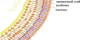
3. Diseases of the retina and optic nerve
– in these cases, the eye normally focuses the image and conducts light, but the entry (“projection”) of the image into the visual fields of the brain is impaired due to problems with the conductive pathways.
Price
| Visometry | 605 rub. |
When performing visometry or studying visual acuity using tables, great attention should be paid to compliance with the standard conditions of the methodology (time parameters for demonstrating optotypes, distance to the table, its level of illumination). Most researchers currently consider an illumination of about 700 lux to be optimal. To best meet standard conditions, a Roth apparatus - a special lighting box with mirrors.
Distance visual acuity testing can be carried out from a distance of 5 meters (in the CIS countries, Germany, France, Japan) or 6 meters (20 feet). It is believed that when the tests are located at such a distance, there is no strain of accommodation and, thus, such a study can be performed at any age, even with the onset of presbyopia.
Near visual acuity is usually assessed at a distance of 33 cm . When selecting optical correction, it may vary depending on the expected distance between the objects in question and the patient’s eye.
During the examination, the patient sits at the required distance from the table. The study is carried out monocularly. First, the right eye is examined, then the left. Next, binocular visual acuity can be assessed, which, due to physiological characteristics, is higher than monocular. It is used when selecting optical correction. If there is a decrease in visual acuity due to a refractive error, then an optical correction is selected that maximizes the visual acuity. If the patient already uses one, then using visometry you can assess its sufficiency.
a pin-hole test can be used for differential diagnosis . If the clarity of the image increases with the aperture, this suggests a refractive cause of decreased vision. Otherwise, damage to the retina or visual pathway may be to blame. However, with high degrees of ametropia, the reliability of the test decreases.
Visometry results are usually recorded in the following form:
Visus OD/OS=1.0/1.0 or Vis OU=0.6 or VA OD 20/200, where Visus, Vis, VA (visual acuity) – visual acuity; OD (oculus dexter) - right eye, OS (oculus sinister) - left eye, OU (oculus uterque) - both eyes.
If the patient is not able to distinguish the optotypes of the table, and his movements are difficult, then approximately visual acuity can be assessed by counting the number of fingers demonstrated by the examiner from different distances. The result is assessed based on the greatest distance from which the patient is able to correctly recognize the object, and is written as:
Visus OD = finger counting from 3 meters.
With more severe vision loss, when the above methods do not allow assessing visual function, light perception . For this, a point light source is used. They are shone alternately into the eye being examined from different directions, assessing the sensitivity of different parts of the retina. If the eye correctly distinguishes the movements of the light source (visual acuity is equal to light perception with correct light projection), then this is written as visus = 1/∞ proectio lucis certa, or abbreviated as plc
Correct light projection indicates the preservation of the functions of the retina and optic nerve. This is necessary to predict treatment results (for example, for cataracts).
If the patient has at least once mistakenly determined the movement of light on at least one side, then such visual acuity is recorded as visus = 1/∞ proectio lucis incerta or plincerta. (light perception with incorrect light projection). An eye that is unable to sense light, does not distinguish light from dark, is assessed as completely blind (visus = 0).
Visometry in children is a rather difficult task . In infants, visual acuity is mainly determined behaviorally. At an early age, up to 2-5 months, the child’s reaction to bright light is examined. A little later, you can use a bright red ball with a diameter of 4 cm, suspended on a thread against the background of a window. It is brought to the child’s eyes and the distance from which he begins to follow him with his eyes or reach out to him with his hand is noted. For children older than 6 months, balls of a smaller diameter can be used. For an approximate assessment of visual acuity, you can also use white balls on a dark floor. The size of those that a child sees from a certain distance will indicate visual acuity. For children over 3 years old, tables with optotypes in the form of animals or other easy-to-recognize symbols are used ( Aleynikova or Orlova tables ).
Indications for visometry
- Preventive examination and medical examination
. Nowadays, a huge number of people constantly work with a computer, exposing their eyesight to excessive stress. Bright blue light “forcibly” dilates the pupil and causes spasm of the extraocular muscles, and an incorrect distance to the monitor, lack of a break to rest the eyes and neglect of visual gymnastics only worsens the situation. Timely prescription of glasses will slow down the decline in vision and restore comfort to life. - The appearance of vision complaints.
Deterioration of vision near and far, blurred vision, the need to squint, difficulty reading, double vision and even constant headaches - all this is a reason to visit an ophthalmologist and signals that the ability to focus the lens may be impaired. - Observation during ongoing vision correction
. Wearing glasses or soft contact lenses, even properly fitted ones, does not guarantee that your current acuity will be maintained forever. Age, degenerative processes and many other reasons can affect the conductive media of the eye, continuing to further reduce vision. Therefore, it is important to visit an ophthalmologist periodically. In such cases, visometry is carried out with correction to ensure that vision close to “unity” is achieved and maintained with these glasses or lenses.
When is the study carried out?
The study of the acuity of visual function is one of the mandatory procedures that is carried out in children when registering for kindergarten and school, as well as in adults when finding employment or during annual preventive medical care. inspection (vehicle drivers).
Other indications for visometry are:
- Presence of complaints of decreased visual acuity;
- When diagnosing diseases, one of the manifestations of which is decreased vision;
- Monitoring the state of vision before surgery;
- Determining the effectiveness of the treatment (medical or surgical).
Diagnosis is not performed in patients who are under the influence of drugs, intoxicated or have acute mental disorders.
Contraindications
There are practically none. Visometry is absolutely safe and can be performed on both adults and children.
However, you need to understand that the organ of vision is sensitive to any changes in the body, so the results may be inaccurate in the following cases:
- during colds and infectious diseases, especially with the presence of conjunctivitis, lacrimation, irritation of the eyelids and cornea
- crisis course of hypertension - due to spasm of blood vessels in the fundus, blurred vision and the appearance of “floaters” before the eyes are possible. In this case, it is better to wait until the pressure stabilizes
- after a recent injury or concussion, as well as after a stroke. For these diseases, it is very important to immediately examine the fundus and retinal vessels, but it is better to postpone the determination of acuity for 1-2 months
- alcohol and other types of intoxication, which may prevent you from strictly following the doctor’s instructions.
No special preparation is required for the examination; the procedure is performed without restrictions at any examination by an ophthalmologist.
Visometry technique
Determination of visual acuity using the visometry method is based on a person’s ability to distinguish specially selected symbols (or optotypes) of various sizes from a fixed distance.
Simply put, the doctor points to the symbols on the poster (vision chart) and asks to name them.
For diagnostic accuracy, the method has been standardized and must be performed in accordance with a number of conditions:
- Distance to the vision table is 5 meters (in Europe and the USA the definition of vision is 6 m)
- Room illumination of at least 700 lux
- Acuity is assessed first on the right eye, then on the left. The second eye is covered with a special shield or palm. After examining each eye separately, both eyes are diagnosed simultaneously (binocularly)
- Using universal vision tables with the same set of optotypes.
There are various modifications of vision testing tables, but in general it is always a poster with lines of letters or symbols, the height and width of which decreases from top to bottom. The color of optotypes is almost always black. In Russia, a domestic version is used - the Sivtsev-Golovin table, in which 7 letters of the Russian alphabet (Ш, М, Н, У, Б, И, К) are arranged in 12 rows. Each subsequent line contains letters of a smaller size and corresponds to a certain severity - 0.1, 0.2, etc. The ability to correctly see all the characters on line 10 means having a visual acuity of 1.0 (or 100%).
During the check, you are given the “right to make mistakes” - for example, you can make a mistake in one character from lines 4 to 6 and 2 times in lines from 7 to 10, which will not affect the result. But from lines 1 to 3, a mistake cannot be made “with impunity.”
People who are able to see lines 11 and 12 have vision exceeding 100% - 1.2 or even 1.5.
This also happens. Most often this is a genetic feature - a high density of visual elements of the retina in the ocular area. There are also other options for optotype sets:
- The Snellen table was proposed back in 1862, and consists of 11 rows of letters of the Latin alphabet, and is used in Europe and the USA.
- Landolt optotypes - the use of rings with breaks on one side. A random set of rings is used in cases where malingering is detected. Thus, when passing the driver's commission, there are occasionally cases of memorizing letters
- Tables by Orlova or Aleynikova - are used mainly for children, and instead of letters they contain simple, generally understandable symbols (typewriter, Christmas tree, etc.)
Minor changes to the technique were made with the advent of the opportunity to use modern projectors instead of vision tables in ophthalmology rooms. Devices displaying optotypes can be calibrated to distances of less than 5 meters, with the optotypes being proportionally reduced in size. Therefore, you should not be surprised if in a small office visometry is carried out from a shorter distance.
Tables for visualization
Eye examination is carried out using a special set of symbols called optotypes. The signs are arranged in 10 mandatory rows, each of which shows the degree of violation.
The key point in constructing an optotype is the ratio of the total width to the width of the individual element of which this symbol consists. The ratio should be 5:1.
Instructions for using Broxinac eye drops can be found here.
Sivtsev-Golovin table
The most common and suitable for most patients are several tables bearing the name of their developers:
- Snellen containing Latin letters.
- Landolt, which depicts open rings. The subject must indicate where the gap is directed.
- Orlova, where black and white drawings of animals or simple objects (mushroom, car, teapot, etc.) are used.
- Sivtseva-Golovin with Cyrillic, although in fact only 7 letters from the entire alphabet are used. This is the most common table in domestic ophthalmology offices.
- Pole, which contains special characters and is used for visually impaired people.
A symptom, the cause of which is best determined immediately, is inflammation of the lower eyelid.
Occasionally, the sharpness can exceed the coveted unit and amount to 110 or 120%. Such a “unique” is able to distinguish between 11 and 12 additional lines.
The choice of one or another diagnostic regimen depends on the patient’s age or the severity of the eye disease. For example, for children optotypes in the form of bright balls are used; for older children these can be animals.
To combat allergies, it is important to know everything - read the instructions for using Vigamox eye drops.
Orlova table
What to do when the upper eyelids swell is described in detail here.
How to measure vision in children
The baby's first meeting with an ophthalmologist usually takes place at the age of 1 month. At this age, the visual system has not yet been formed, and determining absolutely accurate readings of acuity is not only technically impossible, but also unnecessary. As a rule, the doctor limits himself to diagnosing the ability to fix gaze - using a large bright ball, slowly moving it in front of the eyes.
Interestingly, in infancy, periodic convergent strabismus and farsightedness of up to 4-5 diopters are considered normal.
But at the age of 1 year, it is necessary to evaluate visual acuity, since disorders identified at this stage of development without treatment can greatly impair visual acuity in the future.
To determine visual acuity from 1 year to 3 years, siascopy is performed (literally this translates as “observation of shadows”). Directing a beam of light into the child's pupil, the doctor observes the presence of shadows in the fundus through a special ruler with lenses of different diopters. As soon as it is possible to select the required curvature of the lens, the shadow on the fundus will disappear, since the light beam will no longer be refracted.
The method is fundamentally similar to visometry, but in the case of skiascopy, the doctor is not guided by the patient’s own answers, but visually evaluates the passage of light through the lens and its accommodation (the ability to accept the curvature necessary for clear vision). The size of the lens that is necessary to neutralize shadows is an indicator of refractive error.
To direct the baby's gaze in the right direction - above the doctor's ear - a small bright ringing toy is often used. Such an examination is simple, very informative, carried out without expensive equipment, but requires accuracy and speed, as well as significant manual dexterity from the doctor, because babies quickly get tired and can begin to be capricious and resist. The results largely depend on the experience and skill of the doctor.
For children who are already 3-4 years old, they use the classic method of visometry - identifying symbols from a distance of 5 meters. On a poster for children (in Russia these are usually tables by Orlova or Aleinikova), instead of letters, simple symbols well known to children are depicted - a mushroom, a teapot, a car, an elephant, etc. Children at this age do not yet know letters, but they distinguish well-known objects well and can already name them. Before the examination, the doctor and the baby will look at the table closely and agree on what to call each symbol. Autorefractometry can also be used for children over 3 years of age.
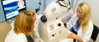
An autorefractometer is a special device that performs computer diagnostics of the eye to determine the ability to focus on an object. During the study, you need to look into the eyepieces of the device, where a glowing picture will be shown (most often it is a house in a field). The eye will automatically “adjust” the clarity of the image, and the device will detect the path of reflection of light rays from the picture to the retina and back, and automatically calculate the approximate visual acuity.
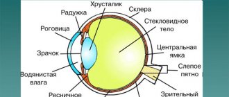
The focal length and ability to project an image onto the retina is determined by the length of the eye and the thickness of the lens.
In children, due to their age, the size of the eye is small and the image is projected behind the retina, that is, young children are always farsighted and this is normal. Normal acuity values for age:
- 3 years – 0.6-0.9
- 4 years – 0.7-1.0
- 5 years – 0.8 – 1.0
- 6 years and older – 0.9 -1.0
As the baby grows, the size of the eyeball also increases, so that in most cases, visual impairment goes away on its own. Vision reaches one at about 5-7 years.
Objective methods of visometry
Such methods may be needed when the doctor suspects the patient is malingering or aggravating (exaggeration of symptoms).
This includes, for example, the method of optokinetic nystagmus. An object moves against the background of the grating, which the eye will involuntarily follow, making involuntary movements. The frequency of these movements is used to judge visual acuity.
Another objective method of visometry is the method of forced selective vision. This diagnostic technology is based on the fact that the child prefers to look more readily at structured objects rather than at homogeneous ones.
Decoding the results
Paying tribute to established traditions, the results obtained are usually described in Latin. The words and symbols in the conclusion mean the following:
Visus – vision OD – oculus dexter – right eye OS – oculus sinister – left eye Sph – spherical component, with a “+” sign means myopia, with a “-” sign farsightedness.
As a result, the sum of the refraction of the eye itself and the lens for correction should ideally be 100% or 1.0
Examples:
- The entry VisusOS = 0.4 means that when testing vision in the left eye, the person recognized 4 lines correctly and vision is 40% of normal.
- The entry VisusOD/OS = 0.2/0.7 means that vision in the right eye is reduced to 20%, and in the left to 70% of normal.
- The entry VisusOD = 0.3 Sph - 1.5 means that vision in the right eye is 30% and myopia is 1.5 diopters.
- Recording ODsph +1.75 means the need for correction in the right eye with a spherical lens with an optical power of 1.75 diopters.
Visometry procedure
The patient undergoing visometry is usually sitting. The table is placed in front of the patient five meters from the eyes (in Germany and the USA the distance to the table is 6 meters). The table should be positioned so that its bottom edge is at eye level with the patient. If you need to evaluate near visual acuity, the table is placed 33 cm from the eyes. First, an examination is carried out for the right eye (the left one is covered with an opaque spatula or palm), after which an examination of the left eye is carried out. You should not close your eyes during the examination, as this may affect the result.
The patient names the indicated symbols in the table. They start visualization from the first line, after which they move to the bottom and so on. The letters in the first six lines must be named accurately. In characters 7-10 of the line you can make an error, but only one. If the patient makes a mistake, then you need to return to the optotypes of the previous row. If the examinee accurately listed all the characters in the line, then the doctor moves on to the underlying line. The result, which is recorded on the card, corresponds to the lowest line, accurately read by the person being examined.