→ Retina
Cones
- (English cone - cone) one of the types of exteroceptors (photoreceptors) of the peripheral processes of light-sensitive nerve cells of the retina. They are called cones because of their shape, similar to a conical laboratory flask.
Cones are a group of receptors consisting of various types of specialized nerve cells that perceive and convert light stimulation into nervous stimulation into bioelectric signals that go to the visual parts of the brain.
Retinal rods
These photoreceptors are cylindrical in shape, with a length of approximately 0.06 mm and a diameter of approximately 0.002 mm.
Thus, such a cylinder is really quite similar to a stick. The eye of a healthy person contains approximately 115-120 million rods. The human eye rod can be divided into 4 segmental zones:
1 - Outer segmental zone (includes membranous discs containing rhodopsin), 2 - Connecting segmental zone (cilium), 3 - Inner segmental zone (includes mitochondria), 4 - Basal segmental zone (nerve junction).
Rods are highly photosensitive. So, for their reaction, the energy of 1 photon (the smallest, elementary particle of light) is enough. This fact is very important for night vision, which allows you to see in low light.
Rods cannot distinguish colors; this is primarily due to the presence of only one pigment in them - rhodopsin. The pigment rhodopsin, otherwise called visual purple, due to the included protein groups (chromophores and opsins), has 2 light absorption maxima. True, one of the maxima exists beyond the range of light visible to the human eye (278 nm - the region of ultraviolet radiation), therefore, it is probably worth calling it the wave absorption maximum. But the second maximum is visible to the eye - it exists at around 498 nm, located on the border of the green and blue color spectrum.
It is reliably known that rhodopsin, present in rods, reacts to light much more slowly than iodopsin, contained in cones. Therefore, rods are characterized by a weak reaction to the dynamics of light fluxes, and in addition, they poorly distinguish the movements of objects. And visual acuity is not their prerogative.
What it is?
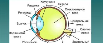
The retina lines the inside of the eyeball; normally its thickness reaches 281 micromillimeters. Moreover, in the area of the macula, the membrane is several times thinner than at the periphery. The element extends from the optical disk to the jagged line. In the optic disc, the retina is attached very tightly; in other areas the connection is loose. This explains the easy development of retinal detachment.
The layers of the shell differ in structure and function, forming a complex structure. Thanks to the close interaction of different elements of the visual apparatus, a person is able to distinguish colors, sizes of objects, and estimate distance.
Penetrating into the eye, light fluxes pass through several refractive media. In the absence of deviations in refraction, people receive a reduced and inverted, but real image on the retina. Subsequently, the impulses are transformed and enter the brain, where the final processing of the image of the external world is carried out.
Cones of the retina
These photoreceptors also got their name due to their characteristic shape, similar to the shape of laboratory flasks. The length of the cone is approximately 0.05 mm, its diameter at the narrowest point is approximately 0.001 mm, and at the widest point - 0.004. The retina of a healthy adult contains about 7 million cones.
Cones have less sensitivity to light. That is, to excite their activity, a luminous flux will be required, which is tens of times more intense than to excite the work of rods. But cones process light fluxes much more intensely than rods, so they perceive their changes better (for example, they better distinguish light when objects move, in dynamics relative to the eye). They also define images more clearly.
The cones of the human eye also include 4 segmental zones:
1 - Outer segmental zone (includes membrane disks containing iodopsin), 2 - Connecting segmental zone (constriction), 3 - Inner segmental zone (includes mitochondria), 4 - Synaptic connection zone or basal segment.
The reason for the above-described properties of cones is the content of the specific pigment iodopsin in them. Today, 2 types of this pigment have been isolated and proven: erythrolab (iodopsin, sensitive to the red spectrum and long L-waves), and chlorolab (iodopsin, sensitive to the green spectrum and medium M-waves). A pigment that is sensitive to the blue spectrum and short S-waves has not yet been found, although the name has already been assigned to it - cyanolab.
The division of cones according to the type of dominance of color pigment in them (erythrolab, chlorolab, cyanolab) is due to the three-component vision hypothesis. There is, however, another theory of vision - nonlinear two-component. Its adherents believe that all cones contain erythrolab and chlorolab at the same time, and therefore are able to perceive colors in both the red and green spectrum. The role of cyanolabe, in this case, is played by the faded rhodopsin of the rods. This theory is also confirmed by examples of people suffering from color blindness, namely the inability to distinguish the blue part of the spectrum (tritanopia). They also have difficulty seeing at night (hemeralopia), which is a sign of abnormal rod activity in the retina.
Features of the transmission of light pulses
Rods and cones perceive the flow of light and direct it to the central nervous system. Both cells are able to work fruitfully during the daytime. The main difference is that cones are more sensitive to light than rods.
Rods and cones are capable of receiving light and transmitting it to the central nervous system. Both types of cells are capable of working during the daytime. The difference is that the light sensitivity of cones is much higher than that of rods. The transmission of received signals is carried out thanks to interneurons, each of which is attached to several receptors.
as well as the ability to detect various movements occurring around us.
Structure
The retina is a multilayered membrane consisting of:
- photosensitive cells;
- pigment layer;
- choroid;
- nerve layer.
Anatomy and physiology of the retina:
Photoreceptor apparatus
Presented by 2 types of light-sensitive cells:
- Sticks. A large number of cells capture weak light rays and provide a person with vision at dusk or at night.
- Cones. They are few in number because they are highly sensitive to daylight. Provides vision in good lighting conditions.
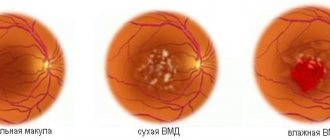
Macular area
The sweet spot, or macula, is the area with the highest concentration of light-sensitive cells. In this place, maximum human visual acuity is achieved. The area is located above the exit zone of the optic nerve.
Optic disc
Alternative name: optic nerve head. This is the outlet for the long processes of the nerve cells of the retina. There are no photosensitive cells in this area, so there is no vision in this area and is called a blind spot.
Blind spot
The diameter is up to 5 mm. It is the area where the optic nerve exits the retina. The cranial-brain pair is formed from 1.2 million nerve cell axons located throughout the surface of the retina. It is also the exit point for blood vessels that supply the retina with oxygen and nutrients.
Blood supply to the retina
Under the pigment layer is the choroid, consisting of choriocapillaris. These are small vessels that provide trophism and respiration of the nerve cells of the fundus. With capillary thrombosis, dystrophic-degenerative changes in the retina develop and its detachment begins.
Prevention
Genetically determined diseases cannot be prevented, but in some cases it is possible to delay the consequences. It is quite possible to avoid acquired pathologies with certain preventive measures.
- Balanced diet;
- Compliance with the visual regime - gymnastics, training, timely rest after stress on the organ of vision;
- Adequate professional selection of corrective glasses for myopia, presbyopia, astigmatism, hypermetropia. And use in accordance with the recommendations of an ophthalmologist;
- Moderate physical restorative load;
- Compliance with light conditions;
- Protect your eyes from UV rays with sunglasses with quality filters.
There are very small parts of our body that play a huge role. The photoreceptors - the cones and rods of the retina - work tirelessly to make our lives bloom with colors.
Notes[edit]
- “Rod-opsin.” Humbio. Retrieved August 16, 2012.
- https://webvision.med.utah.edu/book/part-viii-gabac-receptors/color-perception/
- https://www.conesandcolor.net/home.htm
- https://www.conesandcolor.net/home.htm
- Finn, J. T., Grunwald, M. E, and Yau, K. W. 1996. Cyclic nucleotide-gated ion channels: An ex-tended family with diverse functions. Annu. Rev. Physiol.58: 395‒426.
- Nakanishi, S., Nakajima, Y., Masu, M., Ueda, Y, Nakahara, K., Watanabe, D., Yamaguchi, S., Kawabata, S., and Okada, M. 1998. Glutamate receptors: Brain function and signal transduction.
- https://www.conesandcolor.net/home.htm
- https://www.ghuth.com/
- ↑ “photoreceptors”. Retrieved August 16, 2012.
- V. P. Tyshchenko PHYSIOLOGY OF INSECTS M., “HIGH SCHOOL” 1986, p. 223
- Zagalskaya, E. O. Morphological features of the retinomotor reaction in juvenile masu salmon (oncorhynchus masou) in a magnetic field and red light / E. O. Zagalskaya, V. P. Gnyubkina, A. A. Maksimovich // Morphology: scientific and theoretical medical journal . - 2004. - Volume 126, N 6. — P. 32‒36.
- https://zoometod.com/ixt/ixtiolog_63.html Anisimova I. M., Lavrovsky V. V. Ichthyology
- E. O. Zagalskaya, V. P. Gnyubkina (2006). “Ultrastructure of the retinal pigment epithelium of juvenile masu salmon” (PDF) 32
(1). Marine biology Oncorhynchus Masou. pp. 55–59. Retrieved August 16, 2012.
Developmental abnormalities and diseases
Malfunction of the visual apparatus occurs as a result of injury or is congenital. Some pathologies appear due to the development of allergic, endocrine or parasitic diseases.
Most often, doctors diagnose the following anomalies:
- Myopia. Myopia is characterized by a deviation in refraction, which results in problems viewing objects located at a distance.
- Hypermetropia or farsightedness. Objects at a distance are clearly visible. But nearby objects become blurry.
- Astigmatism. Impaired vision clarity, manifested due to changes in the shape of the eyeball.
- Cataract. Partial or complete clouding of the lens.
- Uveitis. Inflammatory pathology affecting the choroid of the visual apparatus.
| Amblyopia. Lazy eye syndrome is characterized by the fact that the left or right eye ceases to participate in optical function. As a result, the patient develops strabismus. |
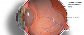
- Detachment of the retina. The structure is disconnected from the vascular ball, which negatively affects the visual process.
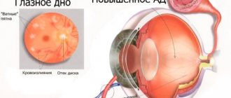
- Glaucoma. Increased intraocular pressure usually passes without pronounced symptoms. May lead to blindness.
- Keratoconus. Changes in the shape of the cornea (from a sphere to a cone), visual acuity decreases.
- Agenesis. Absence or underdevelopment of the eyeball or a certain area of it.
- Retinitis. Inflammatory processes of the retina.
- Atrophy of the eyeball. Accompanied by a decrease in the element's size and disruption of its functioning.
- Diabetic retinopatitis. Pathological processes in the retina caused by increased blood sugar levels.
- Conjunctivitis. Acute inflammation of the eye mucosa.
External structure of the eyeball
When conducting a visual examination of this element of the visual apparatus, only a small part of it is visible (cornea, eyelids, eyelashes). All important structures are reliably protected from external influences by the bones of the skull, fatty tissue, and muscles. These “details” can only be examined using specialized equipment.
The average human eyeball is approximately twenty-four millimeters in size and spherical in shape. From the inside it is filled with aqueous humor. The element includes a lens located opposite the pupil. Its thickness reaches one centimeter.
A horizontal cut visually lengthens the apple into two parts: back and front. The equator of the eye is a circle mentally drawn along the tunica albuginea at a distance equidistant from its poles. The visual apparatus is protected by the eyelids, which also prevent the mucous membrane from drying out.
Symptoms
Ophthalmological diseases are accompanied by the manifestation of characteristic symptoms. If the following symptoms appear, you should immediately contact the clinic:
- Cloudy or blurry vision.
- Pain in the eyeball.
- Dark dots, stripes, and glare appear in the field of vision.
- If you look at the light, a rainbow or cobwebs appear.
- Redness and itching of eyelids, whites.
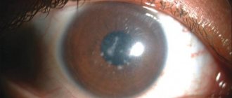
- Changing the shade of the iris.
- Intolerance to bright light.
- Dark spots appear on the surface of the eye.
Also, eye ailments are accompanied by difficulties when moving, a person has to stick to the walls. There are problems with orientation in space.
| When performing everyday tasks, the tilt of the head changes, making it difficult to distinguish faces and surrounding objects. Difficulties in perceiving shades are often accompanied by an absurd choice of things in incompatible colors. |