The optical system of the eye is a complex structure consisting of many important elements. It includes:
- cornea;
- lens;
- vitreous body;
- cameras of the eye;
- retina
The cornea is the completely transparent part of the eyeball, having a convex shape. The cornea occupies the anterior section of the eye and plays the role of the main refractive medium. It reaches 11.5 mm vertically and 12 mm horizontally, its thickness is heterogeneous. The cornea is formed by 5 layers: epithelium, stroma, Descemet's membrane and others.
The lens is the transparent body inside the eye located opposite the pupil. This element is small in size (thickness up to 5 mm, height 7-9 mm). The shape of the lens resembles a biconvex lens with a slightly flattened front side. Its refractive power can reach 20-23 diopters. The transparency of the lens is ensured by special protein enzymes.
The vitreous body is a gel-like transparent substance present in the space between the lens and the retina, occupying up to 2/3 of the volume of the eyeball. The composition of this element is 99% water. There is also a considerable amount of hyaluronic acid in the vitreous body. Inside it there are numerous channels, blood vessels, and an artery that supplies the lens.
The chambers of the eye are closed spaces with intraocular fluid. Normally, they communicate through an opening in the iris (pupil). The eyeball has anterior and posterior chambers. The first is located behind the cornea. The second is located behind the iris and extends all the way to the vitreous. The aqueous humor present in the eye chambers has a composition similar to blood plasma.
The retina (retina, retina) is the inner layer of the eye, formed by highly differentiated nervous tissue. In its central section is the macula (macula), which is the place of greatest visual acuity. The retina consists of many layers containing blood vessels and neurons. The retina contains special light-sensitive receptors called rods and cones.
The parts of the visual organ, represented by the cornea, lens, vitreous body, and eye chambers, are natural lenses with different light refractive indexes. The remaining elements of the eye, such as the iris, pupil, sclera, and ciliary body are not included in the optical system.
The optical apparatus has a reliable device, perfectly suited for visual perception. Despite the fact that the quality of what is visible is lower than in advanced technical systems, it is more than sufficient for the needs of the human body.
Optical system functions
The main purpose of the optical system of the eye is to provide a person with information about the world around him. Its elements are responsible for important features of the vision:
- Binocularity is visual perception with both eyes. This property is supported by a natural reflex, thanks to which the images received by each organ of vision are combined into single pictures.
- Stereoscopicity, which allows you to estimate the distances at which objects are located, as well as perceive them in relief. This function is fully present if objects are viewed simultaneously with both eyes.
The quality of the image is affected by visual acuity, which depends on the size of the cones in the macula area. It is also due to:
- type of refraction;
- transparency of the cornea;
- degree of elasticity of the lens;
- pupil sizes.
Thanks to the natural adaptive abilities of the eye, the optical system adapts to varying degrees of illumination. The sensitivity of the visual apparatus is determined by many factors, among which the intensity of the light source, wavelength, and duration of exposure to the light stimulus predominate.
As the human body ages, the optical characteristics of the organs of vision gradually deteriorate and the sensitivity of the eyeball gradually decreases. Premature development of visual defects can be avoided thanks to high-quality prevention and constant care for eye health.
III. Light-receiving and light-transmitting parts of the eye. Optical power of the eye. Accommodation.
The eye is the perceptive section of the visual analyzer, which serves to perceive light stimuli. Through the eyes, a person receives up to 90% of information about the world around him.
1. The sclera is a fairly strong outer protein shell that protects the eye from damage and gives it a permanent shape.
2. Cornea - the anterior part of the sclera, more convex and transparent; acting as a converging lens with an optical power of +(42-43) diopters. The sclera provides up to 75% of the focusing power of the eye. Its thickness is 0.6-1 mm, and its refractive index
n = 1.38.
3. Conjunctiva - the outer layer of the eye, plays a barrier and protective role.
4. Choroid
- On the inside, the sclera is lined with the choroid. This is a very thin membrane containing blood vessels. In the anterior part it thickens and takes the shape of a ring. This is where the iris and ciliary muscle are attached.
Pigment shell,
containing dark pigment cells that prevent light from scattering into the eye.
5. Iris —
in the anterior part, the choroid passes into the colored iris, the color of which determines the color of the eyes.
6. Pupil
-
a round hole in the iris that allows light to pass through.
The pupil diameter can vary from 2 to 8 mm. The iris and pupil play the role of a diaphragm, regulating the flow of light into the eye. 7. X Rustalik is a natural elastic biconvex lens with a diameter of 8-10 mm and an optical power of +(20-30) diopters. The lens has a layered structure with the highest refractive index n = 1.41; located behind the iris.
8.Front camera —
chamber with an aqueous mass (n = aqueous), which is located in the front of the eye between the cornea and lens, optical power + (2-4) diopters.
9. The vitreous body is a gelatinous substance that fills the space between the lens and the retina (posterior chamber of the eye). Optical power -(5-6) diopters.
10. Optic nerve , which transmits visual information to the brain. Approaching the eye, it branches, forming a light-sensitive layer on the back wall of the choroid - the retina.
11. The retina is a photosensitive layer that perceives light and converts it into nerve impulses. The retina is a branch of the optic nerve with nerve endings in the form of rods and cones. Cones (there are approximately 10 million of them) are used to perceive small details of an object and distinguish colors; The diameter of the cone is 7 µm and the length is about 35 µm.
Rods (120 million cells) do not perceive differences in color and small details, but they are highly sensitive to weak light (responsible for twilight vision). With the help of sticks, a person distinguishes objects at dusk and at night. The diameter of the rod is 2 microns and the length is 6 microns.
Rods and cones are distributed unevenly: cones predominate in the middle part of the retina, and rods predominate at the edges. The sensitivity of the retina is very high: the light of an ordinary candle is visible at a distance of several kilometers.
12. Blind spot —
located where the optic nerve enters the eye. There are no rods or cones, and rays striking this area do not cause the sensation of light (hence the name “blind spot”).
13. The macula (macula) is the most sensitive area of the retina, with an area of about 3 mm2. A person sees clearly those objects whose image is projected onto the macula. Fossa fovea
- the most sensitive part of the macula. This is an area about half a millimeter in diameter in which the retina is recessed. Here there are no rods at all, and the concentration of cones is maximum (the best vision).
14. The ciliary body is the junction of the sclera and cornea, designed to accommodate the eye, supports, fixes and stretches the lens. When the ciliary muscle contracts, the convexity of the lens increases and accommodation occurs to nearby objects (and vice versa).
15. The annular muscle is a muscle that covers the lens and can change the curvature of its surfaces. When the annular muscle is compressed, the optical power of the lens increases.
Thus, the eye includes:
1.Light conducting apparatus —
formed by the cornea, anterior chamber fluid, lens and vitreous body.
The eye is a centered optical system, the main optical axis
(OO) of which passes through the centers of the cornea, pupil, and lens.
2. Light-perceiving (receptive) apparatus - the retina, which contains light-sensitive visual cells (rods and cones).
3. Supporting-mechanical apparatus - sclera, lens capsule and its ligament, vitreous body.
4. Light-regulating apparatus - iris, ciliary body.
The optical power of the eye consists of the optical powers of the cornea, anterior chamber fluid, lens and vitreous body and is calculated as the inverse focal length:
where is the back focal length of the eye, expressed in meters.
With a completely relaxed circular muscle, the optical power of the eye is about +60 diopters, with maximum tension of the circular muscle (examining close objects) D > +70 diopters.
Accommodation (comodus - convenient) is the ability of the eye to clearly see objects located at different distances from the eye.
Accommodation is based on the ability of the lens to change its curvature under the influence of impulses sent from the central nervous system to the ciliary muscle.
Its main characteristics:
1. Near Point of Clear Vision
(punctumproximum) (PP) is
the point corresponding to the min distance at which a strained eye can clearly distinguish an object. It depends on the properties of the accommodation apparatus, on the elasticity of the lens, and with age it moves away. At a young age, it is located at r = 7-10 cm from the eye.
2. Farthest point of clear vision
(punctum remootum) (Pr) is the point corresponding to the maximum distance of clear vision without eye strain. In a normal eye, this point is at conventional infinity. Its position is determined by the anatomical abilities of the eye.
3. Range or volume of accommodation
(L, D, Da) is the distance between Рr and Рр or the difference in the optical powers of the eye when set to the far and near points of clear vision.
Quantitatively:
1. Yes = D = Drr – Drr
2. L= Lpr - Lрp
Yes - accommodation range (in diopters diopter)
Dopter - diopter, a unit of optical power of lenses and optical systems:
1 diopter is the refractive power of a lens with a focal length of 1 meter.
Lpp is the distance from the apex of the cornea to the nearest point (in “ m ”).
Lрr is the distance from the apex of the cornea to the far point (in “ m ”).
At the age of ≈ 20 years, the range of accommodation is 10 diopters, at 40 years old - 2.5 diopters, by 60 years old it is 1-0.5 diopters.
Diseases of the optical system and their symptoms
Common diseases of the optical apparatus include:
- Myopia (nearsightedness), leading to defective perception of distant objects.
- Hypermetropia (farsightedness), which causes the inability to properly visualize objects that are close.
- Astigmatism, causing poor vision at various distances.
- Keratitis, characterized by the presence of corneal syndrome.
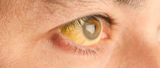
- A cataract that affects the lens, leading to clouding and hardening.
- Glaucoma, which causes an increase in intraocular pressure, a gradual deterioration of peripheral vision, and painful attacks.
- A cataract, which is characterized by the loss of normal transparency by the cornea.
Less common are other pathologies that can directly affect optical structures. The list of such diseases includes keratoconus, keratoglobus, amblyopia, and color blindness.
Each of the ophthalmological disorders has its own characteristic signs. Common symptoms of diseases of optical structures include diplopia (double picture), decreased visual acuity, decreased field of view, increased eye fatigue, abnormal tear production, and photophobia. If there are problems with binocular vision, migraines often develop. If there has been an injury to the organ of vision, pain and a feeling of the presence of a foreign body in the eyeball occur. Infectious diseases provoke redness of the eyes, the appearance of purulent discharge, and blurred images.
Image detail
Visual acuity is responsible for the detail of an image or the ability to distinguish two points separately at a certain distance. First of all, the acuity of visual perception is determined by the angle formed by the rays reflected from the extreme points of the object in question. Moreover, the smaller this angle, the higher the acuity of visual perception.
Such an indicator as acuity is determined by the size of the cones located in the retina, in the area of the macula, as well as some associated factors, such as refraction, pupil size, degree of transparency of the cornea, elasticity of the lens and much more.
The optics of the human eye is a very complex system that requires constant attention, because timely prevention of certain diseases of the visual system will preserve your vision for many years.
Diagnostic and treatment methods
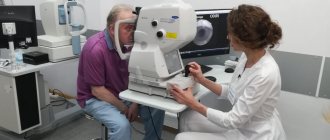
To identify existing diseases of the optical apparatus, patients are prescribed a detailed ophthalmological diagnosis. The examination consists of several informative procedures:
- visometry, which specifies the level of visual acuity;
- ophthalmometry necessary to determine the refractive abilities of the cornea;
- ophthalmoscopy, which examines the condition of the retina and fundus of the eye;
- keratoscopy, which examines the cornea of the eye;
- tonometry, which helps determine intraocular pressure indicators;
- pachymetry, which consists of measuring the thickness of the ocular cornea;
- skiascopy, which helps to obtain information regarding refraction.
- biomicroscopy, which studies in detail the superficial and deep structures of the eyeball.
If necessary, standard examination of the visual apparatus is supplemented with modern methods such as ultrasound, CT, and MRI.
The approach to the treatment of ophthalmological diseases is determined by the type of diagnosis the patient has. In the treatment of myopia, hypermetropia, and astigmatism, spectacle or lens correction methods are predominantly used. In case of infection of the eyeballs, anti-inflammatory drops, antiviral or antibacterial ointments are prescribed. Hardware procedures help enhance the effectiveness of the main treatment course. In the event of the development of serious pathologies, specialists decide on the need for surgical intervention.
The optical system of the eye is the most important part of the human body, allowing us to daily perceive the world around us and enjoy its beauty. It must be remembered that the organs of vision are highly sensitive and require constant care. Even minor disturbances in the functioning of the optical system should not be ignored and require timely medical attention.
Structure and functioning of the cornea
This elastic shell is similar in shape to a convex-concave lens. Cornea
protects the front of the eye. It does not contain blood vessels and consists of 5 layers.
In normal condition, the cornea of the eye is transparent, shiny and smooth, and has a high degree of sensitivity. Diameter of the cornea:
- vertically – 11.5 mm;
- horizontally – 12 mm.
The average thickness of the central part is 500 microns, the peripheral part is up to 1 mm.
The cornea allows light rays to pass through it, resulting in a three-dimensional image being perceived. It is the main refractive medium of the organ of vision
.
The structure of the optical system of the eye
The optical system of the eye includes the following elements:
- Anterior chamber of the eye;
- Cornea;
- Lens;
- Retina;
- Vitreous body;
- Protective systems of the eye (eyelashes, lacrimal gland, etc.).
Moreover, all structural components of the eye have their own characteristic features:
- The shape of the eye is not completely spherical;
- In the outer parts, the refractive power of the lens is less than in the inner layers;
- Eyes may vary slightly in shape and size.
What is not included in the optical system of the eye?
Its structure does not include:
- Sclera. The cornea is transparent and transmits light. The invisible part of the outer shell of the eye is white, which can be compared to the white of an egg. It performs restrictive and protective functions.
- Iris. This part of the eye is a section of the choroid, and the iris is completely devoid of blood vessels. This is the only structure of the human body that is nourished without the intervention of the circulatory system. The pupil is located in the center of the iris, which can expand and contract under the influence of light. This feature is necessary for normal vision, as it ensures the passage of light rays of ideal diameter.
- The ciliary body, which is a connecting link between the choroid and the posterior surface of the iris. The ciliary body contains processes that perform very important functions. Firstly, they have the ability to maintain the lens in a suspended state, and secondly, they produce intraocular fluid.
- The retina is the most complex element of the organ of vision, having many layers. It is a natural sensor, which is a peripheral part of the analyzer. It is in this structure that the perception of light and color occurs. The retina is very sensitive and thin, supported by epithelial ligaments, additionally pressed by the vitreous body. The eye uses it to capture a picture and transmit it along the optic nerves to the brain. The structure of the retina consists of rod and cone cells. Cone cells distinguish color images, and rod cells are responsible for vision in the dark, but they are much more sensitive. Upon closer examination, the retina consists of ten layers, different in structure, and 9 of them are absolutely transparent.
What it is?
The cornea is spherical and the transparent part of the outer layer of the eye. It is an organic lens with a biconvex structure, which is attached to the sclera of the eye through thin fibrous fibers (limb).
Thanks to the cornea and the peculiarities of its structure, light waves easily pass into the deeper layers of the organ of vision and enter the retina.
Functions of the cornea:
- protective;
- supporting;
- light conductive;
- refractive.
Normally, its characteristic features are:
- high sensitivity and ability to regenerate;
- transparency and specularity;
- spherical structure;
- strength and integrity;
- absence of capillaries;
- radius of curvature 7.7-9.6 mm;
- horizontal diameter 11 mm;
- the refractive power of light is 41 diopters.
Inflammation, injury or degenerative processes in the cornea lead to a change in its original parameters and properties.
Structure and functioning of the retina
Retina
, or retina, is a highly sensitive tissue consisting of several layers. This is the inner lining of the eye, which is formed by neurons and blood vessels.
The retina contains two types of receptors - rods and cones, so named because of their shape. They are what allow the eye to discern light.
The retina plays a critical role in visual perception. It is responsible for central and peripheral vision, the ability to see colors and shades.
Physiological role of the optical system of the eye
The main functions provided by the optical system of the eye are presented below:
- Required degree of refraction of rays;
- Focusing images and objects strictly in the plane of the retina;
- Creating the required length of the visual axis.
As a result, a person can perceive objects in volume, clearly and in color, that is, signals about a realistic image are sent to the brain structures. At the same time, the eye is able to perceive dark and light, as well as color indicators, that is, it has the function of light perception and color perception, respectively.
The optical system of the human eye has the following characteristics:
1. Binocularity is the ability to perceive a three-dimensional image with both eyes, while objects do not split into two. This happens at the reflex level, one eye acts as a leader, the second as a slave. 2. Stereoscopicity allows a person to determine the approximate distance to an object and evaluate the relief and outlines. 3. Visual acuity is determined by the ability to distinguish between two points that are located at a certain distance from each other.
Structure and properties of the eye
The eye consists of an eyeball with a diameter of 22–24 mm, covered by an opaque membrane, the sclera, and in front by a transparent cornea (or cornea ).
The sclera and cornea protect the eye and serve as anchorage for the oculomotor muscles. The iris is a thin vascular plate that limits the passing beam of rays. Light enters the eye through the pupil. Depending on the lighting, the diameter of the pupil can vary from 1 to 8 mm.
The lens is an elastic lens that is attached to the muscles of the ciliary body. The ciliary body changes the shape of the lens. The lens divides the inner surface of the eye into an anterior chamber filled with aqueous humor and a posterior chamber filled with vitreous humor.
The inner surface of the posterior chamber is covered with a light-sensitive layer - the retina. From the retina, the light signal is transmitted to the brain via the optic nerve. Between the retina and the sclera is the choroid, which consists of a network of blood vessels that supply the eye.
a macula on the retina - the area of clearest vision. The line passing through the center of the macula and the center of the lens is called the visual axis. It is tilted upward from the optical axis of the eye by an angle of about 5 degrees. The diameter of the macula is about 1 mm, and the corresponding field of vision of the eye is 6–8 degrees.
The retina is covered with light-sensitive elements: rods and cones. Rods are more sensitive to light, but do not distinguish colors and are used for twilight vision. Cones are sensitive to colors but less sensitive to light and therefore serve for daytime vision. In the macula area, cones predominate and rods are few; To the periphery of the retina, on the contrary, the number of cones quickly decreases, and only rods remain.
In the middle of the macula is the fovea centralis. The bottom of the pit is lined only with cones. The diameter of the fovea is 0.4 mm, the field of view is 1 degree.
In the macula, individual fibers of the optic nerve approach most of the cones. Outside the macula, one optic nerve fiber serves a group of cones or rods. Therefore, in the area of the fovea and the macula, the eye can distinguish fine details, and the image falling on the rest of the retina becomes less clear. The peripheral part of the retina serves mainly for orientation in space.
The rods contain the pigment rhodopsin, which collects in them in the dark and fades in the light. The perception of light by rods is due to chemical reactions under the influence of light on rhodopsin. Cones react to light through an iodopsin reaction.
In addition to rhodopsin and iodopsin, there is a black pigment on the posterior surface of the retina. When exposed to light, this pigment penetrates the layers of the retina and, absorbing a significant part of the light energy, protects the rods and cones from strong light exposure.
At the site of the optic nerve trunk there is a blind spot. This part of the retina is not sensitive to light. The diameter of the blind spot is 1.88 mm, which corresponds to a field of view of 6 degrees. This means that a person from a distance of 1 m may not see an object with a diameter of 10 cm if its image is projected onto a blind spot.
Optical system of the eye
The optical system of the eye consists of the cornea, aqueous humor, lens and vitreous body. Refraction of light in the eye occurs mainly at the cornea and lens surfaces.
Light from the observed object passes through the optical system of the eye and is focused on the retina, forming a reverse and reduced image on it (the brain “inverts” the reverse image, and it is perceived as direct).
The refractive index of the vitreous body is greater than one, so the focal lengths of the eye in the outer space (anterior focal length) and inside the eye (posterior focal length) are not the same.
The optical power of the eye (in diopters) is calculated as the inverse of the back focal length of the eye, expressed in meters. The optical power of the eye depends on whether it is at rest (58 diopters for a normal eye) or in the state of greatest accommodation (70 diopters).
Accommodation is the ability of the eye to clearly distinguish objects located at different distances. Accommodation occurs due to changes in the curvature of the lens when the muscles of the ciliary body are tense or relaxed. When the ciliary body is taut, the lens stretches and its radii of curvature increase. As muscle tension decreases, the curvature of the lens increases under the influence of elastic forces.
In a free, relaxed state of a normal eye, clear images of infinitely distant objects are obtained on the retina, and with the greatest accommodation, the closest objects are visible.
The position of an object that creates a sharp image on the retina for an unstrained eye is called the far point of the eye.
The position of the object at which a sharp image is created on the retina with the greatest possible eye strain is called the near point of the eye.
When the eye accommodates to infinity, the back focus coincides with the retina. At the highest voltage on the retina, an image of an object located at a distance of about 9 cm is obtained.
The difference in the reciprocal values of the distances between the near and far points is called the range of accommodation of the eye (measured in diopters).
With age, the eye's ability to accommodate decreases. At the age of 20, for the average eye, the nearest point is at a distance of about 10 cm (accommodation range of 10 diopters), at 50 years of age, the nearest point is already at a distance of about 40 cm (accommodation range of 2.5 diopters), and by the age of 60 it goes to infinity , that is, accommodation stops. This phenomenon is called age-related farsightedness or presbyopia.
The distance of best vision is the distance at which the normal eye experiences the least strain when viewing the details of an object. With normal vision, it averages 25–30 cm.
Adaptation of the eye to changed lighting conditions is called adaptation. Adaptation occurs due to changes in the diameter of the pupil opening, movement of black pigment in the layers of the retina and different reactions to light of rods and cones. The pupil contracts in 5 seconds, and its full dilation occurs in 5 minutes.
Dark adaptation occurs during the transition from high brightness to low brightness. In bright light, the cones work, but the rods are “blinded”, the rhodopsin has faded, the black pigment has penetrated into the retina, shielding the cones from the light. With a sharp decrease in brightness, the pupil opening opens, allowing more light to pass through. Then the black pigment leaves the retina, rhodopsin is restored, and when there is enough of it, the rods begin to function. Since cones are not sensitive to low brightness, at first the eye does not distinguish anything. The sensitivity of the eye reaches its maximum value after 50–60 minutes of exposure to darkness.
Light adaptation is the process of adaptation of the eye during the transition from low to high brightnesses. At first, the rods are greatly irritated, “blinded” due to the rapid decomposition of rhodopsin. The cones, not yet protected by grains of black pigment, are also too irritated. After 8–10 minutes, the feeling of blindness stops and the eye sees again.
The field of view of the eye is quite wide (125 degrees vertically and 150 degrees horizontally), but only a small part of it is used for clear discrimination. The field of the most perfect vision (corresponding to the fovea) is about 1–1.5°, satisfactory (in the area of the entire macula) is about 8° horizontally and 6° vertically. The rest of the visual field serves for rough orientation in space. To view the surrounding space, the eye has to make a continuous rotational movement in its orbit within 45–50°. This rotation brings images of various objects to the fovea and makes it possible to examine them in detail. Eye movements occur without the participation of consciousness and, as a rule, are not noticed by a person.
Treatment regimens
Let's consider the principles of treating diseases depending on their type:
- Erosive changes and minor injuries are treated by using local anesthetics and healing ointments. To prevent infection, your doctor may prescribe an antibiotic ointment. Preparations with hyaluronic acid and drops such as artificial tears speed up recovery. With proper, timely treatment, erosions are well epithelialized and no complications arise.
- Severe wounds are treated surgically in a specialized ophthalmology department. In parallel, therapy with antibiotics, enzymes, and local healing drops is indicated. If you let the problem go, you can go blind.
- Burns and damaged tissue are surgically excised, antibacterial, anti-inflammatory, healing ointments are used, and enzyme therapy is performed.
- If foreign bodies enter, they must first be removed with a cotton swab or a special tool. If this is not possible, the body is left in the eye for a certain time until it moves to the superficial layer. You can only leave glass and plastic that are neutral from a chemical point of view. To speed up healing, the doctor prescribes drops and ointments.
- Keratitis is a pathological measurement in the structure of the cornea, which leads to clouding of the cornea and reduces visual acuity. The nature of the disease is infectious or traumatic. Treatment as for injuries and infections.
- Changes in the shape and size of the shell, the cornea can be too large, small, protrude, or take on the shape of a cone. In severe forms, surgery is indicated; in childhood, conical changes are accompanied by the development of astigmatism, which cannot be cured.
It is important to eliminate the damaging, traumatic factor, otherwise the therapy will not be effective. When this is done, regenerative treatment can be prescribed.
Diagnostic methods for damage to the optical system of the eye
When assessing the operation of the optical system as a whole, it is necessary to clearly determine which eye is the master and which is the slave.
This can be easily determined by a simple test. In this case, you need to look through the hole in the dark screen alternately with your right and left eyes. If the eye is dominant, then the picture does not move. If the eye is a slave, then the picture shifts.
To diagnose diseases, it is necessary to perform a number of techniques:
- Visometry is necessary to determine visual acuity. It can also be carried out against the background of spectacle correction in order to select lenses.
- Skiascopy helps to obtain objective data on the value of refraction.
- Automatic refractometry.
- Ophthalmometry allows you to determine the refractive power of the cornea.
- Pachymetry measures the thickness of the cornea in different areas.
- During keratoscopy, the doctor looks at the cornea through a lens.
- Ultrasound of the eyeball.
- Photokeratotopography.
- Ophthalmoscopy examines the fundus and retina.
- Biomicroscopic examination.
It should be recalled once again that the optical system of the eye is the most important in the structure of this organ. It allows you to obtain a high-quality image on the retina. This is possible due to the implementation of several mechanisms, which include binocularity, refraction, stereoscopicity and some others. If at least one structure of this complex system is damaged, its operation is disrupted. This is why early diagnosis is so important. Only under this condition can you maintain rich and clear vision.
Harmful factors affecting the cornea
The eyes are regularly exposed to the following harmful effects:
- contact with mechanical particles suspended in the air;
- chemicals;
- air movement;
- temperature changes.
When foreign particles enter the eye, a person’s eyelids close according to an unconditional reflex, tears flow intensely and a reaction to light is observed. Tears help flush foreign agents from the surface of the eye. As a result, the protective functions of the cornea are fully demonstrated. No serious damage to the shell occurs.
The same protective reaction is observed during chemical exposure, strong wind, bright sun, cold and heat.
Symptoms of astigmatism
The main manifestation of astigmatism is visual impairment, but over time, other symptoms from the central nervous system and other systems and organs may develop.
With astigmatism, symptoms may vary from patient to patient, but most often patients complain of:
- decreased visual acuity;
- blurry, cloudy, distorted perception of objects;
- irritation, pain, discomfort in the eyes;
- difficulties in reading, looking at small pictures, lines;
- headache, more pronounced in the superciliary areas;
- feeling of tiredness in the eyes;
- fatigue when reading, visual strain;
- To see better you have to tilt or turn your head. A particularly important symptom for attentive parents who may suspect astigmatism in their child if he begins to turn half a turn in order to better examine the object;
- inability to clearly see both near and distant objects.
Interesting to know
According to scientific research by scientists, children in infancy have weak refraction. Vision in children in the first years of life is characterized by farsightedness and gradually transforms into indicators of normal (emmetropia) or myopia (myopia).
The eyeball grows until the age of 15 (intensively up to 3 years), due to which the refraction constantly increases. With age, the length of the main optical axis increases, reaching 22 mm by the age of 7 years (95% of the axis of a healthy adult eye).
Lens
The eye as an optical system is equipped with a refractive element that performs the function of refraction. This is the lens. It can be considered as an independent organ, complex in structure and most important in function.
The lens has the appearance of a semi-solid substance without blood vessels. It is located immediately behind the iris and is responsible for transmitting a clear display of the image seen within the boundaries of the macula to the retina.
The lens has several different layers and a capsular bag that can thicken over time and cause opacification on the surface of the body.
The eye as an optical instrument
The human eye is a complex optical system, which in its action is similar to the optical system of a camera. The schematic structure of the eye is shown in Fig. 3.4.1. The eye has an almost spherical shape and a diameter of about 2.5 cm. On the outside, it is covered with a protective shell of 1 white color - sclera . The anterior transparent part 2 of the sclera is called the cornea . At some distance from it there is an iris 3, colored with pigment. The hole in the iris represents the pupil . Depending on the intensity of the incident light, the pupil reflexively changes its diameter from approximately 2 to 8 mm, i.e. acts like a camera diaphragm. There is a clear fluid between the cornea and the iris. Behind the pupil there is a lens 4 - an elastic lens-like body. A special muscle 5 can change the shape of the lens within certain limits, thereby changing its optical power. The rest of the eye is filled with vitreous humor. The back of the eye is the fundus, it is covered with a retina 6, which is a complex branching of the optic nerve 7 with nerve endings - rods and cones , which are light-sensitive elements.
| Figure 3.4.1. Human eye |
Rays of light from an object, refracted at the air-cornea boundary, pass further through the lens (a lens with varying optical power) and create an image on the retina.
The cornea, clear fluid, lens and vitreous body form an optical system, the optical center of which is located at a distance of about 5 mm from the cornea. With a relaxed eye muscle, the optical power of the eye is approximately 59 diopters, and with maximum muscle tension it is 70 diopters.
The main feature of the eye as an optical instrument is the ability to reflexively change the optical power of the eye optics depending on the position of the object. This adaptation of the eye to changes in the position of the observed object is called accommodation .
The area of accommodation of the eye can be determined by the position of two points:
• the far point of accommodation is determined by the position of the object, the image of which is obtained on the retina when the eye muscle is relaxed. In a normal eye, the far point of accommodation is at infinity.
• near point of accommodation – the distance from the object in question to the eye at maximum tension of the eye muscle. The near point of a normal eye is located at a distance of 10–20 cm from the eye. With age, this distance increases.
In addition to these two points that define the boundaries of the accommodation area, the eye has a distance of best vision , i.e. the distance from the object to the eye at which it is most convenient (without excessive strain) to examine the details of the object (for example, to read small text). This distance in a normal eye is conventionally assumed to be 25 cm.
When vision is impaired, images of distant objects in the case of an unstrained eye may appear either in front of the retina ( myopia ) or behind the retina ( farsightedness ) (Fig. 3.4.2).
| Figure 3.4.2. Image of a distant object in the eye: a – normal eye; b – myopic eye; c – farsighted eye |
The distance of best vision in a nearsighted eye is shorter, and in a farsighted eye it is longer than in a normal eye. Glasses are used to correct vision defects. For a far-sighted eye, glasses with positive optical power (converging lenses) are needed, for a near-sighted eye - with negative optical power (divergent lenses).
To observe distant objects, the optical power of the lenses must be such that parallel beams are focused on the retina. The eye must see through the glasses an imaginary direct image of a distant object located at the far point of accommodation of a given eye. If, for example, the far point of accommodation of a myopic eye is at a distance of 80 cm, then using the thin lens formula we obtain:
d = ∞, f = –0.8 m, therefore diopters.
It should be noted that in a far-sighted eye, the far point of accommodation is imaginary, i.e., an unstrained eye focuses a converging beam of rays on the retina. Therefore, when viewing distant objects, glasses for far-sighted eyes must transform a parallel beam of rays into a converging one, i.e., have a positive optical power.
Glasses for “near vision” (for example, for reading) should create a virtual image of an object located at a distance d0 = 25 cm (i.e., at the distance of best vision of a normal eye), at the distance of best vision of a given eye. Let, for example, a myopic eye have a best vision distance of 16 cm. Using the thin lens formula, we obtain: d = d0 = 0.25 m, f = –0.16 m, therefore, diopter. Due to the narrowing of the area of accommodation for many people, glasses for near vision must have greater (in absolute value) optical power compared to glasses for viewing distant objects.
Rice. 3.4.3 illustrates the correction of farsighted and nearsighted eyes using glasses.
| Figure 3.4.3. Selection of reading glasses for farsighted (a) and nearsighted (b) eyes. Object A is located at a distance d = d0 = 25 cm of the best vision of a normal eye. The virtual image A' is located at a distance f equal to the distance of best vision of a given eye |
| Model. The eye as an optical instrument |
Wonders of vision in nature
Snakes have unique eyes that can perceive infrared radiation. Thanks to this ability, they successfully hunt warm-blooded animals even in zero light conditions.
Butterflies have a different feature: wonderful creatures perceive part of the ultraviolet sector, so it is not difficult for them to detect pollen in flowers.
Geckos are famous for their excellent night vision. Moreover, they see in the same spectral range as people. It’s just that their retina is three hundred and fifty times more sensitive to light rays. A real night vision device!
The chameleon deserves special attention. He doesn’t need to turn his head to see all three hundred and sixty degrees of the environment. He is able to measure the distance to an object with one eye.
The giant squid boasts the largest eyes on the entire planet. He lives in the depths of the ocean, at its very bottom. There is almost never sunlight here, but the mollusk is able to see its enemy at a distance of a thousand meters.
Chamber moisture
The optical system of the eye includes the most important biological medium, aqueous humor. It is a colorless viscous liquid that fills the anterior and posterior ocular chambers. Every day a new portion of intraocular fluid is produced, and the spent amount is released into the bloodstream through Schlemm’s canal.
Chamber moisture, in addition to its refractive function, also performs a nutritional function, saturating all elements of the eye with amino acids. Difficulty in leaving the chamber leads to the development of glaucoma.
Orbicularis oculi muscle and its functions
The orbicularis oculi muscle is one of the facial muscles of facial expression. This muscle, paired with the levator pallidum, is responsible for the movement of the eyelids. It is mainly involved in lowering the eyelids and closing the palpebral fissure.
The orbicularis oculi muscle consists of three parts:
- Orbital
- Century-old
- Tearful
Orbital part of the muscle
starts from the frontal process of the upper jaw. Next, it extends along the upper and lower edges of the orbit. As a result of fusion, a ring is formed.
Secular part of the muscle
continues the circular muscle and is located under the skin of the eyelids, respectively. The muscle has two parts - upper and lower. These muscles begin at the medial ligament of the eyelids of the upper and lower edges of the eyelids and go to the corners of the eyes. In these places they are attached to the lateral ligament of the eyelids.
lacrimal part,
also called Horner's muscle, is a deep network of muscle parts. It starts from the posterior crest of the lacrimal ossicle, where it divides into two parts.
Each part covers the lacrimal sac from the front and back. Then they are “lost” among the muscle bundles of the periphery. It is the peripheral part of one of the parts of the divided muscle that narrows the palpebral fissure and smoothes the transverse folds of the skin on the forehead.
The inner part of this muscle closes the palpebral fissure. In addition, the lacrimal muscle expands the lacrimal sac.
Thus, it should be concluded that the optical system of the eye, its muscular apparatus and other anatomical features is a very complex and specific system, which in a second of time is capable of carrying out many different movements that provide us with high-quality vision and perception of the world around us.
Top
Optics Basics
Let's remember the school physics curriculum. Many teachers showed students an entertaining trick: two rooms with low levels of light, but one of them has small holes in the walls. Behind them is a strong light source, such as the sun. In some cases, instead of the pinholes used to illuminate the room, a small flashlight was used.
If an object made of opaque material is placed between a point light source and the second hole in the wall, then its image will appear on the partition located behind the second hole, inverted one hundred and eighty degrees.
A converging lens performs a similar trick with light rays. The reason lies in the fact that every microscopic point of any object, when illuminated, itself becomes a source of light, reflecting particles that fall on it in all directions.
White products absorb practically nothing from the visible range; they reflect all the light that hits them into the environment. Black objects, on the contrary, use any source of energy for heating.
Treatment
Treatment methods can be used in a variety of ways, and will mainly depend not only on the causes of the disease, but also on the age of the patient and the general clinical picture. For example, infectious diseases require the use of eye drops containing antibiotics. If the problem is caused by more serious reasons, such as thinning of the cornea, the formation of scars and blisters on it, then the patient is observed for a long time and, if absolutely necessary, is operated on.
The main thing to remember is that, regardless of the reasons, if your vision deteriorates, you must immediately consult an ophthalmologist who can make the correct diagnosis and prescribe effective treatment.
Clinical and physical refraction
Refraction of the eye has the following definition: the refractive power of the eye, indicated in diopters. The physical refraction of the eye in humans can vary from 51.8 to 71.3 diopters, this has been determined through numerous studies.
For a clear image of any external picture, not only the refractive power of the entire optical system is important, but also the ability of this power to focus light rays on the retina. Therefore, in ophthalmology the term refraction of the eye is also used.
Clinical refraction of the eye is the relationship that arises between the position of the retina and the refractive power or, in other words, the relationship between the length of the anteroposterior axis of the eye and the posterior focal length of the entire optical system. Clinical refraction of the eye is divided into dynamic and static.
Static refraction allows you to obtain an image on the retina at the moment of maximum relaxation of accommodation. Static refraction can be perceived as a conventional symbol that reflects the structural features of the human eye as an optical camera responsible for forming an image on the retina.
Knowledge of all functional features related to the optical system of the eye allows ophthalmologists to solve many questions about the characteristics of visual activity under normal conditions. Dynamic refraction allows one to judge the optical system of the eye. This term refers to the refractive power in the optical system of the eye relative to the retina during functioning accommodation.
Light refraction
The main refractive media of the human eye are the cornea, which has the highest refractive power, and the lens, which is a biconvex lens. The refraction of light in the eye follows the basic laws that physics studies. Rays passing through the center of the lens and cornea (i.e., through the main ocular optical axis) perpendicular to their surface do not experience refraction. The rest are refracted and inside the camera the eyes converge at a single point - focus. This path of light rays provides a clear image on the retina, and it turns out to be reversed and reduced.
The refractive index of light in the vitreous body is greater than one, so the focal lengths in the outer space (front focal length) and inside (back focal length) cannot be the same. Optical power is calculated as the inverse of the back focal length of the eye, expressed in meters. It depends on whether the organ of vision is at rest or in a state of accommodation. Accommodation is the ability to clearly distinguish objects that are at different distances.