Fundus of the eye - what is it?
This concept refers to the inner part of the eye with the retina, optic nerve head, blood vessels and periphery. Under an ophthalmoscope, a special instrument that is used to examine this area of the eyeball, you can see that the fundus of the eye has a reddish tint. In this case, the optic disc is pale pink, it stands out noticeably against the background of the overall reddish picture. At the very center of the disc are the central retinal vein and artery. They are divided into two branches - lower and upper, after which they diverge into many small branches that spread throughout the entire fundus of the eye. In the very center of the retina is the macula, or macula. Compared to other described structures, the macula has a darker color. Any changes in the color, shape, outline of the disc, macula, or retina may be a consequence of the development of a pathological process. It can be detected at an early stage through a detailed examination of the fundus. The procedure used to examine it is called ophthalmoscopy.
Normal fundus. Ophthalmoscopic picture of a normal fundus
Description
The most noticeable and prominent part of the fundus is the optic nerve papilla with vessels running from it in all directions (Table 5, Fig. 1). In order to see the papilla, it is necessary to invite the examinee to look slightly towards his nose or, as mentioned earlier, the examinee should direct his gaze when examining the left eye to the doctor’s left ear, and when examining the right eye, look slightly past the examiner’s right eye.
If the papilla does not fall into the field of vision, then, with direct ophthalmoscopy, it can be easily found through the retinal vessels.
To do this, it is necessary to notice in which direction the vessels go after branching, and if it turns out, for example, that in a given area of the fundus the vessels go upward, the papilla should be looked for at the bottom, if the vessels go to the side, the papilla is located on the right, etc. This is clearly visible in Fig. . 36, where the fundus of the eye is schematically depicted, part of which, outlined in a circle, fell into the ophthalmoscopic field of view. In the indicated area of the fundus of the eye, after branching, the dishes go to the right, therefore, the papilla is located on the left. In other words, the top of the eye formed by the branching of the retinal vessels, like an arrow, indicates the direction in which to look for the optic nerve papilla.
If the patient is forced to gradually move the eye in the direction indicated by this “anatomical arrow,” the sought-after papilla will appear in the field of vision.
This method of searching for the papilla through the vessels can also be used for reverse ophthalmoscopy; you just need to remember that when examining this method, the patient must turn the eye not in the direction where the papilla is located, but in the opposite direction (against the anatomical arrow).
However, the location of the papilla can be determined simply by ophthalmoscopic transillumination, namely: if a beam of light is directed into the eye with an ophthalmoscope from a distance of 40-60 cm, then, as is known, the reflex from the fundus of the eye will be brightest when the light is reflected from a lighter surface of the papilla. If we sang, now, without losing this area of the fundus, proceed to an ophthalmoscopic examination, then the optic nerve papilla will be in the ophthalmoscopic field of view.
The papilla appears as a yellowish-red or grayish-red circle, which is always lighter than the rest of the bottom of the eye and especially stands out in pigmented eyes. The shape of the papilla is either completely round or slightly oval. The inner edge of the papilla is usually less clearly defined than the outer edge. This is explained by the fact that in the inner part of the papilla there are more nerve fibers and blood vessels.
For the same reason, the inner half of the papilla is redder than the outer half, where the layer of nerve sills is thinner and therefore the white reflex of the cribriform plate is more clearly visible. The papilla is quite often surrounded by a narrow yellowish-white stripe, which is adjacent in the form of a sickle to its outer part or encloses the papilla in a continuous ring. This so-called scleral ring depends on the fact that the hole in the choroid through which the optic seal passes is larger than the hole in the sclera, which is visible in the form of a yellowish-white crescent or ring.
An accumulation of pigment is often observed near the outer border of the scleral ring, which is explained by the stronger pigmentation of the edge of the hole in the choroid. The dark strip bordering some part of the nipple is called the choroidal ring; its width in some cases can reach 14 PD (PD is the diameter of the papilla).
The retinal vessels emerge either from the center of the papilla or slightly inward from the center. The further direction of the vessels and their division is described above in the anatomical sketch. It is quite easy to distinguish arteries from veins: arteries are somewhat thinner than veins, have a lighter (orange-red) color and are less tortuous.
The veins always appear thicker because they are compressed (flattened) by the pressure of the vitreous, are cherry-red in color, and are more tortuous. Arteries also differ from veins by a characteristic reflex in the form of a light central stripe, which is clearly visible on large vascular trunks. On the arteries, the ventral light stripes are light pink, their width is about 14 times the diameter of the vessel.
In those places where the vessel bends so that it is no longer in a plane perpendicular to the line of the observer, the light stripe either becomes poorly visible or disappears completely. Light reflexes on the arteries depend on the reflection of light by the central part of the blood column moving in the vessels. The veins have light central white stripes, their width is much smaller than on the arteries, and is equal to 110 to 112 - the diameter of the vessel.
It disappears at the slightest bend of the vein in a plane that is not perpendicular to the observer’s visual line. Light reflexes on large vein trunks in the area of the papilla and in the immediate vicinity of it may often be absent. The veins of the vessels are almost completely transparent, but in some cases there is a double-circuit of the arteries in the form of delicate, white light strips that accompany the vessel on one side and the other, parallel to the central light strip.
These additional light stripes can be observed in large trunks: -: arteries only in the area of the papilla or close to it. In some eyes with a sharply pigmented fundus, around the papilla over several PDs, the retina has a Serov, it seems as if; The posterior part of the fundus is covered with a light veil. Upon detailed examination (in direct form), one can notice that the retina around the papilla is, as it were, striated with many radially located stripes, which depends on the presence of developed supporting tissue in it, which is located mainly along the nerve fibers.
In eyes with a sharply pigmented fundus, wavy, shiny white stripes can be observed, which are located mainly along the vessels, but can also cross the latter. Sometimes they have the shape of a wide variety of shapes: a sickle, an irregular opal, etc. Whatever shape these wavy stripes have, they are nothing more than light reflexes of the retina.
This can be easily verified if, during the examination, the illuminated area of the fundus of the eye is shifted in different directions by slightly rotating the ophthalmoscope; the observed stripes change their shape, position, and some disappear completely. Such an unusual picture of the fundus often confuses inexperienced researchers and they are inclined to explain the observed phenomenon by the presence of an inflammatory process in the retina, i.e. they consider such a fundus not as normal, but as pathologically altered.
Light reflexes of the retina arise due to the fact that the fundus of the eye does not actually have a strictly spherical surface, since the membrana limitans interna above the retinal vessels is slightly forward and, as a result, concave-cylindrical surfaces are formed between the vessels, which reflect the light of the ophthalmoscope in the form of bright reflexes. All these reflexes with a dilated pupil are less noticeable or even disappear altogether.
The fundus of the eye, illuminated by an ophthalmoscope, in which the papilla and retinal vessels are visible, can have not only a different color in different eyes, but also a unique pattern. In blondes, the fundus of the eye is light and has a light red color, in brunettes it is dark red, and in people with sharply pigmented skin (blacks) the fundus is almost black (the color of a raven's wing).
The color of the fundus is determined by the choroid, visible through the transparent retina, which is red. But, since the outermost layer of the retina is covered with pigment, depending on the amount of retinal pigment and its physiological color, the color of the fundus also changes. In cases where the outer layer of the retina is poorly pigmented and, as a result, the choroid is clearly visible, the fundus of the eye has not only a bright red color, but also a variegated pattern: it appears to consist of wide, looped orange-red ribbons, with dark stripes and spots between them .
These are visible choroidal vessels, which differ from the vessels of the retina primarily in that they look like wide, densely intertwined mites, which is explained by the presence of a large number of anastomoses in these vessels. The chorionic vessels pass under the retinal vessels; they do not have light reflexes; it is impossible to distinguish arteries from veins. When examining the fundus of the eye in the equatorial region, it is sometimes possible to see the vorticose veins, to which the choroidal veins approach from all sides, in the form of radially located ribbons (Table 30, Fig. 1).
In some eyes, especially in those with pronounced pigmentation of the skin and hair, due to the accumulation of pigment in the stroma of the choroid, the intervascular spaces between the choroidal vessels are sharply distinguished by their pigmentation and may have a dark brown or even black-brown color. The fundus in such cases has a peculiar spotted, almost marbled appearance (fundus labulatus).
Anyone who sees such an eye for the first time can easily mistake the detected changes in the fundus as pathological, but if you pay attention to the fact that dark spots are located on the fundus in a certain pattern, corresponding to the distribution of choroidal vessels, and also to the fact that as approaches to the equator become narrower and less convoluted, there can be no doubt that this fundus is normal (Table 5, Fig. 2).
In albino eyes, in which pigment is absent, both in the retinal pigment epithelium and in the choroid, white, shiny areas of translucent sclera are visible between the choroidal vessels, which look like light red stripes.
A very important and most difficult part of the eye fundus to study is the macula area. In order to find the yellow spot during a reverse study, the patient is inserted to look at the hole in the ophthalmoscope, since when looking in the ophthalmoscopic field of view there will be an area of the fundus corresponding to one pole of the eyeball, where, as is known, the yellow spot is located spot.
It must be remembered that with this method of examination, the optic nerve papilla is located outside the macula (reverse image), approximately at a distance of 2 PD.
When using direct ophthalmoscopy, it is most convenient to find the macula by focusing on the outer part of the papilla. To do this, first of all, they look for the optic nerve papilla, take the outer edge of the papilla as the starting point, and by slightly rotating the ophthalmoscope, move the illuminated area outward, where they look for the macula.
If you cannot find it, it is better to return again to the edge of the papilla and from there again go outward, since otherwise it is easy to deviate downwards or upwards from the actual location of the macula. The main difficulty in examining the macula is that this area is most sensitive to light, and when the fundus is illuminated with an ophthalmoscope, a sharp constriction of the pupil occurs.
In this regard, it is sometimes advisable to use a flat mirror for ophthalmoscopy, which directs less light into the eye, and when using an electric ophthalmoscope, you should simply lower the intensity of the light bulb.
The macula ophthalmoscopically is characterized primarily by the fact that small arterial branches are directed towards it from all sides. This area, the size of the C papilla, is located approximately 2 PD lateral to the optic papilla (Table 6, Fig. 1-e). When examined in reverse, it is surrounded by a bright light reflex, which has the appearance of a horizontal oval (Table 6, Fig. 1-c). The vertical diameter of the oval is equal to the diameter of the papilla, the horizontal one is slightly larger.
The inner border of the oval is sharply outlined, the outer border is unclear. The described light reflex, which is called the macular reflex, is especially clearly visible in individuals with a sharply pigmented fundus of the eye, as well as in hypermetrols. The area limited by the macular reflex is darker than the surrounding part of the fundus and has a slightly matte tint. In the center of this area, you can often see a round red-brown spot, corresponding to the fovea centralis and depending on the fact that the choroid is better visible through the thinned retina in this place (Table 6, Fig. 1-a).
Its diameter is approximately equal to 13-16 PD, but sometimes the speck can be larger and have an irregular triangular shape. The spot is especially clearly visible in eyes with a weakly pigmented bottom, where it is red and somewhat resembles a hemorrhage. When examining directly, the macular reflex is usually absent, but if, with this method of examination, strong illumination of the fundus is performed, which often happens when using an electric ophthalmoscope, then it is almost as clearly visible as when examining in the reverse view.
The dark spot corresponding to the fovea centralis is more clearly visible when examined directly and, in addition, when examined by this method, a light reflex, the so-called foveal reflex, is clearly visible in the center of the spot, which in some cases resembles a luminous point, and in others has the shape of a sickle or ring (Table 6, Fig. 1-d). And if you eat and make slight rotations with an ophthalmoscope, as is done during skiascopy, you will notice that the fosal reflex somewhat changes its shape and position.
The macular and foveal reflexes, as well as other retinal reflexes, are less visible with a dilated pupil. The reason for reflexes in the macula area has the following explanation. The macular reflex occurs when light is reflected by a ring-shaped thickening of the retina around the macula.
The darker color and matte tint of the area surrounded by the macular reflex depends on the fact that the inner slope of the ring-shaped thickening of the retina around the macula refracts rays more strongly than the adjacent part of the fundus, and therefore less light reaches the examiner from this area. The foveal reflex depends on the reflection of light by the strongly concave spherical surface of the fovea centralis and is nothing more than a reverse, reduced image of the light source that is located in front of the pupil.
It is quite clear that this image is located in the vitreous body in close proximity to the fovea centralis. When the fundus is illuminated with a mirror with a hole in the center, the reflex has the shape of a sickle or a ring shape, and when examined with an electric ophthalmoscope, an image of a hot filament of a light bulb is obtained in focus and the reflex has the appearance of a luminous point.
_______ Article from the book: Ophthalmoscopic diagnosis (with ophthalmoscopic atlas) | Radzikhovsky B.L..
Indications for ophthalmoscopy
This procedure is standard. It is carried out both separately and during any preventive examination along with other types of research. In addition, ophthalmoscopy is prescribed if there are suspicions of various diseases. With its help, you can identify the following ailments that affect the fundus of the eye:
- macular degeneration - a degenerative disease of the central region of the retina;
- occlusion and thrombosis - circulatory disorders in the central artery and central vein, respectively;
- retinitis - inflammation of the retina;
- peripheral retinal dystrophy;
- cataract - clouding of the lens;
- melanoma of the eye is a malignant neoplasm that affects the eyelid, choroid or connective tissue;
- damage to the optic nerve head.
These are serious pathologies that are important to begin treating on time, at the first stage. Ophthalmoscopy makes it possible to detect them at the very beginning of their development. If symptoms such as “floaters” and “lightning” appear before the eyes, impaired color vision, decreased visual acuity, deterioration of visual functions in low light, you should definitely make an appointment with an ophthalmologist. Ophthalmoscopy will be performed as one of the main examination methods.
The condition of the visual organs is affected not only by eye diseases, but also by other pathologies. The doctor will be able to identify certain disorders in the body by the color and structure of the fundus. In this regard, ophthalmoscopy is prescribed for such ailments as hypertension, renal failure, diabetes mellitus, tuberculosis, and epilepsy. Reasons for examination may include head injuries, persistent headaches, or a stroke. Premature newborns must be sent for ophthalmoscopy.
Women during pregnancy should be seen by an ophthalmologist. Examination of the fundus makes it possible to detect retinal detachment. This disease can lead to a ban on natural childbirth. In other words, ophthalmoscopy is one of the most basic and important diagnostic methods. At the same time, there is no need to prepare for it and it has practically no contraindications.
Who is recommended for regular fundus examinations?
This procedure is needed for patients who are at risk due to the presence of certain diseases or conditions:
- cataracts;
- type 2 diabetes, since this disease often causes retinal detachment;
- pregnancy, especially if there is myopia;
- increased intracranial pressure;
- hypertension and atherosclerosis;
- with impaired color vision and hemeralopia (“night blindness”);
- children born prematurely;
- after various injuries to the organs of vision.
In all these situations, there is a risk of detachment of the retina from the choroid, so it is necessary to monitor its condition. This will allow timely detection of disturbances in the structures of the eye, if any, and take timely measures.
When is fundus examination contraindicated?
There are no absolute contraindications that would forever exclude the possibility of prescribing ophthalmoscopy. However, there may be temporary obstacles to its implementation. If the patient currently has an infectious or inflammatory eye disease, accompanied by excessive lacrimation, increased sensitivity to light and other symptoms, an examination of the fundus will be postponed until complete recovery. Also, ophthalmoscopy will be complicated by opacities of the optical media: lens, cornea, vitreous. The ophthalmologist will not be able to fully examine the fundus. In some cases, the procedure is canceled for angle-closure glaucoma. Examination may result in increased intraocular pressure. But usually this symptom is relieved with appropriate medications.
Features of fundus examination in children
The fundus of the eye in children is checked to identify diseases of the organs of vision, pathologies of blood vessels, brain, as well as signs of hypertension.
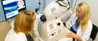
Several methods are used for ophthalmoscopy. Most often, the doctor uses a mirror ophthalmoscope; some specialists prefer an electronic device. In some cases, the study is carried out using fluorescence antiography (FAGD). Ophthalmoscopy is performed in a dark room. A frosted lamp with a power of about 100 W is installed to the left and slightly behind the patient. The doctor is at a distance of 30–40 cm from the patient and looks with his right eye through the hole of the eye mirror-ophthalmoscope, while directing a beam of light into the patient’s pupil. Light enters the eye and is then reflected from the pigment epithelium and choroid.
When checking the fundus of the eye in children, a number of difficulties arise, since small patients are not able to overcome the reflex and close their eyes to protect themselves from light. Therefore, in order to dilate the pupil, a 1% solution of “Homatropin” is instilled before the procedure. If the child cannot cope with squinting, the doctor uses an eyelid dilator. Older children are asked to focus their gaze on some picture or object.
Fundus ophthalmoscopy - how is the procedure performed?
The first ophthalmoscope was created back in the 19th century. Since then, several methods of examining the fundus have been developed. Today, various types of devices are used: Goldmann lenses, electronic and laser, monocular and binocular. The two main types of ophthalmoscopy are direct and reverse (indirect). The first is carried out as follows. The ophthalmologist sits opposite the patient. The light beam is directed directly into the pupil. The doctor holds an ophthalmoscope in his right hand and brings it closer to the eye of the patient. The retina can be seen in detail from 3-4 cm. The image is magnified approximately 16 times. This procedure lasts no more than 10 minutes. Direct ophthalmoscopy requires instillation of mydriatic drops into the patient's eyes, which dilate the pupil. The technique is very accurate due to the strong magnification of the image. But it is not without its shortcomings. The picture is not very voluminous. It is not possible to study the periphery of the fundus. To do this, you need to resort to another method - reverse ophthalmoscopy. How is this procedure carried out?
The oculist holds an ophthalmoscope in his right hand near his eye. The light source is located behind the patient's back. A little to the left of him. The ophthalmologist holds a lens in his left hand. The distance from the cornea to the optical product is 5 or 7 cm, depending on the dioptres with which the lens is equipped. The doctor directs a beam of light into the patient's eye. It is reflected from the fundus of the eye and hits the lens. The stream of light rays is refracted and forms an image from the side of the ophthalmologist. It seems to hover in the air and represents a several times enlarged picture of the inside of the eye. The magnification here is not as strong as in direct ophthalmoscopy. But it is possible to quickly check the condition of not only the optic nerve head, blood vessels and arteries, but also the peripheral area.
In some cases, both reverse and direct ophthalmoscopy are performed. The first method helps to quickly identify the pathological process, and the second helps to find out details about the disease: the nature of development, stage, causes.
What is indirect ophthalmoscopy?
Reverse ophthalmoscopy (indirect, mirror) is performed using a converging lens equipped with optical power. It can have from 10 to 30 diopters. The patient sits on a chair opposite the ophthalmologist. He holds the device at a distance of 7-8 cm from the eye of the subject. The light source is located behind the doctor. Rays of light pass through the eyeball, are reflected from its bottom and are collected at a point in front of the lens. The optometrist sees an image suspended in the air. It is mirrored and inverted.
The doctor can use lenses with greater optical power. The image becomes larger, but its quality decreases. Typically, several different lenses are used depending on the medical indication. The advantage of the procedure is that it is possible to examine the periphery of the fundus. But it is impossible to see all its structures in detail.
An ophthalmologist's office may have several ophthalmoscopes. In this case, a series of inspections are carried out. Each of them lasts only a few minutes. There are also more accurate methods for studying the fundus of the eye, which are successfully used in ophthalmology. One of them is biomicroscopy with a Goldmann lens. Let's find out what it is.
Ophthalmochromoscopy - what is it and how is this procedure performed?
As noted earlier, the inside of the eye has a reddish tint. The optic disc, vessels, arteries, veins, and macula stand out against its background due to a lighter or darker color. Ophthalmochromoscopy is a direct method of examining the fundus of the eye using color filters that are mounted on an ophthalmoscope. This technique was developed by the Soviet ophthalmologist Vodovozov. It is more informative, as it allows you to see the most minor changes that cannot be detected using white light. During the procedure, the doctor can switch the color. For example, a green filter will show whether there is optic nerve atrophy, and if disc edema is suspected, a red filter is used.
What is direct ophthalmoscopy and how is it performed?
Direct ophthalmoscopy allows you to study the fundus of the eye in detail under multiple magnification. The device magnifies the image 13-16 times. The ophthalmoscope is equipped with a light source. The doctor holds the device in his hand and points it at the internal structures of the eye. Thanks to this, it is possible to see even the smallest changes. But the method also has disadvantages. It cannot be used to examine the periphery of the retina.
More accurate information about the condition of the eyeball can be obtained during ophthalmochromoscopy. It is also direct research. Diagnostics uses an ophthalmoscope equipped with color filters - orange, yellow, green, purple. Under one shade or another, it is possible to see certain pathological changes.
Biomicroscopy with slit lamp
Ophthalmoscopy that uses a slit lamp is called biomicroscopy. This lamp is a combination of a lighting system and a binocular microscope. The patient places his chin on the device stand. The ophthalmologist sits opposite him. He examines the condition of the eye through an ophthalmoscope. To clearly see the fundus of the eye, it is necessary to instill a mydriatic drug. There are several types of biomicroscopy: reverse, direct, focal and others. To examine the inside of the eye, you will also need a converging lens. This type of ophthalmoscopy can be contact or non-contact.
There are other methods for examining the fundus. The most modern is laser ophthalmoscopy. Its advantage is that it is performed using a computer. The image is visualized on the monitor. It is possible to make a video recording. Such an examination gives the most accurate results. But there is also a disadvantage to laser examination. Not everyone can afford it financially.
Summarizing
A person who has no pain should still visit an ophthalmologist once a year and undergo an examination.
For people with vision problems, hypertension or other chronic diseases, this procedure should be done even more often - at least once every six months.
The fundus of the eye is a mirror of many ailments. It gives the very first information about them. Early diagnosis of such diseases is very important, because it will contribute to their rapid cure or reduction of symptoms.
Benefits of ophthalmoscopy
The accuracy of the examination is 90-95%, depending on the technique chosen by the doctor. The procedure lasts 10-15 minutes. It does not cause complications. It is carried out on an outpatient basis. The study does not require specific preparation. Women are advised not to wear makeup on their face before going to the clinic. Doctors advise taking sunglasses with you. Mydriatic lasts approximately 3-4 hours. During this time, photophobia and poor near vision may bother you. It is also better not to drive a car. It may be dangerous.
Some patients after ophthalmoscopy complain of dry mouth, dizziness, and nausea. This doesn't happen often. As a rule, the reason for this is the body's reaction to a drug to increase the diameter of the pupils. These symptoms go away quite quickly.
Ophthalmology today has a wide variety of examination methods. Many of them are fully automated. The principle of examining the fundus was invented in the century before last, but ophthalmoscopy is still the most common diagnostic procedure.