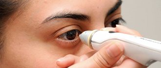
The fundus is the inner surface of the eyeball, which consists of the optic disc, retina and choroid. It, like other parts of the visual apparatus, is susceptible to pathologies. They can be identified at a doctor's appointment using ophthalmoscopy.
The fundus of the eye differs in appearance in children and adults. In newborns, it is normally light yellow in color. After two years, pigmentation becomes more intense.
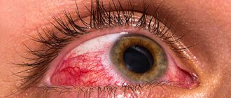
In adults, the fundus of the eye is normally reddish, and the color of the optic disc can vary from pink to yellowish-red.
When examining the fundus of the eye, the ophthalmologist takes into account not only its color, but also other equally important parameters:
- Condition of the vascular network. Enlarged blood vessels may indicate high blood pressure and other diseases.
- Retina color. The appearance of spots on it and uneven coloring often indicates the presence of a hereditary eye disease - retinitis pigmentosa.
- Optic disc size. Too small a size is revealed when it atrophies. Enlargement of the optic nerve head is characteristic of neuritis or edema.
After the consultation, the doctor makes a diagnosis and, if necessary, refers the patient to other specialists. The most common fundus pathologies detected during examination will be discussed below.
Diabetic retinopathy
Diabetic retinopathy is a disease that affects the vessels of the retina of the eyeball. This disease is a common complication of diabetes mellitus.
The disease is characterized by the following symptoms:
- floating spots and spots before the eyes;
- deterioration of near vision;
- the appearance of a veil before the eyes.
Treatment of diabetic retinopathy consists of taking antioxidants and vascular strengthening drugs. Laser treatment is also possible, aimed at cauterizing bleeding and newly formed vessels.
What happens with angiopathy?
If a patient has angiopathy, the tone of the blood capillaries, as well as the outflow of blood, is disrupted. Constriction of the fundus vessels does not occur on its own, but is a harbinger of other diseases.
It signals the occurrence of various pathological processes that lead to eye damage. Doctors pay great attention to the treatment of angiopathy, as it can cause complete loss of vision.
Narrowing of the fundus vessels occurs in both adults and children. However, it is more often observed after 30 years. In children, the severity of angiopathy depends on the position of their body (standing or sitting). And in adults, the disease makes itself felt against the background of increased intracranial pressure. If angiopathy is not treated, cerebral microangiopathy may appear, which often leads to irreversible processes.
Why does it appear?
The reasons why vasoconstriction of the fundus occurs can be different. As noted above, angiopathy is not an independent disease, but signals another, more serious eye disease.
If the causes of angiopathy are not eliminated, another disease may appear. This disease can lead to damage to the walls of the blood vessels of the eyes and their modification.
The following causes of angiopathy are distinguished:
- Hypertension. Constant high intracranial pressure negatively affects the wall of the blood vessels of the eyes, as a result of which its inner layer is damaged. In addition, capillaries burst due to pressure. If you start hypertension to the third stage, then the probability that a narrowing of the vessels of the fundus will occur is 100%.
- Diabetes. With this disease, the vascular wall of not only the eyes, but also the entire body is damaged. This is caused by an increase in blood glucose levels. As a result of this pathology, the diameter of the vessel decreases, and therefore blood circulation in the eye worsens. If treatment is not started in time, the consequences of such a cause can be fatal - complete loss of vision.
- Injuries to the eyes, spine or head. This leads to a rapid increase in intracranial pressure, rupture of fundus vessels and hemorrhage.
- Hypotension. This disease leads to noticeable pulsation in the eye area and causes the formation of blood clots in them.
The reasons for the development of angiopathy lie not only in the above diseases.
A number of other factors can lead to its appearance:
- smoking or alcohol abuse;
- osteochondrosis;
- food or chemical poisoning;
- age over 30 years (but the disease also occurs in children);
- congenital defects of the blood vessels of the eyes.
Kinds
The causes of angiopathy almost completely coincide with its types. This is how they distinguish:
- Hypertensive. If angiopathy of this type is started, changes in the retinal tissue may occur. At the initial stage of hypertension, the symptoms of angiopathy can be eliminated.
- Diabetic. There are two types: microangiopathy and macroangiopathy. In the first case, the walls of the capillaries are affected, and in the second, the larger eye vessels are affected.
- Hypotonic, in which the vessels appear tortuous.
- Traumatic, occurring under the influence of various injuries.
Hypertensive angiopathy
Juvenile angiopathy also occurs. It is considered one of the most unfavorable diseases. Otherwise called Eales disease.
Inflammation of the retinal venous vessels occurs. Connective tissue may form inside the eye. Some complications are also possible (cataracts, glaucoma).
Symptoms
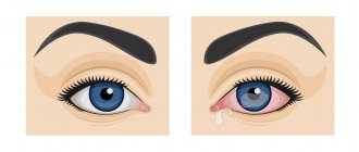
Narrowing of the blood vessels in the eyes manifests itself in a number of symptoms. The patient begins to gradually lose vision: at first the image becomes cloudy, and then the patient cannot distinguish objects. Lightning appears in the eyes.
Nasal and even gastrointestinal bleeding occurs. With advanced disease, blood is observed in the urine.
Yellow spots, pinpoint hemorrhages are observed on the surface of the eyeball, and small vessels are clearly visible.
Retinal disinsertion
Retinal detachment is a condition in which the retina of the eye separates from the choroid. In this case, you need to consult an ophthalmologist as soon as possible, since even a slight delay can lead to blindness.
Symptoms of retinal detachment:
- decreased visual acuity;
- the appearance of floating spots, “spots” and blurred vision;
- sudden loss of lateral vision.
The main treatment for retinal detachment is surgery. The goal of the procedure is to achieve adherence of the retina to the underlying tissues.
Why does it appear
The causes of narrowing of the vessels of the fundus include the following physiological and pathological conditions:
- arterial hypertension;
- atherosclerotic lesions (characterized by the formation of plaques blocking the lumens of large and small vessels);
- vegetative-vascular dystonia;
- spinal and head injuries;
- diabetes;
- elderly and senile age;
- genetic predisposition;
- abnormalities in the structure of the fundus vessels (excessive tortuosity or bends);
- arterial hypotension (persistent decrease in pressure);
- degenerative-destructive changes in the cervical spine (osteochondrosis, spondyloarthrosis);
- chronic intoxication of the body;
- bad habits (smoking, alcohol abuse);
- poor nutrition;
- being overweight;
- increased intracranial pressure;
- exhaustion of the body;
- physical and psycho-emotional overload;
- sedentary lifestyle;
- consequences of prolonged work at the computer;
- oncological diseases of the hematopoietic system (leukemia);
- autoimmune pathologies (systemic vasculitis, periarteritis nodosa);
- infectious lesions of the central nervous system.
Treatment methods
To treat patients with narrowed vessels in the eyes, the following methods of conservative therapy are used:
- Use of medications. Microcirculation correctors (Solcoseryl, Cavinton) help restore blood supply to the retina. The permeability of vascular walls is reduced by drugs based on extracts of ginkgo biloba, diosmin and hesperidin (Detralex, Phlebodia, Ginkor forte). In case of increased blood clotting, antiplatelet agents (Lospirin) are prescribed. To eliminate the underlying disease, hypoglycemic and antihypertensive drugs are used. The use of drops that normalize blood circulation (Taufon, Emoksipina) helps improve vision.
- Physiotherapeutic procedures. Vascular spasm is relieved through laser exposure, acupuncture, magnetic therapy and massage of the cervical region.
- Folk remedies. It is recommended to treat mild retinopathy with infusions of hawthorn, chamomile or lemon balm. 20 g of raw material is poured into a glass of water and left for 3 hours. Take 50 ml infusion 3 times a day.
If conservative therapy is ineffective and the disease develops rapidly, surgical interventions are used - laser coagulation of the retina, photocoagulation, removal of the vitreous.
- 1. General diagnostic issues
- 2. Pigmentary dystrophy (degeneration) of the retina (retinitis pigmentosa)
- 3. Retinal macular degeneration (degeneratio maculae luteae)
- 4. Macular degeneration in Tay-Sachs disease
- 5. Retrolental fibroplasia
- 6. External exudative hemorrhagic retinitis, or Coats disease (retinitis exsudativa haemorrhagica externa)
- 7. Acute obstruction of the central retinal artery (embolia s. thrombosis arteriae centralis retinae)
- 8. Thrombosis of the central retinal vein (thrombosis venae centralis retinae)
LECTURE No. 20. Pathologies of the fundus (part I)
1. General diagnostic issues
If patients complain of deterioration in visual acuity, changes in peripheral vision, loss of visual fields (scotomas), deterioration of vision at dusk (hemeralopia, or night blindness), impaired color perception, then a pathology of the retina can be assumed.
When clarifying the data on the picture of the fundus in children and adults, it should be borne in mind that in newborns the fundus is light, the optic nerve head is grayish-pink, its boundaries are not entirely clear, there may be pigment deposits at the optic nerve head, the macular reflex is absent, the ratio the caliber of arteries and veins is 1:2.
In pathological conditions of the retina due to various reasons, various changes can be observed in the fundus of the eye:
1) swelling and retinal detachment;
2) opacification and ischemia;
3) atrophic foci;
4) tumor-like formations;
5) change in the caliber of blood vessels;
6) preretinal, retinal and subretinal hemorrhages.
To judge the location of hemorrhage foci in the retina, one should focus on their shape, size, color, and location in relation to the retinal vessels.
The lesions located in the layer of nerve fibers have the appearance of streaks or an oblong (linear) shape. In the area of the macula, in the layer of Henle, the lesions are located radially, form a “star” pattern, and have a yellowish-white color. In the middle layers of the retina, lesions are round or irregular in shape and have a yellowish or bluish tint. If the lesion is located in such a way that it covers the retinal vessels, then it is located in the inner layer of the retina. In cases where the lesion is located behind the retinal vessels, it is located in the middle or outer layers of it.
The presence of pigment in the lesion speaks in favor of damage to the neuro-epithelial layer and choroid. Damage to the inner layers of the retina is accompanied by swelling of the optic nerve head and its hyperemia. In this case, the disc tissue is opaque, its borders are blurred, the retina in the peripapillary zone is opaque.
Retinal hemorrhages take the form of lines or streaks located radially around the optic nerve head. Larger hemorrhages are triangular in shape with the apex facing the optic disc.
Dotted hemorrhages of round and oval shape are located in the middle and outer layers of the retina. Hemorrhages in the area of the optic nerve head or in the area of the macula, which are ovoid, dark in color, or cup-shaped with a dark lower half and a lighter upper layer, are characteristic of preretinal localization. Subretinal hemorrhages appear as a vague red spot behind the retinal vessels.
The ischemic retina is matte white, without a sharp border. The edematous retina looks dull, cloudy, and the retinal vessels are not clearly visible. For a detailed examination of the fundus, patients should dilate their pupils with mydriatics.
2. Pigmentary dystrophy (degeneration) of the retina (retinitis pigmentosa)
The anamnesis of patients with retinal pigmentary dystrophy reveals characteristic complaints of poor vision at dusk. A concentric narrowing of the field of vision and a violation of dark adaptation are objectively determined. The optic disc may have a waxy tint, and the retinal vessels, especially the arteries, are narrowed. On the periphery of the fundus there is an accumulation of pigment in the form of “bone bodies”. The field of vision gradually narrows to a tube, the process always ends in blindness due to the death of photoreceptors and atrophy of the optic nerve. The disease is often bilateral and can manifest in early childhood; in half of the cases it is hereditary. The presence of non-pigmented lesions in the fundus is characteristic of non-pigmented retinal degeneration. Functional dysfunctions in patients are the same as in retinal pigmentary dystrophy.
3. Retinal macular degeneration (degeneratio maculae luteae)
Patients complain of decreased visual acuity and note that they orient themselves better in the evening than during the day. With ophthalmoscopy, pigmented and yellowish pinpoint lesions and blanching of the optic disc on the temporal side are determined in the area of the macula. When examining the visual field, a relative central scotoma is determined, as well as a violation of color vision for red and green colors.
The disease is bilateral and often has a family-hereditary nature.
4. Macular degeneration in Tay-Sachs disease
This systemic disease, which is detected in children four to six months old, is manifested by mental retardation, convulsions, muscle weakness due to fatty degeneration of ganglion cells of the retina and brain. In the area of the macula in such children, a degenerative white lesion with a bright red round spot in the center is detected. The child, as a rule, goes blind and dies before reaching the age of two.
Treatment. Due to the fact that the etiological factors for the development of retinal dystrophies are not yet known, treatment is aimed at improving retinal trophism. It consists mainly of prescribing vasodilators, vitamins B, A, E, C, PP, intermidin, pilocarpine, cysteine, heparin, glucose, etc.
5. Retrolental fibroplasia
Every premature newborn, especially if he was in an oxygen tent, should be examined by an ophthalmologist. Such children may develop retrolental fibroplasia – retinopathy of prematurity.
It occurs in both eyes of children in cuvées already in the second to fifth week after birth.
There are three periods of the disease. The first period (acute phase lasting from three to five months) is characterized by the presence of retinal edema, vasodilation, and mild diffuse opacification of the vitreous body on the periphery of the fundus.
The second period (regression period) is characterized by the spread of newly formed vessels with their supporting tissue from the area of retinal changes into the vitreous body, the appearance of hemorrhages, and retinal detachment.
In the third period (scar phase), a significant mass of opaque tissue and foci of proliferation are detected in the vitreous body, covering most of the retina. Fibrous masses gradually fill the entire vitreous body up to the lens. The anterior chamber becomes shallow, posterior synechiae appear due to developing iridocyclitis, then complicated cataracts, secondary glaucoma, and microphthalmos occur.
Often ocular changes are accompanied by microcephaly, hydrocephalus, tissue hypoplasia of the hemispheres and cerebellum, leading to mental retardation.
Treatment is symptomatic: resorption therapy, corticosteroids.
6. External exudative hemorrhagic retinitis, or Coats disease (retinitis exsudativa haemorrhagica externa)
The disease is characterized by varicose dilation of small vessels in the central zone of the fundus. Near the optic nerve head, exudative and hemorrhagic foci of white, gray or yellowish (golden) color with unclear contours, a bumpy or smooth surface are found. The retina in these places is damaged.
The vessels “climb” onto the lesions, make bends, form loops, aneurysms, resembling the picture of angiomatosis. Exudation can be so pronounced that it appears as if there is a retinal detachment, as in retinoblastoma. Exudate accumulates between the choroid and the retina and penetrates the vitreous body. The fundus becomes poorly visible due to exudate and repeated hemorrhages. Gradually, the vitreous body takes on the appearance of a homogeneous grayish mass with hemorrhagic inclusions. Over time, the exudate is replaced by connective tissue, powerful moorings are formed that extend from the retina into the vitreous body and lead to retinal detachment. Gradually, starting from the posterior sections, the lens becomes cloudy. Visual functions decrease depending on the severity of the process. In the later stages, when there is retinal detachment, diaphanoscopy, radioisotope studies and echography help to exclude retinoblastoma.
Treatment. The etiology of the disease is not yet known, so treatment is usually ineffective. Given the similarity of the process with retinal tuberculosis, specific anti-tuberculosis therapy is often used in combination with nonspecific anti-inflammatory and absorbable agents (corticosteroids and enzymes).
7. Acute obstruction of the central retinal artery (embolia s. thrombosis arteriae centralis retinae)
Patients complain of a sudden sharp deterioration in vision, a typical picture is determined in the fundus: against the background of an ischemic milky-white retina, the central fovea stands out in the form of a cherry-red spot (the “cherry pit” symptom), the arteries are sharply narrowed, intermittency of blood flow is determined in narrow retinal arteries. Gradually, the optic disc becomes pale and atrophies, and the patient becomes blind.
In children, embolism is rare, but spasm or embolism sometimes occurs in the presence of autonomic vascular disorders or rheumatic endocarditis. In adults, the causes of embolism are hypertension, rheumatism, and endarteritis.
8. Thrombosis of the central retinal vein (thrombosis venae centralis retinae)
The disease develops more often in older people due to sclerotic changes, vascular changes due to hypertension, and in children due to blood diseases.
Patients complain of a rapid decrease in vision in one eye, the appearance of fiery flashes, and fog before the eyes. Fundus picture: the optic disc and retina are swollen, the size of the disc is enlarged, it is red, protrudes into the vitreous body, the vessels are lost in the edematous tissue, the veins are dilated, tortuous, reminiscent of leeches. Throughout the fundus of the eye, especially in the disc area, hemorrhages of various sizes and shapes are detected.
Treatment of acute obstruction of the central retinal artery, as well as thrombosis of the central retinal vein: treatment of the main suffering, prescription of heparin, fibrinolysin, antispasmodics and vasodilators, and subsequently - absorbable drugs, physiotherapy. For the prevention of secondary glaucoma with thrombosis of the central retinal vein, instillation of pilocarpine is indicated.
Table of contents