Types and degrees of anisometropia
The types of anisometropia are as follows:
- Refractive type.
In this case, the same axis length and different refractive power indicators are observed. - Axial type.
Both eyes have the same refractive power, but the ocular axes are different. - Mixed type.
In such cases, different indicators of refractive power and axis length are observed.
If you do not consult a doctor in a timely manner and do not undergo a course of treatment, convergent or divergent strabismus may occur.
The degrees of anisometropia are as follows:
- Weak.
In this case, the difference between the left and right eyes can be from 0 to 3 diopters. - Average.
This degree has values from 3 to 6 diopters between the eyes. - Strong.
These are all cases where the difference exceeds 6 diopters.
Causes of anisometropia
Anisometropia is a congenital disease that is most often inherited. There are cases when the disease begins to actively develop not in childhood, but in older age. The sudden development of anisometropia can lead to vision loss. If one or both parents have this disease, then the risk of developing anisometropia in the child is much higher. If parents have a pathology in the right eye, the child may develop the disease in both the right and left eyes.
There are cases when the disease develops without a genetic predisposition.
This is rather an exception to the rule. The cause of this condition may be the consequences of cataracts or surgery on the organs of vision. The main causes of anisometropia are the following:
- Unilateral high degree hypermetropia. Due to the fact that after 40 years, some people develop glaucomatous changes, asymmetrical farsightedness may appear.
- Cataract. As a result of clouding of the lens, visual dysfunction occurs in one eye.
- Congenital structural features of the eye.
- Unilateral myopia.
- Astigmatism.
- Surgery.
Possible complications
Undesirable side effects and complications with scleroplasty are quite common.
The main types include an individual allergic reaction to an implanted trans- or implant (the frequency of occurrence varies for different materials), as well as insufficient fixation of the artificial frame with its displacement anteriorly (swelling and swelling of the conjunctiva is observed). In the first case, the symptoms are relieved with standard antihistamines, in the second, corrective re-operation is required (the statistical risk of such a development is 1-2%).
Failure to comply with the most stringent restrictions prescribed by the doctor for the period of rehabilitation - in particular, systematic and long-term neglect of the ban on visual overload - can result in the development of astigmatism or strabismus.
Symptoms of anisometropia
The symptoms of anisometropia are that in the first stages there is slight discomfort, which is not always noticeable. In these cases, correction with glasses is possible.
At later stages, the picture may lose clarity and appear in two. Visual acuity decreases. If you close one eye, the problem disappears. A small child may squint or close an eye when watching TV or reading a book.
In the final stages, binocular vision disturbances occur. The holistic perception of the size and brightness of an object is disrupted.
With heavy loads, fatigue sets in very quickly, and a severe headache occurs, which radiates to the superciliary ridges.
Hepatitis A (jaundice) and liver disease
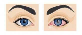
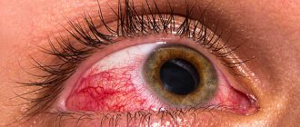
Yellowing of the sclera (whites of the eyes) is one of the main symptoms of liver disease - most often jaundice (disease of dirty hands, hepatitis A). Yellowness is explained by high levels of bilirubin (a yellow substance that is part of bile) in the blood.
If the whites of the eyes are constantly yellow, this indicates that the liver is working under increased load. This can be caused by chronic inflammation of the liver, gallbladder, cirrhosis (a chronic liver disease in which liver tissue is replaced with fibrous tissue).
In such cases, doctors refer for tests (general and biochemical blood tests, liver tests, liver elastography).
Article on the topic
Pressure, apathy, lethargy. How to understand that the thyroid gland is sick
Diagnosis of anisometropia
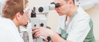
Most often, anisometropia is diagnosed during a routine examination by an ophthalmologist.
The patient himself usually applies in the later stages, when the visual defect causes discomfort. To diagnose anisometropia use:
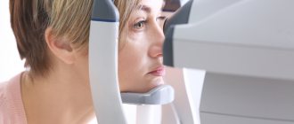
- Ultrasound of the eye - this examination measures the anteroposterior axis of the eyeball. Ultrasound is also used to visualize the vitreous body, optic nerve and retina in case of opacification of the optical medium;
- ophthalmoscopy, or examination of the fundus - thus determining the condition of the optic nerve head and inner membrane;
- visometry - thus, visual acuity is determined;
- perimetry - in this way the asymmetric narrowing of the visual field is determined;
- biomicroscopy of the eye - this examination makes it possible to detect secondary inflammation of the cornea in the first stages;
- Skiascopy of the eye - using this method, clinical refraction is studied, the ratio of the anteroposterior size to the refractive power of the optical system is measured. In patients with anisometropia, darkening movement is observed.
Anisometropia and aniseikonia
Often, anisometropia causes the development of aniseikonia, when the same object in the retina of each eye has unequal sizes. If binocular vision is almost normal, and the difference between the perception of objects with the left and right eyes is approximately the same, then this does not cause discomfort. But if the difference is large, the two objects cannot merge into one image, and binocular vision disturbances occur.
A patient with aniseikonia complains of a significant decrease in visual acuity, images of objects lose clarity and blur, and if work requires constant visual strain, fatigue occurs.
If you equalize the refraction of the left and right eyes with the help of glasses, this will not help eliminate aniseikonia, and may also worsen it. Vision can be corrected only if the difference in visual perception does not exceed two diopters. Only for children who have strabismus is it recommended to completely correct anisometropia.
If anisometropia has reached a high degree, then it is appropriate to use iseikonic glasses, which consist of two lenses, or special contact lenses. They are placed under the eyelid on the eyeball. Between the cornea and the lens there is a thin layer of tear fluid, which creates an optical system that helps reduce aniseikonia. Lenses can be used to correct high refractive errors.
The lenses are very convenient for athletes, actors and people of other professions who cannot always use glasses.
Lenses are made according to an individual prescription. The use of such a product has the following disadvantage - long-term wearing can lead to irritation of the cornea and inflammatory processes.
If organic damage to the eye occurs and visual acuity has decreased significantly, then telescopic glasses are used. They consist of a system of lenses, and the action of such glasses is equivalent to the action of binoculars. Menisci glued together do not change the refractive power of the eye, but simply enlarge the image. Aniseikonic glasses are calculated in percentage magnification rather than dioptres.
The prescription for such glasses specifies the type of lens separately for each eye. The type of astigmatism and the direction of the cylinder axis are also often indicated separately for the left and right eyes. The prescription must also indicate the distance between the pupils in millimeters. If the glasses are very complex, then the doctor should point out the individual characteristics of the patient’s face. This includes the distance from the eye to the lens, from the center of the glasses to the bridge of the nose, the distance between the temples and other parameters.
Methods for correcting anisometropia
Correction of anisometropia is possible only in the initial stages of the disease. It is also possible in severe cases to partially restore visual function. If the first symptoms of the disease appear, you should not self-medicate; you should immediately contact a specialist. It is important to know that after contacting a specialist, glasses or lenses with corrective properties are prescribed. Wearing such devices causes some discomfort.
Vision correction involves wearing glasses or contacts. They must be selected by a specialist. Self-selection can lead to deterioration of visual perception, microtrauma of the cornea, inflammatory processes and swelling. Telescopic glasses have two lenses – converging and diverging. They help restore vision.
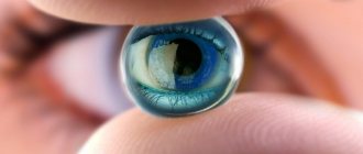
If the difference between the eyes is less than two diopters, then vision correction with lenses is prescribed. They are not suitable for everyone. The specialist selects lenses individually for each patient and monitors the dynamics while wearing them. For those who lead an active lifestyle at night, there are special night lenses. The material from which the lenses are made allows oxygen to pass through, which allows the eye to breathe.
Treatment of anisometropia
The main method of treatment is surgery. It is resorted to only in cases where the correction has not produced results or the disease is in a very advanced stage. Laser vision correction occurs. Such measures are taken only after the recommendation of a specialist.
It is also worth considering that the operation has contraindications and limitations. If corneal diseases are present, laser vision correction is not possible. During the operation, vision is corrected using special equipment. After surgery, you should not put strain on your eyes. Concussions and injuries should be avoided, otherwise the disease will progress.
Before starting treatment for anisometropia, you should consult a specialist and get tested. It is very important to follow all recommendations of specialists in the postoperative period. This will increase your chances of recovery.
Material for scleroplasty
Most parents are especially concerned about the nature and origin of scleroplastic tissue, in particular, whether it is an implant or a graft, i.e. synthetic material or donor material. In the classical version of the technique, as a rule, the corresponding tissues of large animals were used; after complex, several-stage processing, such material acquires the properties of replacement collagen, which is lacking in the myopic eye, which achieves strengthening of the posterior wall. However, in various modifications of scleroplastic surgery (see below), it is possible to use both donor and synthesized biocompatible materials.
Anisometropia in children
Very often, anisometropia is found in children. If treatment is not started immediately, the disease may progress. Often anisometropia provokes the development of refractive amblyopia.
If the difference in indicators reaches two or more diopters, then a correction is prescribed, which includes wearing glasses constantly. In childhood, the difference in glasses lenses from 2 to 5 diopters can be overcome without much effort. In younger children, adaptation is much easier.
But in order to constantly use glasses with different glasses, the child will need some time. The correction is being introduced gradually. Every four months the dioptric difference in the glasses lenses is increased.
If you use modern treatment methods, anisometropia can be easily cured. Very often, glasses are replaced with contact lenses. This method can be used to correct vision even for a child as young as one year old.
Very often, myopic anisometropia is accompanied by impaired binocular vision and amblyopia. In this case, the attending physician uses treatment methods such as orthoptics and pleoptics. With the help of such treatment, visual functions can be strengthened and fixation can be normalized. These are the most effective ways to correct asymmetrical myopia.
Surgery is also often used to solve vision problems in childhood. If the disease is in a mild or moderate stage, then there is a chance of complete restoration of visual function. If the case of the disease is advanced and accompanied by complications, then it is possible to partially compensate for sweating of vision.
Laser has also been used quite successfully to treat anisometropia in childhood. The laser correction procedure takes approximately two weeks. The child does not require hospitalization during the procedure. This method of correction is considered the most painless and humane.
general characteristics
When a person’s visual functions are impaired, effective correction methods are selected. This refers to the use of glasses and lenses.
But if different vision is detected in the eyes, corrective optics are not always able to help. It's all about the reasons that cause anisometropia - a disease that is characterized by the presence of different vision in the eyes.
In order for a correct and unblurred image to be formed, it is necessary that the parallel rays emanating from the object intersect at the retinal focus. If this process is disrupted, a decrease in visual acuity is observed.
When the difference in refractive power between the eyes is one or two diopters, binocular vision will not be particularly affected. But if the indicators differ significantly more, then the development of refractive anisometropia should be expected. Moreover, the refraction in one eye may be normal, but in the other it will be abnormal. But, basically, the pathology affects both eyes.
It is advisable to eliminate anisometropia in time, otherwise the patient may face dangerous consequences:
- squint;
- amblyopia (when, due to inactivity of the eye, its visual functions are lost).
Complications of anisometropia
In the early stages of anisometropia, complications arise in the form of amblyopia - this is a decrease in vision in one or two eyes, which is not associated with pathology of the visual analyzer. This defect cannot be corrected and is often asymptomatic. Sometimes color perception and orientation in space are disrupted, vision decreases, and it is difficult to focus on one object for a long time. If the disease is not diagnosed in a timely manner and treatment is not started, strabismus (convergent and divergent) may develop.
If you wear lenses for a long time, microdamage to the cornea, edema, keratitis, rubeosis of the iris (new vessels form on the surface, causing discomfort), and neovascularization of the cornea may occur. Also, long-term use of lenses increases the risk of various inflammatory processes.
Prevention of anisometropia
If there is a genetic predisposition, this does not mean that anisometropia will necessarily manifest itself, but prevention in this case is mandatory. Do not put too much strain on your eyes. It is also imperative to take eye vitamins and do special exercises. If the work involves a computer, then you need to give your eyes rest and do gymnastics. Once a day you need to take an hour break. At this time, it is better to take an hour-long walk in the fresh air. {banner_horizontalnyy3}
Sports such as football, volleyball and others are exercises for the eyes. They help strengthen the eyes.
Proper food is also a preventative measure for eye diseases. It should include fiber, vitamins A, E, and group B. It is also worth controlling cholesterol intake.
It is also important to regularly monitor visual acuity and clinical refraction. After surgery in the eye area, it is necessary to contact an ophthalmologist for examination and undergo the procedure of visometry and refractometry. This must be done once every six months for two years.
If a child has been diagnosed with a clinical refractive error, it is very important to immediately begin correcting the visual dysfunction to prevent strabismus from developing. If the child is not yet one year old, then special methods are not used.
Types of operations
In the simplest version, small incisions are made in the eyeball, through which a composition of a fairly liquid consistency is injected with a syringe; The implanted material in this case can be either transformed donor tissue or specially developed synthetics. Soon after injection into the desired area, the composition hardens to the required degree. This intervention algorithm is called simplified scleroplasty.
Simple scleroplasty means the formation of a kind of bandage or reinforcing mesh from strips of scleroplastic material, which, as in the previous version, can also be either treated donor or artificial tissue (silicone or metal-plastic).
Complex scleroplasty involves excision of muscle fibers to provide the necessary access and insertion of scleroplastic strips. This technique requires a slightly larger volume of fixing materials. Many descriptions of scleroplasty emphasize that this technique has a minimal list of contraindications, but this is not entirely true. It would be more accurate to say that scleroplasty has no specific contraindications unique to this operation, since the list of possible restrictions, absolute or relative, in this case is almost universal for pediatric ophthalmic surgery. In particular, intervention is not performed under the age of eight years and/or in the presence of infectious and inflammatory processes, concomitant ophthalmopathology, contraindications to anesthesia, scar changes, thinning of the sclera.