If there is a refractive error (farsightedness, nearsightedness, astigmatism), a person experiences serious discomfort. However, these conditions can be corrected quite well. Much worse is complete blindness, which often becomes irreversible. In this regard, it is necessary to be very careful about any changes in vision that may signal the onset of the disease.
In the human body, all systems and organs are interconnected and any deviations can be noticed by an attentive patient. Small changes often warn a person of much more significant deviations. One of these changes in the operation of the optical system is a violation of the visual field. This issue is discussed in more detail below.
Loss of visual fields
Normally, a person can perceive the fingers of a hand that is moved to the side by 85 degrees. If this angle is smaller, then the patient experiences a narrowing of the field of vision.
If the subject can perceive only half of the space, then there is a loss of half of the visual field. This symptom often accompanies serious diseases of the central nervous system, including the brain.
In order to more accurately diagnose the pathology in a patient with loss of visual fields, it is necessary to consult a doctor. Various techniques are used to examine these patients.
When half of the visual fields or even quarters are lost, we are talking about hemianopsia. Usually this pathology is bilateral, that is, the visual field is damaged on both sides.
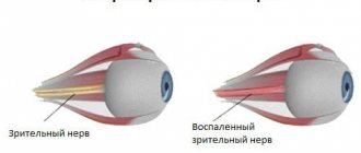
Sometimes the loss of visual fields is concentric in nature. In this case, the condition may worsen to the point of tube vision. A similar symptom occurs with optic nerve atrophy or severe glaucoma. Sometimes this narrowing of the visual field is temporary and is associated with psychopathy.
With focal loss of the visual field, we are talking about a scotoma, which is characterized by the appearance of shadows or islands of absent or decreased vision. In some cases, scotoma can be detected only during a special examination of the patient, that is, he himself does not notice the visual impairment.
If the scotoma is located in the central zone, then most likely it is associated with macular degeneration and age-related changes in the area of the macula. Due to the fact that very effective methods of treating these serious diseases have recently appeared, you should follow all the instructions of your doctor.
Treatment Options
It all depends on your diagnosis. Diagnosis and identification of the cause of problems with visual fields play a significant role.
If these changes are caused by a disease, then you need to go to a clinic, where you will be relieved of the disease itself, and then they will deal with the symptoms and consequences. Treatment can be medicinal, often surgical, but modern medicine can offer very effective methods for eliminating such problems.
If the reasons for the narrowing or loss of your visual fields are diseases that are in no way related to the visual system, then be sure to deal with them first, and only then work on returning your vision to normal.
Don’t forget - first of all, you need a diagnosis from a specialist. Diseases of the visual system are very dangerous, and even such minor changes as "spots" on the retina can be signs of very serious diseases that lead to irreversible blindness in most cases.
The sooner you contact the clinic for help, the faster doctors will identify your problem and prescribe the necessary timely treatment.
Reasons for violations
Depending on the cause of visual field loss, the nature of the pathology may be different. Usually there is a malfunction of the receiving apparatus of the optical system. If the pathology manifests itself as a so-called curtain on one side, then most likely the cause of the disease lies in disruption of the conduction pathways or retinal detachment. In the latter case, the disturbance of the visual field is accompanied by distortion of the shape of objects and a break in straight lines. The size of the visual field defect may also differ between the morning and evening hours. In some cases, the patient perceives surrounding objects in the form of floating figures. Retinal detachment often develops against the background of severe myopia, traumatic injury to the eye, or degeneration of the cells of this layer.
If there is bilateral loss of visual fields from the side of the temples, then we are probably talking about a pituitary adenoma.
If the field of vision is disturbed in the form of a translucent or dense curtain, which is located on the nasal side, then this indicates high intraocular pressure. Also, with glaucoma, rainbow circles appear when looking at point sources of light or fog in front of the eyes.
A translucent curtain on one side may appear when the transparency of the optical media of the eye decreases. These include cataract, cataract, pterygium, vitreous opacification.
When the central part of the visual field is lost, the cause of the disease is most often caused by a malnutrition of this area due to macular degeneration or pathology of the optic nerve and its atrophy. With macular degeneration, there is also a violation of the perception of the shape of objects, uneven changes in image size, and curvature of lines.
With concentric (up to tubular) narrowing of the visual field, we are usually talking about pigmentary degeneration of the retinal substance. At the same time, central visual acuity remains normal for quite a long time. Also, a concentric narrowing of the visual field is observed in glaucoma, but in this case the acuity of central vision is also reduced.
Typically, a concentric narrowing of the visual field is manifested by the fact that a person spends a very long time looking for the keyhole in the door, cannot navigate in an unfamiliar environment, etc.
With sclerotic changes in the arteries of the brain, the nutrition of nerve cells in the cortical visual centers is disrupted. This condition can also cause a concentric narrowing of the visual field, but the acuity of central vision also decreases, and there are other symptoms of brain malnutrition (forgetfulness, dizziness).
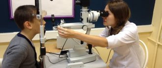
How is the verification done?
To determine whether a patient has visual field defects, a complete examination is necessary. In this case, the doctor will be able to determine the area of the lesion, as well as the level of change in the structure of the optical system. This will help establish the diagnosis of the disease or lead to the need to conduct a number of additional examinations.
To evaluate visual fields, you can use one of the generally accepted techniques.
An experiment that is easy to carry out will allow you to approximately assess the state of your vision. In this case, you need to look into the distance and extend your arms to the sides (at shoulder level). After this you need to move your fingers. With normal peripheral vision, a person can easily notice the movement of the fingers. If the patient cannot notice the movement of the fingers, then he has lost peripheral vision.
Some people think that only central vision is important, but this is not true. For example, in the absence of peripheral vision, it is impossible to navigate in space, drive a car, etc.
The quality of vision can be affected by various diseases, including glaucoma. In this case, the field of view gradually decreases, that is, its concentric narrowing. This symptom is a reason to immediately seek medical help.
When carrying out diagnostic manipulations, the doctor can accurately determine the localization of damage in the optical system (before or after the optic chiasm, directly in the chiasm zone).
If the ophthalmologist identified a scotoma on only one side, then the damage is located up to the chiasm, that is, it affects either the retinal receptors or the optic nerve fibers.
Visual disorders can be present independently or combined with other pathologies of the central structures of the nervous system, which include disorders of consciousness, motor activity, speech, etc. Sometimes they are a consequence of impaired blood flow in the arteries that supply blood to the visual centers of the brain. Most often, young or middle-aged patients are susceptible to this condition.
With vegetative-vascular disorders, the first thing that appears is loss of the visual field. After a few minutes, these defects move left and right. They can also be felt when the eyelids are closed. This leads to a significant decrease in visual acuity, and then to severe headaches.
You can help a patient in this condition if you let him rest in his own bed, after unbuttoning the tight clothes. In addition, you can use receptor drugs, for example, give the patient a validol tablet to dissolve. If this condition recurs, then in addition to the ophthalmologist, you should definitely visit a neurologist.
To assess the patient's condition, you need to use special computerized installations. In them, against a dark background, light points flash unevenly, which can have the same or different brightness and size. After this, the installation registers those areas that did not fall into the field of view.
Focal defects (scotomas)
If vision is reduced or absent in a certain area, the boundaries of which are not adjacent to the outer contour of the visual field, then we are talking about a scotoma. In this case, visual defects may not be perceived by the patient, because the image is completed by the second eye. Such scotomas are called negative. With positive scotomas, the patient perceives the defect as a spot or shadow located in the field of view.
The shape of scotomas can be different (sector, arc, oval, circle, irregular polygon). Depending on the location of scotomas relative to the central point of fixation, they also have different names (peripheral, sectoral, pericentral, paracentral, central). If vision is completely absent in the area of the defect, then the scotoma is called absolute; otherwise, it is called relative (only the clarity of perception is impaired).
An interesting fact is that in one patient the scotoma can be both relative and absolute (when examining the visual field using markers of different colors).
In addition to various pathological scotomas, each patient also has so-called physiological scotomas. These include the blind spot and vascular pattern.
In the first case, we are talking about an oval-shaped absolute scotoma, which is located in the temporal zone of the visual field. This scotoma corresponds to the projection of the optic nerve head. There is no light-receiving apparatus in the blind spot area. A physiological scotoma has clear dimensions and location. If a change in these parameters occurs, for example, an increase in size, then the scotoma becomes pathological. In particular, an increase in the size of the blind spot is observed with papilledema, glaucoma, and hypertension.
To identify scotomas, doctors previously resorted to rather labor-intensive visual field studies. Recently, mainly automatic perimeters are used, as well as testers for central vision, which greatly simplifies the procedure and reduces its execution time to several minutes.
Diagnostics
Diagnosis of hemianopsia is carried out taking into account the study of visual fields using computer perimetry and fundus readings. The presence of clinical symptoms of the disease is confirmed by additional laboratory tests. Especially if a volumetric process of the pituitary gland is suspected. Typically, symptoms of hemianopsia indicate serious brain damage. To clarify the diagnosis, computed tomography, MRI, and skull radiography are performed.
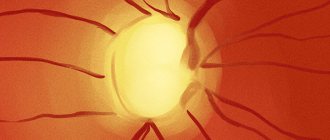
Perimetry allows you to determine the type of visual field loss
Changing the boundaries of the field of view
The narrowing of the boundaries of the visual field can be concentric, that is, global, or local. In the latter case, the formation of a defect occurs in a certain area, while on the rest of the perimeter the boundaries of the visual field are not broken.
Concentric narrowing
With concentric narrowing, much depends on the degree of this process. Thus, in severe cases, so-called tube vision is formed, in which peripheral perception is almost completely lost.
Concentric narrowing of vision can be associated with various pathologies, including neuroses, neurasthenia, and hysteria. In such conditions of the nervous system, the narrowing of the visual field is functional in nature.
However, concentric narrowing of the visual field is more often associated with organic pathology, for example, peripheral chorioretinitis, atrophy or neuritis of the optic nerve fibers, retinitis pigmentosa, and glaucoma.
To accurately determine the nature of the narrowing of the visual field (functional or organic), it is necessary to conduct a series of studies. They use objects of different sizes, colors, and brightness. In case of functional deviations, the size of the object and its other characteristics do not affect the result of the study. In addition, the patient’s ability to navigate in space is used as a distinguishing feature. If this property is violated, then most likely we are talking about an organic lesion.
With local narrowing of the visual field, the process can be bilateral or unilateral. With bilateral lesions, the defects can be located symmetrically or in different areas of the visual field.
In this case, some characteristic areas of vision loss, for example, hemianopia (half loss of visual fields), are of important diagnostic importance. In this condition, we are talking about damage to the visual pathway in the chiasm zone or closer to the central structures.
Hemianopsia can be diagnosed independently, but more often such visual impairments are identified during examination of the patient.
Hemianopsia can be homonymous (loss of the temporal half on one side and the nasal half on the other) or heteronymous (simultaneous loss of the nasal or temporal halves on both sides). There is also quadrant hemianopsia, when the beginning of the defect coincides with the fixation point.
What is “field of view” in ophthalmology?
The field of view is what we see in front of us at a certain moment, without moving our head and concentrating on one object in space. To put it simply, our vision can be divided into 2 types - central and peripheral.
Central vision is responsible for what we intentionally or instinctively focus our attention on. It grabs central objects and helps us focus in case of danger or any other need. An example of the importance of central vision: a person looking for a coffee cup on the table in front of him.
Peripheral vision is everything that does not fall within the radius of central vision, but is still visible to the eye (at least normally). If the entire surface of the retina read and transmitted information to the brain at the same speed, a person would never be able to focus on anything visually, or would achieve this through unimaginable effort.
The uniqueness of peripheral vision is that it transmits information more blurred and not as clearly as central vision. This allows us to do something, but at the same time also notice the events that occur around it (within a normal radius for a person, of course), helps to navigate in space.
An example of the action of peripheral (side) vision: a person crosses the road and notices a car coming around the corner. The visual field for all people is normally approximately the same, and their differences create diseases, of which there are quite a lot in practice, including loss of visual fields.
Hemianopsia
Homonymous hemianopsia most often occurs as a result of pathological space-occupying formations in the brain (tumor, abscess, hematoma) or with retrochiasmatic lesions of the optic pathway (opposite side). In such patients, hemianoptic scotomas may be detected, which are located in symmetrical areas of the visual field.
In heteronymous hemianopsia, the defects can be located on the outer side (bitemporal hemianopsia) or on the inner side (binasal hemianopsia). In the first case, the visual pathway in the chiasm zone is affected, which is characteristic of a tumor process in the pituitary tissue. With binasal hemianopsia, there is damage to the uncrossed fibers of the optic pathway in the chiasm zone. This can occur when the internal carotid artery aneurysm puts pressure on the external nerve fibers in the decussation area.