Drusen are very small yellow or white spots that appear in Bruch's membrane (one of the layers of the retina).
They are globules of mucoproteins and mucopolysaccharides that gradually calcify the optic nerve head. Retinal pigment epithelial cells accumulate damaged cells.
The remaining damaged cells (called lipofuscin) from the oxidative process accumulate in Bruch's membrane and create hyaline bodies (drusen), which are the earliest visible signs of dry macular degeneration. This is a cluster of proteins and oxidized lipids that are not destroyed.
The color is white or yellowish-pink. Over time, they undergo calcification.
Symptoms:
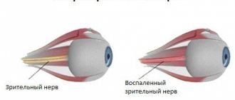
- swelling of the optic nerve head;
- blurred disc boundaries.
During diagnostics, shiny particles are found on the surface of the optic disc. These are the Druze.
They have a pathological effect on nervous tissue, which is characterized by a deterioration in the acuity of visual perception. Over time, the field of vision narrows, the boundaries of the blind spot expand, and arcuate scotomas appear.
Globules of mucoproteins and mucopolysaccharides are located deeply, simulating stagnation and swelling of the optic disc. Therefore, the diagnosis is often made incorrectly.
Changes occur in one or both eyes. Often the second visual organ is affected several years after the disease is diagnosed.
There are several types of drusen:
- hard - round, have clear edges, small and scattered at a far distance from each other;
- soft - large, close, blurred edges.
Soft hyaline bodies can disrupt the layers of the retina and lead to detachment of the pigment epithelium.
Diagnostics
Drusen are easy to diagnose if they are located on the surface of the optic nerve. FAGD plays an important role in diagnosis. Fluorescein angiography of the fundus does not require special preparation.
The examination takes no more than 2 minutes for each organ. To carry out diagnostics, a dye is used that is easily washed off with cold water. The dye does not penetrate beyond the vessels into the surrounding tissue. It begins to fluoresce when the source of exciting light is turned on.
FAGD shows scalloped marginal hyperfluorescence of the disc; tissue contrast does not occur beyond its boundaries.
In some cases, during the diagnostic process, hemorrhage is detected in the retina and vitreous body.
Additionally, ultrasound and ophthalmoscopic examinations are performed. If hyaline bodies have become calcified, computed tomography is prescribed.
Visual fields are also determined to identify defects in peripheral vision.
Differential diagnosis
It is necessary to distinguish drusen from a number of diseases that are accompanied by white or yellow deposits in the posterior pole of the eyeball. All these diseases are united by Flecked retina syndrome:
- Fundus punctatus albescens is a white-spot abiotrophy of the fundus, which is accompanied by the formation of white lesions resembling drusen in appearance. They are located in the middle periphery of the eye. The disease is accompanied by a sharp decrease in visual acuity and progressive nyctalomia, which has similar features to mild and early forms of retinal pigment abiotrophy.
- Fundus albipunctatus (fundus albipunctatus) is a bilateral hereditary pathology that has similarities with the previous disease. The main difference is a non-progressive course, no decrease in visual function and stationary night blindness. With ophthalmoscopy, you can detect whitish round spots that are located at the level of the pigment epithelium and cover the entire fundus of the eye, especially the macula and equatorial region. Dominant drusen have a different size and are more dominant in the macula area.
- Fundus flavimaculatus (yellow-spotted fundus) is a bilateral process in which there are yellowish macular deposits in the pigment epithelium. During angiography, blockade of choroidal fluorescence in the periphery and posterior pole can be detected. With dominant drusen, such changes are not recorded. Also, this pathology is often combined with bull’s eye macular degeneration. Visual acuity decreases with the development of Stargardt disease. There is also a narrowing of the field of vision, which does not happen with drusen.
- Bietti's dystrophy (crystalline dystrophy) is accompanied by whitish deposits in the retinal area, which have a polygonal shape and a brilliant-white shine. At the same time, vision loss and marginal retinal dystrophy are progressive.
Treatment
There is no proven and standard treatment. The number of drusen will gradually increase and they will increase in size.
Visual perception can be improved with glasses or contact lenses.
To prevent the growth of abnormal blood vessels, it is necessary to have regular eye exams with an ophthalmologist. This anomaly requires surgery to prevent bleeding.
Regular examinations are necessary to study the progression of peripheral visual field loss.
It is impossible to prevent the deterioration of the condition, since doctors do not know the exact mechanism of development of the pathology, as well as methods of prevention.
Forecast
Optic nerve damage is progressive and insidious. Ultimately, 75% of patients will develop peripheral field defects. These may include nasal step defects, enlarged blind spots, arcuate scotomas, sectoral field loss, and altitude defects.[6] Clinical symptoms correlate with the visibility of drusen.[13] Loss of central vision is a rare complication of peripapillary hemorrhage. choroidal neovascular membranes. Anterior ischemic optic neuropathy (AION) is a potential complication.[19]
Useful video
Vision is restored up to 90%
Poor vision significantly worsens the quality of life and makes it impossible to see the world as it is.
Not to mention the progression of pathologies and complete blindness.
Optic disc drusen occur as a result of a hereditary abnormality in the genome or exposure to various factors that contribute to the deposition of protein inclusions in nerve fibers. In this case, the patient may not feel any manifestations of the disease for a long time, but in severe cases, loss of visual fields, impaired color sensitivity and other manifestations appear. There is no specific treatment for this pathology; only supportive therapy is possible.
Pathological inclusions can be detected using ultrasound.
Causes and course of pathology
ONH drusen can occur as a result of a genetic abnormality and are inherited in an autosomal dominant manner. That is, the disease is transmitted from a sick parent to all children. Pathology can also be observed as a result of exposure to any pathological factors. Drusen appear in both eyes and this disease mainly affects representatives of the European race. At birth, this anomaly is not visible, and its first signs appear at approximately 12 years of age. In this case, the optic disc changes its appearance and many vessels grow into it, and pathological inclusions appear. Drusen are protein inclusions in nerve fibers, as a result of which the functional activity of nerve endings is disrupted. Over time, calcium salts are deposited in these lesions, which causes systemic eye diseases.
The pathology is most often diagnosed in young people under the age of 30.
Drusen on the retina and optic nerve occur as a result of exposure to the following factors on the human body:
- intoxication;
- the presence of a focus of chronic bacterial infection;
- diabetes;
- intrauterine infections;
- eclampsia during childbirth;
- intrapartum hypoxia;
- injuries;
- long-term inflammatory process of the eyeball.
Symptoms
The color is white or yellowish-pink. Over time, they undergo calcification.
Symptoms:
- swelling of the optic nerve head;
- blurred disc boundaries.
During diagnostics, shiny particles are found on the surface of the optic disc. These are the Druze.
They have a pathological effect on nervous tissue, which is characterized by a deterioration in the acuity of visual perception. Over time, the field of vision narrows, the boundaries of the blind spot expand, and arcuate scotomas appear.
Globules of mucoproteins and mucopolysaccharides are located deeply, simulating stagnation and swelling of the optic disc. Therefore, the diagnosis is often made incorrectly.
Changes occur in one or both eyes. Often the second visual organ is affected several years after the disease is diagnosed.
There are several types of drusen:
- hard - round, have clear edges, small and scattered at a far distance from each other;
- soft - large, close, blurred edges.
Soft hyaline bodies can disrupt the layers of the retina and lead to detachment of the pigment epithelium.
Diagnostic methods
The study is absolutely safe and does not require prior preparation.
The presence of optic disc drusen can be suspected accidentally during a routine ophthalmoscopic examination or when the patient develops complaints characteristic of this disease. To confirm the diagnosis, it is recommended to conduct an ultrasound diagnosis of the eyeballs with visualization of the optic nerve. You can also use computed tomography, which will reveal smaller elements of inclusions. Visiometry and establishment of visible fields of vision are recommended. A general and biochemical blood test is indicated.
Facts about age-related macular degeneration (AMD)
What you need to know about AMD
What is AMD?
AMD is a common eye disease that occurs in people 50 years of age and older, although AMD sometimes affects younger patients. AMD is the leading cause of vision loss in adults. The disease causes changes in the macula, the part of the eye that provides the central, sharp vision needed to see objects clearly. In some cases, AMD progresses so slowly that vision may not change for a long time. In other cases, rapid progression of AMD leads to vision loss in one or both eyes. Decreased vision causes difficulty recognizing faces, reading, driving, and close-up work such as sewing. The macula is made up of millions of light-sensitive cells that provide sharp and detailed central vision. This is the most sensitive part of the retina, which is located in the back of the eye. The retina quickly turns light into electrical signals and sends them to the brain through the optic nerve. The brain then turns the electrical signals into pictures that we see. When the macula is damaged, details become unclear.
Who is at risk for developing AMD?
AMD usually develops in people 50 years of age and older. As you age, your risk of developing AMD increases. Other risk factors include: · Smoking. According to research, smoking increases the risk of AMD by 2 times. · Family history. People who have family members with AMD have a higher risk of AMD. · Overweight and obesity. · Presence of cardiovascular diseases. · Working with an intense light source. · Previous surgery for cataracts.
Does lifestyle matter in the development of AMD?
Some lifestyle habits, such as smoking, are associated with the risk of developing AMD, although it is not known how much of an effect quitting smoking has on AMD. However, the following components of a healthy lifestyle may influence the development of AMD: Stop smoking Exercise Maintain normal blood pressure and cholesterol levels Eating a healthy diet rich in greens, vegetables and fish
How is AMD diagnosed?
Early and intermediate stages of AMD are usually asymptomatic.
Only a thorough examination of the eye with a dilated pupil can help diagnose AMD. The examination includes the following methods: · Visual acuity examination
.
Using special tables, it is determined how you see at a distance. · Examination of the eye with a dilated pupil
.
The doctor puts drops into the eyes to dilate the pupils, which makes it possible to better examine the back of the eye. Using special magnifying lenses, it is possible to better examine the retina and optic nerve to identify signs of AMD and other eye pathologies. · Amsler grid
.
The doctor may also ask you to look at a special mesh. Changes in central vision may cause lines to disappear or become distorted. · Optical coherence tomography
.
A non-contact and painless method that allows you to obtain a microscopic image of the retina with clear visualization of its elements. · Fluorescein angiogram
. To conduct this type of study, a contrast agent is first injected into a vein and, as the contrast passes through the vessels of the eye, photographs are taken. This test allows the doctor to identify areas of vascular leakage and select the appropriate type of treatment.
Here are questions you can ask your doctor:
—
What is my diagnosis and how do you spell its name?
—
How is AMD treated?
—
How does this disease affect my vision now and will it affect it in the future?
—What
symptoms should I watch for?
-
Should I change my lifestyle?
What forms of AMD can lead to vision loss?
There are two forms of AMD: dry and wet. The appearance of the wet form is in all cases preceded by the dry form.
DRY AMD
What is dry AMD?
Dry AMD is the most common form of AMD. It develops in 90% of patients with AMD. In dry AMD, the light-sensitive cells in the macula are slowly destroyed, gradually blurring the central vision of the affected eye. As AMD progresses, you may notice spots in the center of your visual field. Your doctor may call this condition "geographic atrophy."
What symptoms accompany dry AMD?
In the early stages, dry AMD has few symptoms. It is important to have your vision examined regularly before the disease progresses. In advanced stages, the most common symptom of dry AMD is blurred vision. Objects do not appear as bright as before. As a result, it may be difficult to recognize faces and more light may be required for reading. Dry AMD can occur in both eyes or may initially affect one eye.
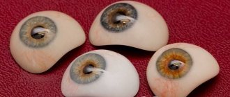
What are drusen?
Drusen are an early symptom of AMD. These are orange deposits under the retina. They can be both small and large. A doctor can identify drusen by examining the retina with a dilated pupil. Drusen by themselves do not cause vision loss. But the appearance of large drusen poses a risk of developing a more severe form of AMD, which in turn leads to severe vision loss.
Stages of AMD
· Early AMD. In early AMD, either small drusen or a small number of medium-sized drusen are observed. At this stage, there may be no symptoms or decreased vision. · Intermediate AMD. At this stage, medium-sized drusen or one or more large drusen may be detected. Many patients also have no symptoms. Some note the appearance of a blurred spot in the central field of vision, some may require more light when reading, etc. · Late changes in dry AMD. Along with drusen, at this stage there may be destruction of the light-sensitive cells of the macula with the development of so-called “geographic atrophy,” which leads to the appearance of a blurry spot in the central field of vision, which may increase in size and become darker over time.
Can dry AMD turn into wet AMD?
In all patients, the development of a more severe, wet form of AMD was preceded by an intermediate stage of dry AMD. Wet AMD can develop rapidly even in early-stage AMD.
WET AMD
What is wet AMD?
Wet AMD affects 10% of all patients with AMD. However, this form is more severe than the early or intermediate stage of the dry form. With wet AMD, pathological blood vessels grow under the retina in the macula area. Newly formed vessels are characterized by increased fragility and permeability, which causes fluid and blood to leak with the development of macular edema and damage to the retina. Although vision loss occurs quickly in wet AMD, there are medications that can stop the progression of wet AMD and even improve vision if treatment is started before significant vision loss occurs.
What symptoms accompany wet AMD?
During the early stages of wet AMD, there may be waviness/curvature of straight lines, impaired distance vision, "fogging", and lack of image sharpness. Wet AMD can also develop a blind spot with loss of central vision. When reading, the curvature of letters or lines and letters falling out become noticeable. If these symptoms appear, you should consult a doctor immediately!
Treatment for wet AMD
With early diagnosis and timely treatment, it is possible to stop the progression of AMD. The earlier AMD is diagnosed, the higher the chances of maintaining vision. It is very important to remember that vitamins and dietary supplements cannot improve or preserve vision in wet AMD. One of the options to help slow the progression of AMD and even improve vision is injections of anti-VEGF drugs into the eye (into the vitreous body). In Russia, this is the only drug “Lucentis” registered for wet AMD. With wet AMD, increased levels of VEGF, an endothelial vascular growth factor, are formed in the eyes, which causes the growth of pathological blood vessels and an increase in their permeability. Anti-VEGF blocks these effects. Treatment is not limited to one injection. Monthly injections may be required over a period of time; The number of injections and frequency of observation is determined by your doctor.
Central retinal dystrophy is the main cause of visual impairment in people over forty years of age. As a result, this disease is often called senile dystrophy.
In order to promptly treat a disease such as central type of dystrophy or peripheral retinal dystrophy, it is necessary to remember the first symptoms of the development of the pathological process at the initial stage of its course.
Central dystrophy - is the disease dangerous?
Macular degeneration, or otherwise central dystrophy, is a dangerous disease. The disease may occur in one eye but quickly spread to the other. Moreover, it can occur either independently or under the influence of other diseases. Loss of vision, if macular degeneration occurs, can even occur at a young age. The first period of the disease may be asymptomatic. Therefore, to prevent the onset of the disease, it is necessary to undergo examination by an ophthalmologist as often as possible.
The retinal dystrophy that appears in the patient indicates that in this case there are serious disturbances in the organization of nutrition, as a result of which the retina of the eye suffers. And the definition of “senile” will tell you that peripheral retinal dystrophy, or dystrophy of the central zones, is directly related to the effects of age-related changes in the body, including the human eyes. Central dystrophy develops as a consequence of pathological vascular changes or hypertension, atherosclerosis.
Human eyes are designed like a camera. Lenses that are in front of the camera transfer images to the back wall of the camera. The eyes perform the same actions. The retina of the eye is a visual tissue that contains certain zones: the macular, that is, the central, and the peripheral zone. The macular region occupies the central region together with the optic nerve. A small part of it, namely the retina, helps us see.
Types of retinal dystrophy
Retinal dystrophy is conventionally divided into the following categories:
- congenital or hereditary (for example, pigment variety);
- acquired.
Hereditary dystrophy includes:
- Pigmentary dystrophy. The pigmentary type of dystrophy is associated with poor functioning of the receptors that are responsible for vision at dusk. Pigmentary dystrophy is a fairly rare disease nowadays.
- Spot-white retinal dystrophy, which appears in childhood and seriously progresses as a person grows.
Pigmented and dotted white eye dystrophies, as a rule, are age-related, i.e. are caused by aging of the body and appear in combination with cataracts.
Retinal dystrophy is characterized by the possibility of developing such categories as:
- central dystrophy;
- peripheral type of disease (otherwise known as pvhrd).
Peripheral dystrophy more often appears with myopia or eye injury (not including the central region of the retina). In this case, specific “floaters” may appear in the eyes.
If a patient has a seriously damaged retina, treatment should be carried out after a thorough study of the causes of such a disease. The reasons may be:
- Low insulin production in the patient’s body, high blood pressure, development of certain kidney diseases.
- Myopia of the patient.
- Received eye injuries (or as a result of tissue degeneration).
- Congenital predisposition.
Symptoms of the disease: description and features
Central retinal dystrophy can be expressed by the appearance of such a symptom as a feeling of pain and discomfort when looking “close” and “far”. The most alarming outcome of this retinal disease is the curvature of straight lines, when the patient feels as if he is looking through hot air. A dark spot may appear in front of the affected eye; often you cannot distinguish, simply not see, some letters in the text. Often, objects that are at a distance may have a different shade or appear reduced in size when viewed with the affected area of the retina.
If at least one of these alarm bells is present, it is necessary, without delay, to contact an ophthalmologist, undergo a high-quality examination, after which the doctor will be able to prescribe the correct and necessary treatment in each specific case.
It should be noted that you can test your vision yourself, for which you just need to use a simple method using a checkered sheet. The technique is usually called the “Amsler lattice”. This test will allow you to see even small deviations in a person’s vision.
An ophthalmologist who examines the fundus of the eye at an early stage of the development of a particular disease may find that the retina of the eye has fairly small (yellow) foci, which, in turn, are called “drusen.” They do not harm vision, but require specialist supervision to prevent the occurrence of dry and wet forms of the disease.
Treatment of retinal dystrophy
If retinal dystrophy is detected, treatment will not be able to completely restore the patient’s lost normal vision. To date, effective ways to eliminate dystrophy have not yet been created. However, regular visits to an ophthalmologist and proper monitoring prevent the occurrence of severe complications, while preventing the progression of the disease and thereby preserving visual function.
If treatment is not started in a timely manner, dry central dystrophy can gradually develop into a wet form. Drusen, in turn, begin to mix with one another, as a result of which the retina of the eye begins to delaminate, and fluid accumulates in the freed space. If the course of the disease occurs this way, then the risk of vision loss increases significantly.
The disruption of blood supply occurs due to swelling of the patient's eye, which, as a rule, appears in the central region of the retina. The development of pathological vessels of the circulatory system leads to the fact that the retina of the eye rises. As a result, these vessels may bleed or leak fluid, causing further retinal detachment. The longer this disease progresses, the faster the functions of the eye decline; treatment in this situation is simply necessary.
Laser therapy applied in time significantly stops vision loss if the retina of the eye is damaged, but treatment does not guarantee its complete preservation. The main goal of such therapy is to promptly remove the swelling that appears and stop the occurrence of pathological blood vessels.
Retinal laser treatment can help make the black marks in front of the eyes disappear. For complex treatment, medications are prescribed that relieve swelling and hemorrhage. However, there is one thing: although the retinal area of the eye has been restored, vision becomes somewhat worse. The decrease in vision is milder than if no treatment was carried out, and central dystrophy continued to progress.
It should be noted that, whatever the form of central dystrophy, it is necessary to register with an ophthalmologist and undergo an examination at least once every six months. Treatment may include avoiding physical activity beyond the permissible level. A patient with eye dystrophy should:
- avoid hot baths or showers;
- be sure to follow a dietary diet;
- fried and fatty foods should be avoided;
- consume the required amount of vegetable oil;
- limit yourself in the consumption of salt and sugar;
- if the day is sunny, you need to use glasses with tinted lenses;
- if the work involves eye strain, then it is necessary to apply ten-minute eye exercises during breaks, preferably every half hour.
A person who has relatives in their family who have been diagnosed with central dystrophy or peripheral dystrophy should be very careful about the health of their eyes. This disease can be hereditary. If the area of the retina has undergone dystrophic changes, then the reverse process is very difficult to treat (virtually impossible). Much depends on the person himself: the sooner you seek help from a doctor if central dystrophy is detected, the sooner treatment will be started that will help preserve vision and a full life.
Pathogenesis of dominant drusen
Today, three theories of drusen formation are being considered:
- The transformation theory suggests that drusen are formed due to direct transformation of pigment epithelial cells.
- The depository or secretory theory suggests that drusen are formed as a result of the secretion and accumulation of abnormal pigment epithelial cells.
- The chorovascular theory suggests that drusen are a product of hyaline degeneration of the chorocapillaries or the organization of chorocapillary hemorrhages. This theory of the origin of drusen is currently not confirmed by histological studies.
As a result of histological studies, it was found that the structure of drusen includes two main components: mucopolysaccharide (sialomucin) and lipid - cerebroside. Some researchers believe that the formation of both substances occurs in degeneratively changed pigment epithelium. In the inner portion of Bruch's membrane there are drusen, which are adjacent to the pigment epithelium. They are formed due to its autophagic destruction, which is associated with abnormal lysosomal activity. Subsequently, most likely, lysosomes located in pigment epithelial cells are transformed into an amorphous material that fills the internal collagen regions of Bruch's membrane. Drusen can vary in size and some become calcified. The retinal pigment epithelium that covers the drusen undergoes a number of changes.
Important! Dot-whitish (pigmentless) retinal dystrophy: causes, symptoms and treatment
At the initial stages of the pathological process, pigment dissipates in the cytoplasm of pigment epithelial cells, and mitochondrial degeneration and nuclear displacement also occur. At the final stage, the degenerated pigment epithelial cells merge with drusen. This leads to the formation of areas where there is no pigment epithelium. Photoreceptors are also displaced and dystrophically changed.
In the process of further development of drusen, non-exudative predisciform macular degeneration or its exudative disciform form with choroidal or subretinal neovascularization may develop. Such transformations are more typical for the fifth and sixth decades of a person’s life. In the presence of hard drusen, atrophic changes develop, and if there are soft drusen located bilaterally, exudative detachment of the pigment epithelium and other complications occur, which determine the further progression of the pathological process.
Retinal dystrophy - treatment, classification, symptoms
Retinal dystrophy - what is it? This question is asked by every person who has certain pathological disorders in the visual organ. In fact, this disease includes a whole group of pathologies of the retina, which is the functional structure of the eyeball. Retinal dystrophy is characterized by the death of tissue in the retina, that is, the organ begins to rapidly degenerate. The retina is considered the most important element of the visual organ, as it is responsible for visual acuity. After all, it is she who transmits light impulses to parts of the brain. It consists of a thin layer of nerve and light-sensitive cells, which are photoreceptors.
Classification features of retinal dystrophy
Dystrophy can be congenital or acquired. It is classified as follows:
Symptoms of retinal dystrophy
The main symptoms of retinal dystrophy:
- Blurred image of objects and partial loss of color vision.
- The presence of dark spots and spots before the eyes.
- Deterioration of lateral perception and loss of visual acuity.
- Formation of bright flashes and distortion of objects.
- The blur before the eyes and the importance of good lighting when reading.
- The patient cannot distinguish a moving object from a stationary one.
Reasons for the development of retinal dystrophy
- Pathological changes in the functionality of the retinal circulatory system, which leads to significant scarring.
- Weakened immune system and poor diet. It turns out that low-quality products can lead to retinal deformation.
- Bad habits: smoking and drinking alcohol.
- Viral infection that was not treated promptly or competently.
- Diabetes mellitus, heart disease, hypertension, pathologies of the endocrine apparatus.
- Complication after surgery.
- Poor metabolism.
How to treat retinal dystrophy, diagnosis
To make an accurate diagnosis of retinal dystrophy, a hardware examination is prescribed. This can be viziometry, ultrasound examination, perimetry, fluorescein angiography. In addition, samples for laboratory testing and instrumental examination are collected. Electrophysiological research methods are required to determine the degree of functioning of visual and nerve cells. Treatment of retinal dystrophy is carried out as comprehensively as possible and, first of all, it is drug therapy, which includes the following:
- Vasodilator and angioprotective drugs strengthen and expand the circulatory system, and also eliminate symptoms. These may be drugs such as Ascorutin, Papaverine, Complamin. The dosage and duration of treatment is determined solely at the individual level.
- Antiplatelet agents prevent the formation of blood clots. This could be Acetylsalicylic acid, Ticlopidine, Clopidogrel.
- Vitamin premixes are required.
- To improve microcirculation, Pentoxifylline is prescribed.
- Lucentis can prevent the growth of capillaries.
- Eye drops: “Taufon”, “Ophthalm-Katachrome”, “Emoxilin” and others. These drugs lead to the regeneration of damaged cells and acceleration of metabolism in the area of the visual organ.
- Electrical stimulation of the retina.
- Photostimulation of the eye.
- Low-energy laser radiation.
- Magnetotherapy, electrophoresis and so on.
Surgical intervention
In the most complex and advanced forms, surgical intervention is used. These could be the following methods:
- Laser coagulation of the retina can stop the progression of the disease. Using a laser unit, targeted cauterization of damaged tissue is performed.
- The operation can be performed using the vasoreconstructive method.
- It could also be revascularization.
- Vitrectomy involves removing the damaged areas using suction and installing an implant.
IMPORTANT! After drug therapy, you must follow all the doctor’s instructions. First of all, you should not expose your eyes to overstrain and exposure to sunlight. It is important to take additional vitamins and minerals and give up bad habits.