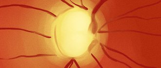
Optic nerve structure
The optic nerve is made up of retinal nerve cells, each of which has more than 1 million axons. Axons carry visual information from the retina to the cerebral visual centers. All retinal nerve fibers are collected in the optic disc.
The part of the nerve located inside the orbit is called intrabulbar. Extending beyond the orbit, the optic nerve penetrates the cranial cavity (retrobulbar part). The end of the nerve is located in the visual centers of the midbrain and diencephalon.
Throughout its entire length, the optic nerve is protected by membranes that are connected to the structures of the orbit, brain and cerebral membranes. Therefore, inflammation from these structures easily penetrates the optic nerve. This is how optic neuritis develops.
Classification of optic neurites
The first signs may appear unexpectedly and abruptly. Usually the process begins with a decrease in visual acuity or pain in the orbital area. Depending on which part of the visual pathway is affected, retrobulbar and intrabulbar neuritis are distinguished.
Kinds:
- intrabulbar (true) neuritis, when the inflammatory process affects the optic nerve and optic disc inside the orbit;
- retrobulbar neuritis, when inflammation is concentrated behind the eyeball and affects the axial bundle of nerve fibers.
Diagnostics
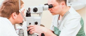
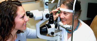
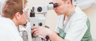
You need to start the examination by visiting an ophthalmologist. During the appointment, the doctor asks the patient about complaints and examines anamnestic data to identify risk factors. Visual acuity is assessed. Additional signs can only be detected with the help of special examinations.
Ophthalmological examinations:
- Assessment of color perception using special tables. A decrease in brightness and intensity of color perception is detected. This is a very important diagnostic criterion in the absence of objective changes in the fundus of the eyeball.
- Assessment of visual fields using a special apparatus. Patients exhibit loss of visual fields.
- Direct ophthalmoscopy is an examination of the fundus structures using a hand-held optical instrument (ophthalmoscope). Exudative edema, hemorrhage and tissue atrophy are detected. In some forms of the disease, no changes are detected.
After ophthalmological examinations, the patient may need to consult doctors of another profile.
Neurological examination
A consultation with a neurologist also begins with questioning. The physician then evaluates the patient's neurological status to look for reflex, cognitive, and mental changes that indicate the presence of serious cerebral pathology. Additional examinations help clarify all the features of the disease.
Ongoing research:
- Assessment of visual evoked potentials is an objective diagnostic method that allows one to determine the localization of the pathological process. Electrodes are attached to the patient's head in the area of the occipital cortex of the cerebral hemispheres to assess electrical activity in all areas of the nerve fibers. While collecting information, the patient looks at the monitor screen to stimulate activity in the studied areas of the central nervous system.
- Ultrasound examination of the eyeball and its nervous structures. This safe and accurate visual diagnostic method allows the doctor to detect swelling and tissue atrophy.
- Magnetic resonance imaging of the brain, which produces three-dimensional images of different parts of the brain. MRI helps to detect even minor changes in the organ, indicating the presence of demyelinating, infectious or vascular pathology.
- Laboratory examination of cerebrospinal fluid. A puncture using a needle is performed in the lumbar region. Using PCR and serological tests, the material is checked for infection that can affect various structures of the nervous system.
An important role in the examination is played by differential diagnosis, which is necessary to exclude diseases with symptoms similar to neuritis. For this purpose, patients are prescribed a study of the retinal vessels.
Causes
The exact reasons for the development of neuritis remain unknown. Research shows that under certain conditions, the immune system begins to mistakenly attack myelin, the substance that covers the optic nerve. This leads to its damage and the spread of inflammation. Myelin serves to conduct electrical impulses (visual information) from the eyes to the brain. Neuritis is dangerous by disrupting this process and, accordingly, vision itself.
What diseases are associated with optic neuritis:
- Multiple sclerosis. In multiple sclerosis, the autoimmune system begins to attack the lining of the brain and spinal cord. It is believed that after one case of optic neuritis, the risk of developing sclerosis increases by 50%. An MRI scan can detect brain damage after neuritis.
- Neuromyelitis optica. This is an inflammation that simultaneously affects the optic nerve and spinal cord. A condition similar to multiple sclerosis, but with less damage to the nerves in the brain.
- Inflammatory processes in the brain and its membranes (meningitis, encephalitis, arachnoiditis).
- General diseases (diabetes mellitus, gout, blood diseases, sarcoidosis, lupus).
The development of the disease is often associated with acute or chronic general infections (syphilis, Lyme disease, measles, herpes, mumps, tuberculosis, brucellosis, gonorrhea, malaria, erysipelas, smallpox, typhus, diphtheria, tonsillitis, influenza). Even nasopharyngeal infections such as tonsillitis, otitis, sinusitis, sinusitis, and pharyngitis can play a role.
Ophthalmological reasons:
- inflammation of the orbit (phlegmon, periostitis);
- inflammation of the eyeball (iridocyclitis, keratitis, retinitis, panophthalmitis).
Often the cause of the onset or recurrence of neuritis is intoxication due to alcoholism. Women with pathological pregnancies and people with traumatic brain injuries are also at risk. Sometimes inflammation of the optic nerve is associated with taking medications (especially quinine and some antibiotics).
Treatment
Treatment usually gives positive results with partial atrophy. However, it is impossible to completely restore vision with its help. The main task in this case is to stop the progressive degradation of tissues and fibers, and subsequently the decrease in severity.
By medication
Medicines can play a big role in the treatment of pathology. As a rule, in this case, ophthalmologists prescribe the following categories of medications to their patients:
- Drugs that dilate blood vessels (Dibazol, Papaverine). Necessary to restore normal blood circulation.
- Improves metabolism (Pyrogenal). Promotes resorption and elimination of pathological processes.
- Stimulating the restoration of tissues and fibers (vitamins, Peat, ginseng).
Surgically
In advanced stages of complete or partial nerve degradation, two types of surgical operations are used:
- Tissue transplantation. In this case, healthy human tissue with good capillaries is transplanted to the site of the affected areas. As a result, metabolism and blood circulation improves.
- Reduced blood flow to healthy tissue. The purpose of this operation is to redistribute the blood supply. Some healthy vessels are blocked, resulting in blood flowing to the affected tissue areas.
Hardware treatment methods are also used as additional therapy:
- Magnetic stimulation. With the help of a special device that generates a magnetic field, blood supply, oxygen transfer and tissue nutrition are improved.
- Oxygen therapy. The method is aimed directly at improving metabolic processes and oxygen nutrition.
- Electrophoresis. The method involves dilating blood vessels in order to improve metabolism, using a low current.
- Laser therapy. Uses laser beams to stimulate the affected areas.
- Ultrasound. Used to improve blood flow.
What happens with optic neuritis
Optic neuritis begins with infiltration and proliferation. The inflammatory process passes from the soft membrane of the brain to the nerve fibers. Inflammation can develop in the optic nerve trunk or sheaths.
Tissue inflammation occurs with the involvement of leukocytes, lymphocytes and plasma cells. Infiltration and swelling will compress the fibers, impairing their functions and causing vision impairment. Subsequently, neovascularization develops and connective tissue is formed. Secondary damage to nerve fibers occurs.
After the acute phase, individual fibers can recover, but severe inflammation leads to total breakdown of the fibers and proliferation of glial tissue. Persistent neuritis leads to optic nerve atrophy with irreversible deterioration of visual function.
If the cause of neuritis is multiple sclerosis, the disorder is based on demyelination of the fibers, that is, destruction of the myelin sheath. Despite the fact that the process of demyelination is not inflammation, such a lesion is identical to neuritis in clinical symptoms. Therefore, destruction of the optic nerve in multiple sclerosis is equated to retrobulbar neuritis.
Basic information
Optic neuritis is an inflammation of the nerve fiber that carries visual information from the retina to the brain. As a rule, inflammation is accompanied by destruction of the insulating (myelin) sheath. The disease usually begins to appear at a young age. Patients develop blind spots in their field of vision. Timely drug therapy eliminates the inflammatory process and restores visual function, but irreversible changes may develop.
According to epidemiological data, the disease is most often diagnosed in people suffering from multiple sclerosis and may be the first complication of this disease. Therefore, when characteristic symptoms of the disease are detected, special examinations are prescribed. Neuritis of the oculomotor nerve and damage to adjacent nerve fibers is also often diagnosed with an infectious lesion.
Clinical picture
Symptoms of neuritis usually appear in only one eye. Inflammation is characterized by impaired visual functions and a narrowing of the visual field. The degree of visual impairment with neuritis depends on changes in the papillomacular bundle. Typically, the narrowing of the visual field is concentric, and central and paracentral scotomas (blind spots) occur. There may be a complete lack of color perception. The optic disc protrudes only when neuritis is combined with edema.
Symptoms of intrabulbar neuritis
Intrabulbar neuritis lasts 3-6 weeks. If treatment is started in time, complete restoration of vision is possible. In severe and advanced cases, there is a risk of complete loss of vision in the affected eye.
Damage to the optic nerve in the area located inside the eyeball always begins acutely. The first symptoms appear within 1-2 days, the patient’s condition deteriorates very quickly. The severity of symptoms will depend on the degree of damage to the optic nerve.
Characteristic symptoms of intrabulbar neuritis:
- Scotomas (blind spots in the field of vision). This symptom is observed in most patients with neuritis. With inflammation of the optic nerve, central scotomas predominate.
- Deterioration of vision (mainly myopia). Visual impairment is diagnosed in every second person with inflammation. Initially, visual acuity decreases by 0.5-2 diopters, but as the optic nerve is damaged, complete blindness may occur. Depending on the quality of treatment and the aggressiveness of the inflammation, blindness may be reversible or incurable.
- Deterioration of twilight vision. The eyes of a healthy person quickly adapt to poor lighting conditions (40-60 seconds), but with inflammation, the visual system begins to distinguish objects in the dark only after 3 minutes or more. Typically, deterioration of twilight vision occurs on the side of the affected eye.
- Change of color perception. With optic neuritis, patients lose the ability to distinguish certain colors. Due to nerve irritation, colored spots may appear in the field of vision.
Symptoms of retrobulbar neuritis
Inflammation of the nerve outside the orbit is diagnosed less frequently. Since in this zone the nerve is not surrounded by fiber and lies freely, the infection can affect the outer surface or the inner surface - peripheral and axial neuritis, respectively.
Symptoms:
- Axial: decreased visual acuity by 3-6 diopters, one-sided blindness, central scotomas.
- Peripheral: visual acuity remains normal, a decrease in the visual field from the periphery, dull pain in the orbit (intensified by movement of the eyeball, relieved by non-steroidal anti-inflammatory drugs and glucocorticosteroids).
Transverse neuritis, when the infection affects the entire thickness of the nerve, combines signs of two types of retrobulbar inflammation. Retrobulbar neuritis is characterized by the appearance of a central absolute scotoma. At first the spot is large, but as vision improves it becomes smaller and may disappear. Sometimes a central scotoma can turn into a paracentral annular scotoma.
At first, with retrobulbar neuritis, the fundus of the eye remains in a normal state, but slight hyperemia and blurring of the boundaries of the optic disc are possible. Usually the inflammation affects one eye, the disc enlarges, the boundaries are not defined, the veins expand and twist. Sometimes the clinical picture of retrobulbar neuritis resembles a congestive disc.
There are acute and chronic retrobulbar neuritis. In acute neuritis, a decrease in visual acuity occurs over 2-3 days, and in chronic neuritis, it occurs gradually. Acute inflammation is accompanied by pain behind the eyeball. Vision begins to return after a few days, although in rare cases this may not occur.
Toxic neuritis
Typically, toxic neuritis is caused by methyl alcohol poisoning. In addition to the symptoms of poisoning, after 1-2 days the vision in both eyes drops sharply (up to complete blindness). The patient's pupils dilate and the reaction to light is weakened or absent. Mild hyperemia of the optic disc is possible; in rare cases, symptoms of ischemic neuritis occur (pale disc, blurred borders, narrowed arteries).
Serious disorders in the papillomacular bundle develop with chronic alcoholism and smoking strong tobacco with a high nicotine content. Alcohol and tobacco intoxication is diagnosed mainly in men over 30 years of age.
The disease occurs as a chronic retrobulbar inflammation. Changes in the fundus are not observed, only occasionally there is slight hyperemia of the optic disc. A central scotoma appears, but the peripheral boundaries of the visual fields remain normal. By giving up bad habits, vision improves and blind spots decrease.
Neuritis in tuberculosis
In this case, the development of ordinary neuritis or solitary optic disc tubercle is possible. Tubercle is a tumor-like formation on the surface of the disc that spreads to the retina.
Neuritis in diabetes
Neuritis associated with diabetes mellitus is usually diagnosed in men. Chronic inflammation is often bilateral, and vision deteriorates slowly. Central scotomas occur, although the peripheral boundaries of the visual fields remain normal. Initially, the optic disc is normal, but with the development of inflammation, temporal pallor is observed.
Neuritis in neurosyphilis
Against the background of relapse of neurosyphilis, an edematous form of neuritis is usually diagnosed. There are two possible options for the development of inflammation: mild (hyperemia and unclear boundaries) and severe papillitis (sharp deterioration of vision). Rarely, papulous neuritis develops when the disc is hidden behind a massive gray-white exudate that protrudes into the vitreous.
Manifestations of the disease
Symptoms of optic neuritis depend on the degree of tissue damage. An acute course is characteristic, in which a decrease in visual acuity occurs in the first days. In severe cases, complete blindness develops. Patients also complain of the appearance of blind spots in the field of vision, decreased color sensitivity and impaired adaptation to darkness. As the symptoms progress, the symptoms intensify and various complications arise.
Other symptoms:
- pain in the eye area, discomfort intensifies during movement of the eyeball;
- unilateral visual impairment; in most cases, optic neuritis is not characterized by simultaneous damage to two nerves;
- the appearance of flashes of light in the field of view;
- headache and dizziness.
Acute symptoms resolve on their own within 1-2 months. After the blood vessels are restored and pathogens are destroyed, vision is restored.
Diagnosis of optic neuritis
It is easy to recognize a typical case of optic neuritis. It is much more difficult to diagnose mild neuritis or inflammation with edema, which are similar to pseudoneuritis and congestive disc. The main difference is the preservation of visual functions.
If there are symptoms of increased intracranial pressure, a spinal tap is performed to confirm a congestive disc. It is most difficult to distinguish neuritis from edema or complications of a stagnant disc, since in both cases there are visual disturbances.
The diagnosis of neuritis can be confirmed by the presence of small hemorrhages or foci of exudate in the disc tissue or retina. The most informative method for diagnosing optic neuritis is fluorescein angiography of the fundus.
Signs of neuritis according to the severity of the process:
- mild: moderate hyperemia of the optic disc, unclear boundaries of the optic disc, dilatation of arteries and veins;
- pronounced: sharp hyperemia of the disc, the boundaries of the disc merge with the retina, white spots and many hemorrhages appear in the peripapillary zone;
- transition to atrophy: disc blanching, narrowing of arteries, resorption of exudate and hemorrhages.
Medical assistance
Treatment for optic neuritis is selected based on the diagnostic results. If the disease is infectious, broad-spectrum antimicrobial drugs are prescribed. If the cause of the pathology has not yet been established, the drug therapy regimen includes anti-inflammatory drugs, corticosteroids and desensitizing drugs. The listed medications suppress inflammatory processes in tissues, weaken immune activity and improve blood supply to cells. When the examination results appear, the treatment is adjusted. Along with the main treatment, general restorative therapy is prescribed in the form of injections of vitamins, minerals and neuroprotectors.
Treatment of acute forms of the disease is carried out in a hospital. Sometimes patients require emergency care to prevent dangerous complications from occurring quickly. So, in case of poisoning with methyl alcohol, the stomach is washed and an antidote is prescribed in the form of an ethanol solution. Clinical recommendations for the treatment of a chronic form of pathology accompanied by degeneration of nerve fibers include the use of antispasmodics and drugs that improve blood supply to the brain.
The prognosis is conditionally favorable. With timely treatment, most patients regain full vision within a few weeks. Much more dangerous may be the underlying causes of an ophthalmic disorder, such as an acute infection or multiple sclerosis. In severe cases of the disease, irreversible changes are possible that negatively affect the patient’s quality of life.
Treatment of optic neuritis
If neuritis is suspected, the patient must be hospitalized immediately. Until the reasons are clarified, drugs are prescribed to suppress infection and inflammation, as well as measures for dehydration, desensitization, immunocorrection and improvement of metabolism.
Treatment regimen:
- For 5-7 days antibiotics (injections or droppers). Do not use drugs with ototoxic effects (Streptomycin, Gentamicin, Kanamycin, Neomycin).
- Retrobulbar administration of corticosteroids is indicated (dexamethasone solution 0.4%, 1 ml for 10-15 days). Orally Prednisolone 0.005 g 4-6 times a day for 5 days with a reduction in dosage.
- Diacarb orally 0.25 g three times a day (after three days of taking two days off), while taking Panangin 2 tablets three times a day.
- The dosage of Glycerin is calculated from the ratio of 1-1.5 g/kg body weight.
- 10 ml of magnesium sulfate solution 25% is injected intramuscularly.
- Intravenous glucose 40%, hexamethylenetetramine solution 40%.
- Intranasal tampons with 0.1% adrenaline solution every day for 20 minutes with constant monitoring of blood pressure levels.
- B vitamins orally.
- Piracetam up to 12 g per day.
- Solcoseryl intramuscularly.
- Dibazol 10 mg twice a day for 2-3 months.
After determining the diagnosis, the underlying disease is treated and the patient’s condition is normalized for chronic disorders. In the case of toxic retrobulbar neuritis, similar treatment is carried out, excluding antibiotics. Additionally, detoxification of the body is required: taking a 30% ethyl alcohol solution (first 90-100 ml, then 50 ml every 2 hours).
Optic neuritis cannot be ignored. Any inflammation in the visual system without treatment can lead to blindness, so at the first signs of neuritis you should urgently consult a therapist or ophthalmologist.
Sources used:
- Vitreous replacement in eye surgery / G.G. Bordyugova. - M.: Medicine, 2002.
- Ophthalmological manifestations of common diseases / E.A. Egorov, T.V. Stavitskaya, E.S. Tutaeva. - M.: GEOTAR-Media, 2009.
- Penetrating wounds and contusions of the eye / R.A. Gundorova, G.A. Petropavlovskaya. - M.: Medicine, 1975.
- The University of Chicago's Department of Ophthalmology and Visual Science