Causes
The formation of blisters on the eyelids is caused by various pathogens: viruses, bacteria, allergens. The main factors that contribute to their appearance near the eyelashes:
- violation of personal hygiene rules;
- close contact with a person with a viral or bacterial disease;
- burns, other eye injuries;
- weak immune system, vitamin deficiency;
- use of decorative cosmetics of dubious quality;
- regularly being in a smoky room.
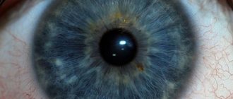
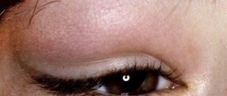
Colorless blisters on the eyelids are observed in people of different age groups and can indicate different diseases.
Conjunctivitis
The membrane that covers the inside of the eyelids is called the conjunctiva. It protects the eyes from small foreign bodies, microorganisms, and maintains the tear film.
Conjunctivitis is an infectious inflammatory process on this membrane. The causative agents are bacteria.
There are acute and chronic forms.
| Sign | Acute form | Chronic |
| Localization | First one eye, then can move to the other | On two eyes |
| Symptoms |
|
|
| Duration of the disease | It appears suddenly. Duration ranges from 5-6 days to 2-3 weeks. In case of severe complications – longer. | Appears unnoticeably. May last for years. |
Ophthalmoherpes
The causative agent is the herpes simplex virus. It affects the skin of the eyelids and mucous membranes. Characteristic symptoms:
- Patients experience severe pain in the eyes.
- Red spots begin to appear, which quickly turn into bubbles under the eyelashes. After some time, they burst and a crust forms in their place. It is possible that the rash may reoccur in the same place.
- Often inflammation is accompanied by fever, chills, and muscle pain.
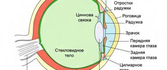
Anatomy of the eyelids
The eyelids (upper and lower) are skin-muscle-connective tissue formations that cover the eyeball from the front. They perform a protective function, protecting the eye from mechanical damage, excessive sudden lighting, and harsh atmospheric influences. Reflex blinking movements of the eyelids cause uniform distribution of tear fluid over the anterior surface of the eyeball, which maintains constant hydration of the eyeball.
The formation of the eyelids occurs in the embryo by the 2nd month, but only from the 4th month does the skin fold split into the upper and lower eyelids. By the 7th month of intrauterine life, there is a formed palpebral fissure, which increases in the first 2-3 years of life and is finally formed at 8-10 years.
If during the period of initiation and further development pathological processes occur in the mother’s body, then the cycle of formation of the eyelids and palpebral fissure can be disrupted at any stage. A child is born with developmental anomalies of the eyelids, such as cryptophthalmos (a rare deformity), microblepharon, ankyloblepharon, eyelid coloboma, ptosis, ablepharia, etc.
Changes in the eyelids can also occur at any age as a result of diseases of the central and peripheral nervous system, injuries, etc. These are lagophthalmos, ptosis, entropion, ectropion, ankyloblepharon.
The border of the upper eyelid is the eyebrows, the lower - a conventional line passing 2-3 mm below the lower edge of the orbit. The free space between the edges of the open eyelids is called the palpebral fissure (rima palpebrarum). Its horizontal length is 30 mm (in an adult), and its height in the central section ranges from 10 to 14 mm. Within the palpebral fissure, almost the entire cornea is visible, with the exception of the upper segment (normally, the edge of the upper eyelid covers the upper limbus by 2 mm), and the white areas of the sclera bordering it. When the eyelids are closed, the palpebral fissure disappears.
The upper and lower eyelids at the medial and lateral corners are connected to each other by means of adhesions (comissura palpebralis medialis et lateralis). Approximately 5 mm before fusion, the inner edges of the eyelids change the direction of their course and form an arched bend. The space outlined by them is called the lake of tears (lacus lacrimalis). There is also a small pinkish-colored elevation - the lacrimal caruncle (caruncula lacrimalis) and the adjacent semilunar fold of the conjunctiva (plica semilunaris conjunclivae).
On the free edge of the eyelids there is a front rib, where the eyelashes grow, and a posterior rib. There are 100-150 eyelashes on the upper eyelid, 50-70 on the lower eyelid. Eyelashes are constantly changing. Their lifespan is on average 150 days. Near each eyelash root there is a sebaceous gland, the excretory ducts of which open into the hair follicle of the eyelash.
There is an intercostal space between the anterior and posterior ribs. Closer to the posterior rib, the excretory ducts of the meibomian glands (glandulae tarsales), which are modified sebaceous glands and have the structure of the alveolar glands, open. There are approximately 30 of them on the upper eyelid, and about 20 on the lower eyelid. They run in parallel rows and open into excretory ducts near the posterior edge of the eyelids. Their function is to secrete a fatty secretion that protects tears from evaporation from the surface of the cornea; it also promotes a tighter closure of the eyelids and their adherence to the eyeball, and prevents the transfusion of tear fluid through the edges of the eyelids, protecting them from maceration.
The glands located at the edges of the eyelids can change to form cysts and tumors.
- meibomian glands - modified sebaceous glands located in the thickness of the cartilaginous plate, produce a fatty secretion that is part of the precorneal tear film;
- Zeis glands are modified sebaceous glands that connect to the hair follicles of the eyelashes;
- Moll glands are modified sweat glands; their ducts open into the hair follicle of the eyelashes or directly into the intermarginal space of the eyelid.
The eyelids consist of a skin layer, a muscle layer, a connective tissue plate (cartilage = tarsus), and a mucous membrane lining the back surface of the eyelid. The skin of the eyelids is thin, soft, and elastic. The underlying tissue is devoid of fat and very loose, which contributes to the rapid spread of edema and hemorrhage in this area. Usually, two orbital-nalpebral folds are clearly visible on the skin surface - upper and lower; as a rule, they coincide with the corresponding edges of the cartilage.
Under the skin there is a circular muscle (m. orbicularis oculi), which causes the eyelids to close. It mainly starts from the internal ligament of the eyelids - a dense fibrous cord formed from the periosteum of the frontal process of the maxillary bone, and only a small part of the fibers originates from the adjacent orbital edge.
The cartilages of the eyelids (tarsus superior el inferior) look like horizontal plates slightly convex outward with rounded edges, about 20 mm long, 10-12 and 5-6 mm high, respectively, and 1 mm thick. They consist of very dense connective tissue. With the help of powerful ligaments (lig. palpebrale mediale et laterale), the ends of the cartilages are connected to the corresponding walls of the orbit. In turn, the orbital edges of the cartilage are firmly connected to the edges of the orbit through fascial tissue (septum orbitale).
The back surface of the eyelids is covered with a connective membrane (conjunctiva), which is tightly fused with cartilage, and beyond them forms mobile vaults - a deep upper one and a shallower, easily accessible lower one.
The free edges of the eyelids are limited by the anterior and posterior ridges (limbi palpebrales anteriores et posteriores), between which there is a space about 2 mm wide. The anterior ridges contain the roots of numerous eyelashes (located in 2-3 rows), into the hair follicles of which the sebaceous (Zeiss) and modified sweat (Moll) glands open. On the posterior ridges of the lower and upper eyelids, in their medial part, there are small elevations - lacrimal papillae (papilli lacrimales). They are immersed in the lacrimal lake and are equipped with pinpoint openings (pimctum lacrimale) leading to the corresponding lacrimal canaliculi (canaliculi lacrimales).
The mobility of the eyelids is ensured by the action of two antagonistic groups of muscles - closing and opening them. The first function is realized with the help of the circular muscle of the eye (m. orbicularis oculi), the second - the muscle that lifts the upper eyelid (m. levator palpebrae superioris) and the lower tarsal muscle (m. tarsalis inferior).
The striated muscle that lifts the upper eyelid, which is innervated by the oculomotor nerve, and the smooth muscle of Müller, which is innervated by the sympathetic nerve, are involved in raising the eyelids and opening the palpebral fissure.
The orbicularis oculi muscle has two parts:
- pars palpebralis - present only on the upper and lower eyelids, causes blinking movements,
- pars orbitalis - makes a circle from the internal ligament of the eyelid and joins there, causing protection of the eyeball during contraction.
The lower eyelid is pulled down by a poorly developed eye muscle (t. tarsalis inferior), which connects the cartilage to the lower fornix of the conjunctiva. The fascial processes of the sheath of the inferior rectus muscle are also woven into the latter.
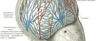
The eyelids have a rich blood supply.
A. takes part in the nutrition of the eyelids. lacrimalis (blood supply to the eyelids from the temporal side) and a. ethmoidalis (blood supply from the nasal side). Each of these arteries gives branches (aa. palpebrarum) to the upper and lower eyelids, which go towards each other, merging and forming the arteries of the marginal arch on the eyelids (arcus palpebrae superior et inferior). This arc is located in the thickness of the eyelids in the loose tissue that separates the skin-muscular part of the eyelid from the conjunctival-cartilaginous part. On the upper eyelid, the arterial arch goes 1-2 mm higher, and on the lower eyelid 1-3 mm below the free edge of the eyelids. Thin branches extend from the arterial arches in all directions, especially to the free edge and to the conjunctiva, where they penetrate by perforating the cartilage at right angles to it.
The veins of the eyelids are much more numerous than the arteries and accompany the arteries of the same name; they drain into the veins of the face and the veins of the orbit. It should be noted that the largest is the angular vein (vena angularis - anastomosis between the anterior facial and superior ophthalmic veins), passing at the internal attachment of the medial ligament of the eyelid. Wounding it can cause significant bleeding. The veins of the eyelids do not have valves, so infectious processes from the eyelids can easily spread along the venous bed into the orbit and cavernous sinus.
In the outer part of the eyelids, the arterial and venous vessels pass relatively shallowly, in contrast to the vessels located in the upper inner part of the entrance to the orbit. The vessels of the eyelids abundantly anastomose with the vessels of the face.
The eyelids also have a well-developed lymphatic network, which is located at two levels - on the anterior and posterior surfaces of the cartilage. In this case, the lymphatic vessels of the upper eyelid flow into the pre-auricular lymph nodes, and the lower - into the submandibular ones. Massive injection of the solution near the temporal edge can lead to a blockage of the lymphatic pathways leading to the preauricular lymph node, which can cause “elephantiasis” of the upper eyelid to develop in the postoperative period.
Sensitive innervation of the eyelids is carried out by the I and II branches of n. trigeminus. The upper eyelid is innervated by n. nasolacrimalis, n. nasociliaris, n. supraorbitalis and n. intratrochlearis. The lower eyelid is innervated by n. infraorbitalis (departs from the second branch of the trigeminal nerve). Motor innervation is carried out: for the round muscle - the facial nerve, for the levator - the oculomotor nerve, for the Müllerian smooth muscles - the sympathetic nerve.
In eyelid surgery, incisions are usually made either along the intermarginal edge or through the skin - along the muscle fibers of the orbicularis muscle and skin folds. Deep incisions can damage the levator.
Types of palpebral fissure:
- Normal - the external commissure is on the same horizontal line with the internal commissure.
- Mongoloid - the external commissure is located above the internal one.
- Antimongoloid - the external commissure is located below the internal one.
When to see an ophthalmologist
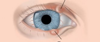
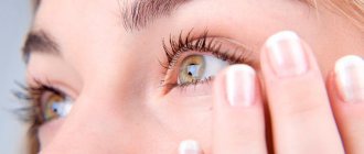
Redness and blisters on the eyelash edge are a reason to seek help from a doctor.
Symptoms:
- severe sudden eye pain;
- rashes on the face, especially around the eyes and nose;
- decreased visual acuity;
- open wounds of the cornea.
Diagnostics by an ophthalmologist includes several stages.
First, the doctor carefully asks about the symptoms of the disease. Then he conducts an external examination of the eyelids and eyelashes. If he suspects a viral infection, he takes secretions from the blisters. Biomicroscopy and skin allergy tests are performed.
The goal of treatment is to rid the patient of the cause of the inflammatory process.
Treatment methods:
- Treatment of bubbles with antiseptics.
- The use of drugs that relieve pain and stabilize the condition of the mucous membrane.
- Prescription of antibiotics, antiviral, antihistamines, hormonal agents.
The drugs must be used in accordance with the prescription of the ophthalmologist.
If Moll cysts or papillomas form under the eyelashes, surgery may be required.
When treating papillomas, laser and cryodestruction are used.
Diagnostics
The accuracy of the diagnosis and, accordingly, treatment depends on how quickly, completely and correctly the diagnosis is made. What may be included in the complex of diagnostic procedures:
- Interviewing the patient and collecting anamnesis. This allows us to understand as accurately as possible what exactly happened to the patient.
- External examination of the eyes, eyelashes and eyelids. An experienced specialist will already suspect a particular diagnosis at this stage, and will subsequently recommend carrying out clarifying research methods.
- Scraping from the affected surface or discharge from blisters. This makes it possible to understand whether the appearance of bubbles is bacterial or viral.
- Enzyme immunoassay blood test. It is necessary in order to detect an increased immunological reaction of the body.
- Examination of individual areas for histology. It is carried out if the tumor is suspected to be of an oncological nature.
Overview of home treatment methods
When treating blisters at home, it is necessary to eliminate the cause of the disease.
Medicinal herbs help cleanse eyelids and eyelashes, relieve puffiness and increased sensitivity.
Folk recipes described in the Herbalist reference book:
- Washing. Chamomile herb is poured with boiling water and left for 1 hour. Proportions 1:20.
- If you are not allergic to honey, you can try diluting it with warm boiled water in a 1:2 ratio. Make lotions.
If the cause of the inflammatory process is viruses or bacteria, you should follow the recommendations of an ophthalmologist.
An important part of treating blisters under the eyelashes is strengthening the immune system. The journal “Basic Research” No. 7 for 2012 emphasizes the need to consume vitamins and minerals for the resistance of the immune system to infections, the herpes virus.
Include vegetables and fruits rich in vitamin C in your diet. Avoid foods with preservatives and additives that cause allergies.
On the recommendation of a doctor, you can take medications to boost immunity.
If the bubbles under the eyelashes are allergic, avoid all contact with allergens.
Carefully read the instructions for use of medications, there are contraindications.
Follow the rules of personal hygiene and keep your home clean.