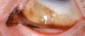
Tumors of the lacrimal glands
– a group of neoplasms of the lacrimal gland, predominantly of epithelial origin. Localized in the outer part of the upper eyelid, painless. Benign tumors of the lacrimal gland develop over many years, and during the growth process they can cause exophthalmos and thinning of the orbital wall. Malignant neoplasms rapidly progress, impair the mobility of the eyeball, provoke pain and increased intraocular pressure. Germination of surrounding tissues and distant metastasis are possible. The diagnosis is made on the basis of an ophthalmological examination and instrumental studies. Treatment is surgical.
Etiology
The exact etiological picture regarding the development of this type of pathological processes has not yet been established. Only a few predisposing factors can be identified:
- personal or family history of cancer;
- frequent relapses of chronic ophthalmological diseases;
- congenital pathologies of the visual organs;
- weakened immune system.
It should be noted that inflammation of the lacrimal glands is very rare, according to statistics only in 12 out of 10,000 patients.
Tumors of the lacrimal glands
Tumors of the lacrimal glands are a group of tumor lesions of the lacrimal gland of heterogeneous structure. They originate from the glandular epithelium and are represented by epithelial and mesenchymal components. They belong to the category of mixed neoplasms. They are rare, diagnosed in 12 out of 10,000 patients. They make up 5-12% of the total number of orbital tumors. The question of the degree of malignancy of such neoplasms is still debatable. Most experts conditionally divide tumors of the lacrimal gland into two groups: benign and malignant, resulting from malignancy of benign neoplasms. In practice, both “pure” and transitional options can be encountered. Benign processes are more often detected in women. Cancer and sarcoma are diagnosed with equal frequency in both sexes. Treatment is carried out by specialists in the field of oncology and ophthalmology.
Classification
The following types of tumors of the lacrimal glands are distinguished:
- Pleomorphic adenoma is more often diagnosed in women than in men, in approximately 50% of the total cases of diagnosing this type of pathological process. It is characterized as a benign neoplasm, but there is a high risk of malignancy.
- Adenocarcinoma is the most common cancer of the lacrimal gland. It is marked by a high rate of development of the clinical picture and a sharp deterioration in vision. The prognosis is unfavorable.
- Cylinder or malignant cyst of the lacrimal gland. In terms of its clinical picture and prognosis, it is almost identical to adenocarcinoma, however, the development of the clinical picture is somewhat slower, but the tendency to hematogenous metastasis is greater.
If the lacrimal caruncle has enlarged due to a benign formation, then there is no threat to human life and health, however, surgical excision is still necessary.
The most unfavorable prognosis for ocular cancer. Even with the timely initiation of therapeutic measures, relapse of the disease cannot be ruled out after a few years.
Benign tumors of the lacrimal glands
Pleomorphic adenoma is a mixed epithelial tumor of the lacrimal gland. Accounts for 50% of the total number of neoplasms of this organ. Women are affected more often than men. The age of patients at the time of diagnosis can range from 17 to 70 years, the largest number of cases of the disease (more than 70%) occur in 20-30 years. Arises from epithelial duct cells. Some experts suggest that the source of the tumor is abnormal embryonic cells.
It is a node with a lobular structure, covered with a capsule. The tissue of the lacrimal gland tumor on the section is pink with a grayish tint. Consists of two tissue components: epithelial and mesenchymal. Epithelial cells form chondro- and mucus-like foci located in a heterogeneous stroma. The initial stages are characterized by very slow progression; the period of time from the appearance of the lacrimal gland tumor to the first visit to the doctor can range from 10 to 20 years or more. The average time interval between the onset of the first symptoms and seeking medical help is about 7 years.
For some time, the tumor of the lacrimal gland exists without causing any particular inconvenience to the patient, then its growth accelerates. Inflammatory swelling appears in the eyelid area. Due to the pressure of the growing node, exophthalmos and an inward and downward displacement of the eye develop. The upper outer part of the orbit becomes thinner. Eye mobility is limited. In some cases, a tumor of the lacrimal gland can reach gigantic sizes and destroy the wall of the orbit. Upon palpation of the upper eyelid, a fixed, painless, dense, smooth node is determined.
A survey X-ray of the orbit reveals an increase in the size of the orbit due to displacement and thinning of its upper outer part. Ultrasound of the eye indicates the presence of a dense node surrounded by a capsule. CT scan of the eye allows you to more clearly visualize the boundaries of the tumor, assess the continuity of the capsule and the condition of the bone structures of the orbit. Surgical treatment involves excision of the lacrimal gland tumor along with the capsule. The prognosis is usually favorable, but patients must remain under clinical supervision throughout their lives. Relapses can occur even several decades after removal of the primary node. In more than half of patients, signs of malignancy are detected already at the first relapse. The shorter the period of remission, the greater the likelihood of malignancy of a recurrent tumor.
Symptoms
The clinical picture will depend on the nature of the pathological process. Common symptoms include:
- in the area of the affected eye, the eyelid swells;
- due to increasing pressure, symptoms of exophthalmos develop;
- limited eye mobility;
- the eyeball shifts;
- palpation of the upper eyelid can reveal a dense, smooth nodule;
- the upper outer part of the orbit becomes thinner;
- increased lacrimation, which leads to the formation of crusts;
- decreased visual acuity;
- hypersensitive reaction to light stimuli.
With lacrimal sac cancer, the general clinical picture may be supplemented by the following symptoms:
- congestion in the conjunctiva.
- hypoesthesia of the lacrimal nerve.
- swelling of the optic nerve head.
- the neoplasm causes the eyeball to shift.
In addition, general symptoms may be present:
- enlargement of regional lymph nodes;
- general deterioration of health;
- low-grade body temperature;
- irritability, frequent mood swings;
- hormonal imbalances;
- exacerbation of existing chronic diseases.
It should be noted that the clinical picture of this pathological process (both benign and malignant) is rather nonspecific, therefore, at the first symptoms, you should seek medical help, and not start treatment on your own by taking medications for no reason and using folk remedies.
Lacrimal gland carcinoma
The main and most important clinical manifestation of the formation of lacrimal gland carcinoma is diplopia, that is, double vision. At first, this symptom will appear temporarily, after which it becomes permanent; there is also a risk of swelling of the eyelids (most often the upper eyelid is affected). In the case of rapid development of a malignant formation, quite strong unpleasant painful sensations begin to appear in the area of the affected eye socket.
Rapid protrusion of the eyeball occurs (this process is called exophthalmos) and can last from several weeks to several months. As a result of the beginning of the growth of a tumor-like formation, quite strong compression of the neurovascular bundle in the orbit appears, and a rapid process of deformation of the eye occurs. As a result of this, over time, the patient cannot close his eyelids due to constantly increasing exophthalmos, as well as chemosis (swelling of the conjunctiva occurs), this leads to a process of very rapid destruction of the cornea of the affected eye.
Most often, between the ages of 50 and 60 years, adenocarcinoma develops, and it is characterized by the formation of areas of mitosis, cellular polymorphism occurs, and a certain tendency to form satellites appears directly in the node of the tumor itself.
In some cases, in order to determine the area of tumor malignancy that has occurred, there is a need to conduct a number of additional clinical studies of multiple sections.
This type of tumor is characterized by rather slow growth; more than five years can pass from the moment the first sign appears until the patient seeks help from a doctor. During this period of time, this neoplasm turns from a mobile and fairly smooth tumor into a non-movable and very lumpy one.
The formation of metastases is quite rare (in approximately 7% of cases), and their occurrence most often occurs in a fairly distant period, after surgery was performed to treat carcinoma (approximately within 7 years). However, there is a possibility that the morphological variant of adenocarcinoma may be characterized by invasive growth at very early stages of development (tumor cells begin to intensively penetrate into the tissues located around the tumor). Ingrowth may occur directly into the gland tissue, as a result of which the tumor begins to grow along the nerve trunks, as well as blood vessels.
Since adenocarcinoma includes a fairly large number of mitoses, there is a possibility of its development taking into account the scenario of squamous differentiation. This neoplasm is characterized by tissue polymorphisms (glandular growths, as well as tubular alveolar growths); quite extensive areas of epithelial growth occur, which will be replete with cribriform structures and ketotic cavities, epithelial complexes (separate), which are sandwiched directly in the hyalinized stroma.
Proliferating cells will block the ductal lumens of the lacrimal gland. The cytoplasm is characterized by less homogeneous and eosinophilic characteristics, in contrast to benign adenomas. This type of adenocarcinoma will differ in its rather intensive growth from adenoma, while the history of its development will be several months and can last up to two years.
Among the main symptoms of the onset of carcinoma growth, fairly early complaints of a certain displacement of the eye, as well as drooping of the lower eyelid, may appear. As a result of such deformation of the eye, the formation of myopic astigmatism can occur. The patient begins to complain of a certain discomfort or unpleasant pain that appears in the area of the affected eye; in some cases, severe lacrimation may begin to form.
As a result of such growth of the tumor, it can grow into the adjacent bones, occupy the temporal fossa, as well as the cranial cavity. Early metastasis may occur.
Approximately a third of the formation of lacrimal gland tumors is due to the appearance of adenoid cystic carcinoma. Most often, its development occurs at a young age (from 25 to 50 years). Tumor cells have a fairly diverse structure, with small cell areas with round and oval acellular fields predominating.
If symptoms of carcinoma appear, you should immediately seek help from a doctor.
Diagnostics
In this case, you need to consult an ophthalmologist, however, you will definitely need to consult an oncologist. First of all, a physical examination of the patient is carried out, during which the doctor must establish the following:
- how long ago the first symptoms began to appear, their intensity.
- there were cases of cancer in your personal or family history (not only regarding the localization of the visual apparatus).
In addition, for an accurate diagnosis, the following laboratory and instrumental diagnostic methods are used:
- blood sampling for general and biochemical analysis;
- tumor marker test;
- CT;
- X-ray examination of the organ of vision;
- biopsy of the tumor for cytological and histological examination;
- contrast dacryocystography;
- neurological research.
It should be noted that histological analysis of the tumor is mandatory and is the main diagnostic method, since only by its results can the nature of the neoplasm be determined.
Based on the results of diagnostic measures, the doctor determines the type and form of the pathology and, taking into account the data that was collected during the initial examination, determines further therapeutic measures.
Treatment
Regardless of the nature of the tumor diagnosed, treatment is only radical, that is, the tumor is removed. If the pathological process is benign, the prognosis is favorable; after surgery, the patient can be prescribed the following drugs:
- antibiotics.
- anti-inflammatory.
- local antiseptics.
If the tumor is malignant, the prognosis is most often unfavorable, since metastasis to the brain and spinal cord, lungs, and other body systems is possible. Treatment in this case will include:
- surgical removal of the tumor and nearby tissues;
- radiation or chemotherapy (can be performed both before and after surgery);
- the use of special corrective agents to improve vision.
In the postoperative period, a course of drug therapy may be prescribed, which includes the following drugs:
- local antiseptics.
- anti-inflammatory.
- painkillers.
- antibiotics.
As for traditional medicine, in this case their use is inappropriate, since they will not provide the desired therapeutic effect.
Herbal decoctions (chamomile, St. John's wort, sage) can only be used as an addition to drug therapy after surgery to relieve swelling and prevent inflammation.
Prevention
Unfortunately, due to the fact that there is no specific etiological picture, specific preventive measures have also not been developed. Therefore, it is advisable to adhere to general recommendations:
- eat right, namely include in your diet foods that provide all the necessary vitamins and minerals;
- stop smoking and excessive alcohol consumption;
- promptly and correctly treat all diseases in order to prevent their chronicity;
- if there is a family history of cancer, you should regularly visit an oncologist for a preventive examination;
- If you feel unwell, do not self-medicate.
These recommendations do not guarantee the elimination of the occurrence of this type of pathology, however, they make it possible to reduce the risk of its development or diagnose the disease in a timely manner.


