Electrical stimulation of the eye muscles and optic nerves through the skin is one of the most common physiotherapeutic procedures used to treat eye diseases. How effective is it and how exactly does it improve vision?
Transcutaneous electrical stimulation of the eyeball improves blood supply to the retina and the muscles that control the movements of the lens. This allows you to avoid dystrophy, which is often observed with a high degree of myopia (myopia).
The main indications for the use of electrical stimulation include the following diseases:
- optic nerve atrophy;
- severe myopia;
- amblyopia (lazy eye syndrome);
- strabismus - both in children and adults;
- retinal dystrophy;
- ptosis;
- spasm of accommodation;
- presbyopia.
Effect on the body
Electrical stimulation affects the body through pulses of different durations - from 0.5 to 300 ms, with a current strength of up to 5 mA (on the face), up to 100 mA (on the body) and a frequency of 10-150 Hz, which operate intermittently. Despite the fact that the patient does not move during the procedure, this effect is identical to the normal work of the muscles during their activity. The current passes through the tissue, excites the cells and stimulates the active work of the muscle, and during pauses it relaxes.
The current, thanks to this rhythm of work, does not irritate the skin under the electrodes and the epidermis is not damaged. When an electric current is applied to muscles or nerves, their bioactivity changes. The impulses provoke contraction of muscle fibers, which strengthens and activates them. If a muscle is overstrained, electrical stimulation relieves such tension well.
Electrical stimulation of the back muscles in the neurology clinic is performed on patients with an immobile or curved spine. It reduces pain, restores sensitivity, strengthens muscles. Procedures during the rehabilitation phase after spinal surgery help strengthen the back muscles.
Electrical stimulation of the muscles of the lower extremities allows you to restore the function of the following muscles:
- biceps femoris muscle – restores flexion of the knee joint;
- calf muscle – helps restore foot flexion function;
- peronalis muscle – strengthens dorsiflexion and abduction of the foot;
- rectus femoris muscle – improves leg extension at the knee joint.
Electrical stimulation of the thigh muscles helps actively fight osteoarthritis and helps recover after joint replacement surgery. Electrical stimulation of facial muscles accelerates oxygen utilization and reduces energy expenditure for contraction. After physical activity, lactic acid accumulates, and the current removes it from the muscles, relieving pain.
Electrical stimulation of the arm muscles restores the function of the following muscles:
- deltoid - helps to resume shoulder abduction to the side, back and forward;
- extensors of the hand and fingers – restores extension function;
- triceps - improves extension of the arm at the elbow joint;
- biceps – will help bend the arm at the elbow joint;
- flexors of the hand and fingers – improves their flexion.
What happens under the influence of current
The operating principle of the technique is based on the positive effect of low-current pulses on the main elements of the visual analyzer. Each time this type of therapy is performed, the speed and response of the neuromuscular impulse improves. The main effect of exposure is an increased response to external stimuli and signal transmission to the brain.
During the procedure, electrical impulses stimulate the rhythmic contraction of the visual muscles and excite the nerve endings. Thanks to the targeted effect of electrical stimulation on the ciliary muscles and the optic nerve, the friendly functioning of the eyelids and eye movements is restored. In addition, the procedure has a positive effect on blood vessels, increases their tone, relieves spasms and reduces the risk of blood clots. As a result, the blood supply to the retina improves and the circulation of aqueous structures increases.
After just 5-7 procedures, the activity of the neural connection between the retina and the visual cortex of the brain improves. The number of nerve endings increases, allowing the analyzer to simultaneously process more signals. The number and functionality of photoreceptors (cones and rods) are normalized, thereby improving visual acuity. After just a few months, the volume of visual cells in the brain increases, so the processing of external signals increases, and new areas of excitation and inhibition appear. You can find out why nystagmus is dangerous and why it occurs here.
Electrical stimulation is a complex method of therapy. A full course of physiotherapy using a beneficial current affects cell metabolism, trophism and the functional efficiency of all elements of the visual analyzer.
Indications and contraindications
Rehabilitation specialists at the Yusupov Hospital perform electrical stimulation for all conditions that are accompanied by paralysis or paresis of muscles, or traumatic nerve damage. Indications for electrical stimulation are:
- spastic paralysis in multiple sclerosis, amyotrophic lateral sclerosis, polyneuropathy and Guillain-Barré syndrome;
- Sensitivity disorders due to radicular syndromes and various traumatic injuries;
- central paresis and paralysis due to acute cerebrovascular accidents;
- prolonged muscle adynamia, accompanied by their hypotrophy.
Electrical stimulation is not used as a monotherapy and is a complementary rehabilitation measure. It is effective for the back muscles in patients who are on bed rest for a long time. During the procedures, the back muscles restore their resistance to stress and their former strength.
There are many contraindications to electrical muscle stimulation:
- acute period of ischemic damage to brain tissue, heart (stroke, myocardial infarction);
- malignant neoplasms;
- severe diseases of the cardiovascular system;
- febrile conditions, including sepsis;
- skin diseases at the site of stimulation.
Electrical stimulation of a patient suffering from epilepsy is contraindicated due to the fact that the procedure can provoke a convulsive attack. In case of traumatic injuries to nerves, muscles and tendons, electrical stimulation can be performed a month after the time of suturing. Otherwise, due to the sharply increasing mechanical load during muscle contraction during the procedure, suture failure may develop. Electrical stimulation is strictly contraindicated during pregnancy, because electrical stimulation can lead to an increase in uterine tone and cause premature termination of pregnancy.
Are there any contraindications for electrical stimulation?
Of course, electrical stimulation of the eyes, like other types of therapy, has a number of contraindications. These include:
- pregnancy and lactation period;
- infectious diseases of the organs of vision;
- epilepsy;
- increased risk of cerebral hemorrhage;
- the presence of malignant tumors in the body.
Due to these and other contraindications, it is advisable to take courses of electrical eye stimulation only after consulting a doctor and receiving appropriate recommendations.
Duration of treatment
The duration of electrical stimulation therapy at the Yusupov Hospital is determined by the attending physician depending on the severity of symptoms, how long ago the lesion was, and the nature of the pathological process. The duration of one procedure does not exceed 40 minutes, while one muscle or nerve is stimulated for a maximum of 2-3 minutes. After this, a pause of about 10 minutes is necessary.
Electrical stimulation is carried out daily or several times a day. The course of treatment ranges from 10 days to a month. A break between bites is made for 30 days, after which repeated treatment is carried out using this method.
Procedure
Electrical eye stimulation for children is a fairly simple and safe procedure, however, the first sessions must be performed in a hospital, under the supervision of qualified medical personnel. For treatment, a device is used that generates an electric current with the necessary characteristics. Electrical stimulation of the eyes requires a mandatory preliminary examination, during which the necessary parameters for the subsequent procedure will be determined.
Then medical workers seat the patient in a special chair and attach one of the electrodes to his arm, and ask him to apply the second to the outer edge of the eyelid of the closed eye. After this, the device is turned on and the procedure begins. It is necessary that the patient is in a calm, relaxed state, therefore, before physical treatment, a person may be recommended to perform a small set of eye exercises. The best option is regular physical therapy performed at home.
If treatment is prescribed to a child, you need to be very careful and careful, because he may get scared. Therefore, you need to explain in detail the procedure and make sure that he correctly understands what needs to be done and is not afraid. Much depends on the age of the child, so it is advisable that his parents be present at the first sessions, which will serve as an additional calming factor.
The procedure lasts 10-20 minutes, depending on the set parameters, and does not cause pain or other unpleasant feelings. It is also absolutely safe and does not cause any side effects. Usually the course lasts 7-10 days, however, if necessary, longer treatment regimens can be used.
Electrical stimulation of the eyes in children gives the following results:
- Improving peripheral vision by stimulating the visual area of the retina.
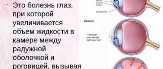
- Stabilization of intraocular fluid pressure (due to activation of blood circulation in the orbital vessels).
- Normalization of the tone of the muscles that control the movements of the eyeball.
- Expansion of accommodation reserve.
- Increased visual acuity and clarity.
What is included in the procedure
Before electrical stimulation, neurologists at the Yusupov Hospital find spasmodic muscles, determine the degree of increase in their tone, and recognize the presence of fibrotic changes. If the procedure is performed on the face, the patient is advised to remove makeup and degrease the skin. The physical therapist then applies a conductive gel to the skin and electrodes.
Electrodes are applied to areas where motor points are located: face, chest, back, abdomen, limbs and secured. Having selected the required current strength, frequency and duration, he conducts a session of electrical stimulation. At the end of the procedure, removes the electrodes and cleanses the skin of the gel.
Indications for electrical stimulation of the pelvic floor muscles
Electropulse therapy of the pelvic muscles involves the use of current with different frequency and power levels. Gynecological indications for electrical stimulation of the uterus and perineum are:
- weakening of the pelvic floor muscles after pregnancy and childbirth;
- hiatus (gaping) of the genital slit;
- enuresis;
- prolapse (prolapse, prolapse) of the anus or uterus;
- cystitis (inflammation of the bladder) in the chronic stage;
- pain syndrome localized in the pelvic region.
Electrical stimulation of the uterus is permissible, but with caution, in case of some gynecological pathologies - fibroids, polyps, endometriosis, etc.
Electrical stimulation of muscles while walking
Treatment of motor neurological deficits involves the use of several treatment regimens. One of the modern ways to increase their effectiveness is artificial correction of movements. In this method, the patient is given electrical stimulation while walking. This is external electrical stimulation of muscles, the parameters of which are selected individually by rehabilitation specialists at the Yusupov Hospital in accordance with the patient’s characteristics, physiological norms and walking dynamics.
The method of electrical muscle stimulation allows you to achieve the following results:
- reduction of fatigue;
- increased range of motion when walking;
- increasing the pace of walking;
- increasing the maximum distance a patient can walk.
Electrical muscle stimulation in Moscow is used by rehabilitation clinic specialists as a method of treating motor neurological deficits. You can undergo a course of treatment by calling the Yusupov Hospital.
Use of lasers in ophthalmology
- Directions for using lasers in ophthalmology
- Lasers in diagnostics
The first branch of medicine in which lasers were used was ophthalmology. The word "LASER" is an abbreviation for the English "Light Amplification by Stimulated Emission of Radiation" - "light amplification using stimulated radiation." The term laser, made up of the first letters of the words “optical quantum generator,” is also used.
Lasers are fundamentally different from other light sources in the properties of the light flux: coherence, monochromaticity, strict directionality (low divergence). The operation of lasers is based on the principle of stimulated radiation in atoms and molecules. This means that the radiation of the atoms of the active medium occurs simultaneously, as a result of which the total radiation has ideal regularity in space and time.
Solid, liquid and gaseous substances can be used as active media in lasers. Solid-state lasers use crystalline or amorphous dielectrics, while liquid lasers use solutions of various substances. The active medium (crystals, gases, solutions, semiconductors) most often determines the type of laser (for example, ruby, argon, diode, etc.).
The monochromaticity and parallelism of laser light allows it to be used to selectively and locally influence various biological tissues.
Existing laser systems can be divided into two groups:
- Powerful lasers using neodymium, ruby, carbon dioxide, carbon monoxide, argon, metal vapor, etc.;
- Lasers that produce low-energy radiation (helium-neon, helium-cadmium, nitrogen, dyes, etc.), which do not have a pronounced thermal effect on tissue.
Currently, lasers have been created that emit in the ultraviolet, visible and infrared regions of the spectrum.
The biological effects of a laser are determined by the wavelength and dose of light radiation.
The following are commonly used in the treatment of eye diseases:
- excimer laser (wavelength 193 nm);
- argon (488 nm and 514 nm);
- krypton (568 nm and 647 nm);
- diode (810 nm);
- Nd:YAG laser with frequency doubling (532 nm), also generating at a wavelength of 1.06 μm;
- helium-neon laser (630 nm);
- 10-carbon dioxide laser (10.6 µm).
The wavelength of laser radiation determines the scope of application of the laser in ophthalmology.
For example, an argon laser emits light in the blue and green ranges, which matches the absorption spectrum of hemoglobin. This makes it possible to effectively use an argon laser in the treatment of vascular pathologies: diabetic retinopathy, retinal vein thrombosis, Hippel-Lindau angiomatosis, Coats disease, etc.; 70% of blue-green radiation is absorbed by melanin and is mainly used to affect pigmented formations.
The krypton laser emits light in the yellow and red ranges, which are maximally absorbed by the pigment epithelium and choroid without causing damage to the neural layer of the retina, which is especially important when coagulating the central parts of the retina.
A diode laser is indispensable in the treatment of various types of pathology of the macular region of the retina, since lipofuscin does not absorb its radiation. The diode laser radiation (810 nm) penetrates into the choroid of the eye to a greater depth than the radiation of argon and krypton lasers. Since its radiation occurs in the infrared range, patients do not feel a blinding effect during coagulation. Semiconductor diode lasers are more compact than lasers based on inert gases, can be powered by batteries, and do not require water cooling. Laser radiation can be delivered to an ophthalmoscope or to a slit lamp using fiber optics, which makes it possible to use the diode laser on an outpatient basis or at the hospital bed.
A neodymium yttrium aluminum garnet (Nd:YAG) laser with near-infrared radiation (1.06 µm), operating in pulsed mode, is used for precise intraocular incisions, dissection of secondary cataracts and pupil formation. The source of laser radiation (active medium) in these lasers is an iridium-aluminum garnet crystal with neodymium atoms included in its structure. This laser is named “YAG” after the first letters of the emitting crystal. The frequency-doubled Nd:YAG laser, emitting at a wavelength of 532 nm, is a serious competitor to the argon laser, as it can also be used for pathology of the macular region.
He-Ne lasers are low-energy, operate in continuous radiation mode, and have a biostimulating effect.
Excimer lasers emit in the ultraviolet range (wavelength 193-351 nm). These lasers can remove specific superficial areas of tissue with an accuracy of 500 nm using a photoablation (evaporation) process.
Directions for using lasers in ophthalmology
- Laser coagulation. Thermal effects of laser radiation are used, which gives a particularly pronounced therapeutic effect for vascular pathology of the eye: laser coagulation of the vessels of the cornea of the iris, retina, trabeculoplasty, as well as exposure of the cornea to infrared radiation (1.54-2.9 microns), which is absorbed by the corneal stroma, in order to change refraction.
Among lasers that allow tissue coagulation, the argon laser is currently still the most popular and frequently used. An increase in the size of the eyeball with myopia is in most cases accompanied by thinning and stretching of the retina and its degenerative changes. Like a stretched delicate veil, it “creeps” in places, small holes appear in it, which can cause retinal detachment - the most severe complication of myopia, in which vision can be significantly reduced, even to the point of blindness. To prevent complications in case of dystrophic changes in the retina, peripheral preventive laser coagulation (PPLC) is used. During the operation, argon laser radiation is used to “weld” the retina in areas of its thinning and around breaks. When the pathological growth of the eye is stopped and complications are prevented (PPLK), refractive surgery for myopia becomes possible. - Photodestruction (photodiscision). Due to the high peak power, tissue is dissected under the action of laser radiation. It is based on electro-optical “breakdown” of tissue, which occurs due to the release of a large amount of energy in a limited volume. In this case, plasma is formed at the point of influence of laser radiation, which leads to the creation of a shock wave and micro-tear of tissue. To obtain this effect, an infrared YAG laser is used.
- Photoevaporation and photoincision. The effect is a prolonged thermal effect with evaporation of the fabric.
For this purpose, an IR CO2 laser (10.6 μm) is used to remove superficial formations of the conjunctiva and eyelids. Photoablation (photodecomposition). It consists of dosed removal of biological tissue. We are talking about excimer lasers operating in the hard UV range (193 nm). Area of use: refractive surgery, treatment of dystrophic changes in the cornea with opacities, inflammatory diseases of the cornea, surgical treatment of pterygium and glaucoma. - Laser stimulation. For this purpose, low-intensity red radiation from He-Ne lasers is used in ophthalmology. It has been established that when this radiation interacts with various tissues as a result of complex photochemical processes, anti-inflammatory, desensitizing, absorbable effects appear, as well as a stimulating effect on the processes of repair and trophism. Laser stimulation in ophthalmology is used in the complex treatment of uveitis, scleritis, keratitis, exudative processes in the anterior chamber of the eye, hemophthalmos, vitreous opacities, preretinal hemorrhages, amblyopia, after surgical interventions, burns, corneal erosions, some types of retino- and maculopathy. Contraindications are uveitis of tuberculous etiology, hypertension in the acute stage, hemorrhage less than 6 days old.
The first four areas of using lasers in ophthalmology are surgical, and laser stimulation is a therapeutic treatment.
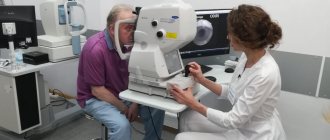
Lasers in diagnostics
- Laser interferometry allows us to make a conclusion about retinal visual acuity in cloudy ocular environments, for example, before cataract surgery.
- Scanning laser ophthalmoscopy makes it possible to examine the retina without obtaining an optical image. At the same time, the power density of radiation incident on the retina is 1000 times lower than when using the ophthalmoscopy method, and there is no need to dilate the pupil.
- Using a laser Doppler velocity meter, you can determine the speed of blood flow in the retinal vessels.