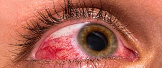
Red eye syndrome - what is it?
This syndrome can be defined as a microcirculatory disorder that occurs due to expansion of the vascular wall of the sclera. It is considered the most common sign in ophthalmology, as it is included in the symptoms of a very large number of diseases. You should definitely be wary of the accompanying symptoms of red eye syndrome, such as pain and decreased quality of vision. The intensity of the vascular pattern on the protein membrane, as a rule, is not related to the degree of any pathology. Let's find out what causes red eye syndrome. Treatment depends on the etiology and nature of the development of the underlying disease.
What causes red eye syndrome?
There are many reasons that can trigger the appearance of this syndrome. They can be combined into several large groups:
- physiological;
- environmental exposure;
- ophthalmopathology;
- diseases that are not related to ophthalmopathologies.
Physiological factors include heavy physical activity, vomiting, prolonged coughing or sneezing, the consequences of drinking alcoholic beverages, working at a computer, etc. This group can also include a factor such as discomfort caused by wearing incorrectly selected optics. The redness of the protein membrane is not accompanied by inflammation. This syndrome does not cause any complications. However, this does not mean that these causes cannot lead to the development of the disease if their effect on the eyes is systematic.
Environmental factors: exposure to sunlight, strong or cold winds, frost, changes in weather conditions, exposure to dust and other foreign objects in the eyes, mechanical injuries, chemical burns to the eyes.
Diseases in which the eyes turn red may have an infectious etiology or relate to aseptic processes. In the first case, we are talking about conjunctivitis (viral, bacterial, chlamydial), dacryocystitis, blepharitis, uveitis, keratitis, episcleritis, iridocyclitis and other diseases caused by pathogenic microorganisms. Aseptic processes of a pathological nature, in which microbes do not play any significant role, include: keratopathy, keratoconus, floppy eyelid syndrome, hemorrhagic tissue lesions, corneal ulcers, lacrimal gland tumors, glaucoma, trichiasis, mucosal detachment and other diseases. Each of these diseases has extensive symptoms and its own specific symptoms. Thus, red eye syndrome can signal relatively mild diseases, as well as pathologies that can lead to blindness.
Another group of factors are diseases that are not related to ophthalmic pathologies. The human body is a single system in which the disease of one organ often affects the condition of other organs. Red eye syndrome most often occurs with diseases that affect blood vessels: hypertension, allergies. Sometimes the sclera turns red due to an overdose of anticoagulants - medications that prevent the formation of blood clots. These causes do not lead to inflammation of the eyes and its structures.
Separately, it is worth listing the risk factors that can provoke red eye syndrome: autoimmune diseases, diabetes mellitus, dry eye membranes that develop with age-related changes, asthenopia, the use of low-quality cosmetics, infectious diseases.
Causes
The vascular network is visible through the sclera, which gives color to the visual apparatus. As the sclera becomes thinner and its transparency increases, a blue discoloration of the whites of the eyes is observed. The main root causes of the manifestation of pathology are:
- genetic predisposition;
- pathological conditions in the body.
If there is a hereditary disorder, the sclera takes on a blue tint after the baby is born. After six months, the proteins should acquire a white tint, which will confirm the absence of pathology in the child’s body. The presence of gray shades of the sclera of the eyes indicates its congenital insufficient pigmentation.
As the child grows, the pigment accumulates, the proteins acquire a white tint. Pathological disorders in adult and pediatric patients are shown in the table.
| Organ system | Pathological conditions |
| Syndromes | Lobstein-Van der Heve. Ehlers-Danlos; Marfana; Lobshtein - Vrolik. |
| Connective tissue | pseudoxanthoma elastic type. |
| Hematological and bone pathologies | Diamond-Blackfan anemia; Paget's pathology; insufficient production of acid phosphatase; anemia. |
| Ophthalmological disorders | scleromalacia; iris hypoplasia; abnormal structure or damage to the cornea; myopia; decreased level of color perception; congenital glaucoma; embryotoxon anterior. |
| Cardiovascular disorders | Congenital heart defect. |
The blue tint of the sclera in pediatric patients provokes disturbances in the healing of fractures.
Pathogenesis of red eye syndrome
All structures of the eye need nutrients that come along with the blood through the vessels. If the walls of blood vessels expand and stretch greatly, they are filled with more blood than required. A vascular pattern begins to appear on the surface of the sclera. Due to the transparency of the mucous membrane, it is very clearly visible. Dilatation of blood vessels can lead to thinning of their walls. This becomes a predisposing factor to hemorrhages. Redness occurs in different parts of the protein membrane. Sometimes they cover the entire sclera, in some cases they are localized in the corners of the eyes.
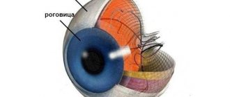
Shell structure
The sclera is the outer tunica albuginea made of dense connective tissue that protects and holds the internal functional elements.
The white of the eye consists of bundle-shaped, randomly arranged collagen fibers. This explains the opacity and different densities of the fabric. The thickness of the shell ranges from 0.3 to 1 mm; it is a capsule of fibrous tissue of unequal thickness.
The white of the eye has a complex structure.
- The outer layer is a loose tissue with a branched vascular system, which is divided into deep and superficial vascular networks.
- The sclera itself consists of collagen fibers and elastic tissues.
- The deep layer (brown plate) is located between the outer layer and the choroid. Consists of connective tissue and pigment cells - chromatophores.
The posterior part of the eye capsule has the appearance of a thin plate with a lattice structure.
Symptoms of red eye syndrome
The syndrome develops in three stages. In the first, superficial, redness is noticeable on the periphery of the conjunctival sac, which indicates hyperemia - overcrowding of blood vessels. You need to visit an ophthalmologist in the next 1-2 days. At the deep (ciliary) stage, a bright red border forms around the limbus, the junction of the cornea and the sclera. This suggests that inflammation is occurring inside the eye. The mixed stage is accompanied by hyperemia of the vessels of the mucous membrane and sclera around the limbus. If you notice a symptom, consult your doctor immediately. This is the big picture. Each pathology has its own symptoms.
With episcleritis, redness is usually one-sided; they develop in parallel with such signs as irritation and lacrimation. Scleritis is accompanied by acute pain in the eye and profuse tear production. An attack of glaucoma is manifested by pain, nausea, the appearance of halos around light sources, clouding of the cornea and decreased visual acuity. With uveitis, photophobia develops and flashes appear before the eyes. The vascular pattern is pronounced. Allergic reactions are almost always accompanied by itching, burning in the eyes, and tearing.
Congenital pathologies
Melanosis (melanopathy) is a congenital disease that is expressed by melanin pigmentation of the skin. Changes appear in the first year of life. The baby's whites have a yellowish tint, and pigmentation appears in the form of spots or stripes. The color of the spots may be gray or light purple. The cause of the anomaly is a violation of carbohydrate metabolism.
Blue sclera syndrome is often accompanied by other eye defects, anomalies of the musculoskeletal system, and the hearing system. The deviation is congenital. Blue sclera may indicate iron deficiency in the blood.
Diagnosis of red eyes
To establish an accurate diagnosis, a complete examination is required. After collecting anamnesis, a primary ophthalmological examination is performed, including:
- visual acuity test;
- assessment of the oculomotor system;
- slit lamp examination;
- eye pressure test;
- fundus examination.
Bacteriological analysis is carried out if an infectious disease is suspected. The composition of the tear film is determined using the Schirmer test - this is a test using special paper strips. Other diagnostic methods may be required: gonioscopy, ultrasound of the eye.
Treatment of red eye syndrome
There is no general approach to treating the syndrome, as it is caused by various reasons. The doctor’s task is to identify the pathology, if any. In accordance with what factor, that is, the disease, manifests itself in the “red eye” syndrome, treatment is prescribed. In case of allergic conjunctivitis, it is necessary to eliminate the allergen. Symptoms of the disease are relieved with anti-allergic ophthalmic drops. Viral forms of the disease require antibacterial and antiviral therapy. For glaucoma, beta blockers, carbonic anhydrase inhibitors, pilocarpine and other drugs are taken. When they do not bring a positive effect, surgery is prescribed.
You almost always need to take vitamins and medications containing antioxidants and mineral complexes. Vitamin A strengthens the immune system, ascorbic acid has a healing effect. Some pathologies are treated using physiotherapeutic procedures. These include ultrasound, laser, and magnetic stimulation. Folk remedies - herbs, infusions, lotions - can only be used after consultation with a doctor. Otherwise, they can aggravate the situation and cause complications. In any case, you should not self-medicate. If the symptoms of the syndrome do not go away, appear too often, or are accompanied by other symptoms, visit the clinic.
healing
There is no single treatment regimen for blue sclera, since transformation of the color of the eyeballs is not a disease. As therapy, the doctor may recommend:
- electrophoresis with calcium salts;
- massage course;
- therapeutic exercises;
- painkillers that will help relieve pain in bones and joints;
- correction of diet;
- use of a course of chondroprotectors;
- buy a hearing device (if the patient has hearing loss);
- bisphosphonates, which prevent bone loss;
- surgical correction (for otosclerosis, fractures, deformation of the bone structure);
- the use of medications containing calcium and other multivitamins;
- antibacterial drugs if the disease is accompanied by an inflammatory process in the joints;
- Women in menopause are prescribed hormonal medications containing estrogen.
What complications does red eye syndrome cause?
Lack of treatment can subsequently cause complete loss of vision. Such complications are rare, but no one is immune from them. The syndrome should not be ignored under any circumstances. Blindness can be caused by keratitis and scleritis. Dry eye syndrome in its advanced form leads to corneal clouding, scarring, and amblyopia. Long-term and ineffective treatment of iridocyclitis causes overgrowth of the pupillary opening. Because of this, the outflow of aqueous humor from the eye is disrupted. This becomes the cause of glaucoma. In most cases, red eye syndrome is not serious. It is only important to identify its etiology in time and begin treatment.