Pathological processes on the retina in ophthalmology are considered the most dangerous. It is the retina that is the element of the visual system that receives images of surrounding objects. Any pathology of the retina carries the risk of a significant decrease in visual acuity up to complete blindness.
Diseases of the retina, depending on the etiology, are divided into:
- inflammatory;
- dystrophic;
- vascular.
Diseases of the inflammatory group arise as a result of infection after injury or surgical treatment, and can also develop without mechanical damage to the eye with reduced immunity and against the background of some common chronic diseases.
Diseases associated with retinal tissue dystrophy are the most common. Depending on the clinical picture they are divided into:
- retinal hemorrhages;
- retinal detachment;
- retinal tears;
- peripheral retinal dystrophy;
- macular degeneration.
The group of vascular dystrophic diseases of the retina includes: angiopathy, diabetic retinolopathy, thrombosis of the central retinal vein. These diseases most often develop against the background of general cardiovascular diseases and diabetes mellitus. Any metabolic disorders deprive the eyes of adequate blood supply. In this case, local death of sensitive retinal cells may develop. This phenomenon does not develop in reverse, so it is worth promptly seeking medical help at the slightest sign of vascular insufficiency of the retina.
Rare retinal diseases constitute a separate group. They are not associated with blood vessels, dystrophy or inflammatory processes. These may be tumor diseases, angiomatoses, aplasia and hypoplasia of the central fovea, as well as other anomalies in the development of retinal tissue (intrauterine or intravital).
Retinal hemorrhages
Hemorrhages in the retinal area can occur as a result of injury to the eyeball, vascular pathologies or dystrophic diseases of the retina. Retinal hemorrhages are found in diabetes mellitus, thrombosis of the central retinal vein, defects of the cardiovascular system, blood diseases, and burns.
The blood in the cavity of the eyeball has a toxic effect on the internal structures of the eye, changes the structure of the vitreous body and reduces the transparency of the optical media of the eye, which is accompanied by a deterioration in the quality of vision. Severe complications of retinal hemorrhage are increased intraocular pressure and secondary glaucoma, retinal detachment, degenerative changes in the retina, etc.
To treat hemorrhages in the retina, conservative methods are used (injections of drugs that accelerate the resorption of hemorrhages), laser treatment (laser vitreolysis, laser coagulation), and surgical techniques (vitrectomy with replacement of the clouded vitreous).
Read more about the treatment of retinal hemorrhages >>>
Causes
The onset of dystrophic abnormalities in the retina is most often associated with its injury. Functional failure or organic disruption of tissue integrity occurs as a result of an accident or as a complication after surgery. Reasons may also be:
- general systemic diseases of the body (diabetes mellitus, hematopoietic diseases, atherosclerosis, rheumatism, hypertension, meningitis, kidney disease);
- background ophthalmological diseases (myopia and farsightedness, inflammatory processes, pathological conditions that impair the nutrition of the eye);
- infectious diseases of the eyes and the whole body (syphilis, toxoplasmosis, tuberculosis, viral and bacterial purulent infections);
- allergic reactions affecting the mucous membranes of the eyes;
- brain and facial injuries;
- poisoning (substances with neurotoxic effects are especially dangerous);
- serious stress and shock.
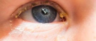
Retinal inflammation
Inflammation of the retina of the eyeball (retinitis) can occur as a unilateral or bilateral process. The causes of retinitis are varied: infectious diseases of a bacterial and viral nature (including tuberculosis, syphilis, AIDS, herpes), toxic eye damage due to other pathologies, allergic diseases, chronic heart and kidney diseases.
Manifestations of retinitis can be different and depend on in which parts of the retina the pathological process is localized. Common symptoms for any form of retinitis include changes in visual fields (scotoma) and a decrease in its acuity. Scotomas are focal loss of visual fields; their size and location directly depend on the location and size of the inflammatory focus. If retinitis is untimely or incompletely treated, persistent vision deterioration occurs, which is especially noticeable in poor lighting conditions.
Treatment of retinitis should be aimed at identifying and eliminating the cause that caused the inflammation of the retina. The most effective is an integrated approach, including drug treatment, injections of anti-inflammatory and antibacterial drugs, and laser coagulation of retinal vessels.
Read more about the treatment of retinal inflammation >>>
Treatment of retinitis
Treatment is prescribed after a full diagnosis and determination of the etiology of the disease. This ensures correct and safe therapy. Treatment is aimed at eliminating the inflammatory process. For this, the following groups of drugs are prescribed:
- antibiotics for internal and local use;
- eye drops and ointments based on corticosteroids;
- anti-inflammatory and antibacterial drops.
Medicines based on acyclovir and interferon are also prescribed. Subconjunctival injections will be helpful. Specific retinitis requires taking medications that eliminate a specific pathogen. Complex therapy consists of taking vasodilators and antispasmodics. Additionally, it is necessary to take medications to improve metabolic processes. Vitamin complexes are prescribed as auxiliary therapy.
In some cases, it is recommended to undergo the procedure with electrophoresis. These pathologies can cause retinal detachment. The only treatment for this complication is laser coagulation.
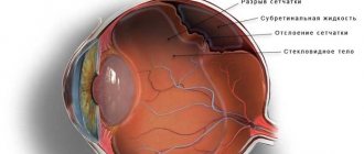
Retinal detachment (detachment)
It is a separation of the retinal layer and the underlying tissues of the choroid. In the event of a tear or rupture of the retina, intraocular fluid leaks between it and the choroid, leading to disruption of the normal nutrition of photoreceptor cells. If you suspect retinal detachment, you must immediately contact an ophthalmologist to provide qualified assistance. The ability to maintain the patient’s visual acuity will directly depend on how quickly and completely assistance is provided for retinal detachment. Delay in this case threatens irreversible loss of vision and blindness!
When retinal detachment is detected, surgical treatment methods are used: extrascleral filling, laser coagulation of tissue around the area of tissue detachment, vitrectomy (replacement of the vitreous body of the eye) to remove blood from the cavity of the eyeball and restore the transparency of the optical media of the eye.
Read more about the treatment of retinal detachment >>>
Types of eye diseases
You might be surprised, but there are more than 2 thousand eye diseases. Most often, people go to the hospital with “dry eye syndrome,” farsightedness, myopia, and conjunctivitis. In older patients, cataracts and glaucoma are most often diagnosed.
Note! Eye diseases are treated by an ophthalmologist or ophthalmologist. There is no difference between these doctors.
The eye is a complex organ of the visual system. A huge number of structures are involved in the process of obtaining a picture of the surrounding world:
- the eyeball, most of which is filled with vitreous humor;
- the outer shell, which includes the cornea and sclera;
- the retina and its central part - the macula;
- choroid, which consists of the iris, ciliary body and choroid;
- lens;
- pupil.
The normal functioning of the eye is ensured by blood vessels, extraocular muscles, the optic nerve and lacrimal glands. The protective function is performed by the eyelids and eyelashes.
Tumors of the retina of the eye
Retinal neoplasms can be benign or malignant, affect one or both eyes, and are detected in young (usually children) and adulthood. Gradually growing, the volumetric formation of the retina occupies most of the eye, and therefore the patient develops an asymmetric protrusion of the eyeball noticeable to the naked eye. Vision in the affected eye disappears, the eye loses mobility.
For timely detection and treatment of retinal diseases, contact the Retina Center. Our specialists will conduct a qualified eye examination using modern diagnostic equipment and will accurately determine whether you require eye treatment. If your doctor recommends surgery, do not delay the decision for long; this will help avoid progression of the disease and preserve your vision.
Treatment of retinal tumors is carried out in specialized medical institutions; delay in seeking qualified help can lead to irreversible consequences.
Diagnosis of retinitis
The following ophthalmological methods are used for diagnosis:
- visometry;
- definition of PZO;
- computer perimetry;
- color perception tests;
- ophthalmoscopy;
- biomicroscopy;
- X-ray.
After the examination results, the etiology of this disease can be established. In this case, it is very important to study the condition of the fundus. The tuberculosis type is accompanied by damage to the retina with a large number of small foci of inflammation. The syphilitic type of the disease causes severe swelling, pigmentation and atrophy of the membrane.
With toxoplasmosis retinitis, almost all eye membranes are affected. During ophthalmoscopy, the doctor observes a yellow lesion. The sunny appearance has red areas of inflammation. During diagnosis, the ophthalmologist often detects the presence of scotomas and a violation of the PZO. Contrast angiography can detect vascular changes. They can expand or contract. It is very important to study their condition.
To diagnose a bacterial or viral type of disease, laboratory blood tests are performed. If the doctor suspects that the pathology has arisen against the background of autoimmune diseases, then it is necessary to conduct immunological tests.
If the disease has an unknown etiology, examination of the following specialized specialists will be required: venereologist, infectious disease specialist, diabetologist, endocrinologist, rheumatologist. Systemic diseases often cause the development of a certain type of pathology that affects the retina.
Retinal dystrophies
The cause of dystrophic changes in the retina is usually disorders in the vascular system of the eye. Most often, dystrophies are found in elderly patients, as well as with moderate and high myopia. The myopic eye is usually extended in the anteroposterior direction, and the resulting overextension of the posterior parts of the eyeball is accompanied by the appearance of dystrophic changes in the fundus. Retinal dystrophies can also occur with various vascular and inflammatory diseases of the eye.
The main method of treating retinal dystrophies is photocoagulation of altered tissues using an argon laser to strengthen thinned areas of the retina and delineate the area of detachment, if one has been identified.
Read more about the treatment of retinal dystrophies>>>
Symptoms
The developing pathology of the retina does not cause pain or other physical discomfort, since its constituent tissue is devoid of sensitive innervation. Most often, gradually developing retinal dystrophy is subjectively felt as a thickening veil before the eyes. Objects become blurry, and even in good lighting the image is blurry, like at dusk. If the pathology is localized in a certain area, then vision is impaired only in the focal area, which, without proper treatment, expands over time.
In some cases, sudden visual phenomena may be observed: lightning, flashes of light, glare, sparking. Black spots and flies appear in the visible area, the field of vision narrows or is lost in a certain area. Some patients note micropsia or macropsia, as well as a sharp deterioration of vision in twilight and dark times of the day.
Age-related macular degeneration (AMD)
The disease is most often detected in people over 50 years of age and is the main cause of irreversible vision loss. The risk of the disease increases with a genetic predisposition, as well as in patients with diabetes mellitus, hypertension, and atherosclerosis. Symptoms of the disease are associated with damage to the macula area of the retina.
The initial manifestations of AMD do not have any clinical manifestations and can only be detected during an examination by an ophthalmologist. As the disease progresses, symptoms such as distorted outlines of objects, blurred vision, and blurred vision appear.
The dry form of AMD does not require special treatment. To treat the wet form of age-related macular degeneration, intravitreal injections (injections into the vitreous cavity) of drugs that block the growth of defective vessels under the retina (anti-VEGF drugs) are used. In addition, photodynamic therapy and laser photocoagulation of the retina are indicated.
Read more about AMD treatment >>>
Main “risk groups”
- People with moderate and high degrees of myopia;
- pregnant women;
- elderly people with diabetes.
The initial stages of the disease may not be accompanied by any symptoms , so if you are at risk, be sure to undergo vision diagnostics using modern equipment. This examination will reliably determine whether you need treatment. If you are scheduled for surgery, do not put off surgery for too long. Before surgery, you need to protect your eye from possible damage in every possible way.
If diseases of the retina of a dystrophic nature are detected, with its thinning and rupture in the periphery, it is strengthened using a laser. Otherwise, any sufficiently strong tension can lead to detachment, requiring immediate surgical intervention. It is better to prevent such a situation. Moreover, detachment can occur when urgent provision of qualified ophthalmological care is impossible (at the dacha, on a trip, etc.)
Diabetic retinopathy
Eye changes in diabetes mellitus cause disruption of oxygen delivery to the retina and lead to the development of diabetic retinopathy. The disease does not develop immediately, but 5–10 years after the onset of diabetes.
Depending on the form of the disease (diabetes type 1 or 2), retinal lesions will differ. A patient with type 2 diabetes often exhibits signs of diabetic maculopathy, leading to decreased visual acuity. In the insulin-dependent form of diabetes, changes in the retina are much more pronounced and are manifested by proliferative changes: the growth of new defective vessels (retinal neovascularization), hemorrhages and scarring of the retina, macular edema.
To treat diabetic retinopathy, laser coagulation of the retina, intravitreal injections of anti-VEGF drugs, and in severe cases, surgical treatment (vitrectomy surgery) are used.
Read more about the treatment of diabetic retinopathy >>>
TsSHRD (TSSKHRP, TsSH)
Central serous chorioretinopathy (CSC) is one of the most common pathologies of the posterior segment of the eye. The cause of the disease is considered to be distress, a situation of “stress” that is extremely common in the modern world. Patients are young people, under 50 years old, mostly men. Complaints: the appearance of a translucent spot in front of the eye, a decrease in the size of objects, impaired color vision - everything seems a little yellowish-brown.
This disease of a non-inflammatory nature, in 50–70% of cases, resolves on its own, but requires dynamic observation and active intervention for a duration of more than 3–6 months.
The observation is based on regular optical coherence tomography of the macular zone in order to assess the dynamics of the height and extent of exudative retinal detachment in the macula.
To identify the point (or points) of filtration - sources of subretinal fluid, FA (fluorescein angiography) is used. The treatment method is laser coagulation of points of fluid leakage under the retina according to FA data. An outpatient painless procedure, after which fluid is reabsorbed under the retina.
Retinal reattachment after laser photocoagulation of leakage points.