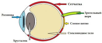
Complex myopic astigmatism is a disease associated with deformation of the cornea in combination with myopia of varying severity in the two main meridians of the eye.
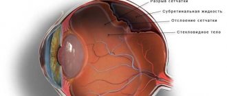
With this pathology, the light beam penetrating the eye is not focused at one point on the retina, but is collected in several beams. Some parts of the image are in focus in front of the retina, while others are on it. In some cases, the entire image is focused at two points located inside the vitreous.
Causes
They distinguish congenital, i.e. hereditary complex myopic astigmatism and acquired due to any factors. Causes of congenital astigmatism:
- uneven curvature of the cornea;
- irregular shape of the lens;
- uneven force of pressure of the eyelids and eye muscles on the eyeball.
These factors are inherited genetically, from generation to generation. If one or both parents suffer from vision problems, it is recommended that the child be seen by an eye doctor as early as possible.
Astigmatism acquired during life is often a consequence of eye injuries or infectious eye diseases.
Types of disease
In medicine, there are several classifications of the disease. Depending on which of the main meridians (horizontal or vertical) the refraction of the eye occurs, there are:
- simple astigmatism - the disorder is observed only along one main meridian;
- complex astigmatism – myopia spreads over 2 eye meridians;
- mixed astigmatism – a combination of myopia and astigmatism, common on the horizontal and vertical meridians.
Depending on the reasons for the development of pathology, there are:
- congenital astigmatism - develops due to the anatomical features of the structure of the cornea or lens;
- acquired astigmatism – develops after surgery or eye injuries.
Degrees of astigmatism
Based on the examination results, 3 degrees of astigmatism are distinguished:
- weak - up to 3 diopters. The distortion of visible objects is not clearly expressed. A weak degree can be equally successfully treated surgically or corrected with glasses and lenses;
- average - from 3 to 6 diopters. It occurs much less frequently, visual function is greatly reduced. The selection of glasses is not recommended; the best solution is lenses and surgery;
- strong – above 6 diopters. As a rule, it is accompanied by destructive changes in the cornea. Correctable with surgery.
The exact degree of astigmatism can only be determined after diagnostic studies have been performed.
Symptoms
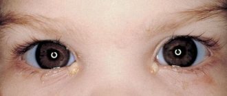
Astigmatism can lead to very interesting changes in visual perception: the patient can see objects in an elongated shape, for example, a square is perceived as a rectangle. In addition, myopia takes away the clarity of images and makes it impossible to see objects in the distance.
Often weak degrees of myopic astigmatism go unnoticed, because a person is already accustomed to seeing objects somewhat differently, and therefore only an ophthalmologist can identify deviations. Very often, myopic astigmatism is accompanied by periodic headaches and blurred vision. If it is mild, the patient’s complaints about weakened vision after sitting at the computer or reading for a long time can also help to suspect it.
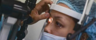
Diagnostics
A set of studies for diagnosing complex myopic astigmatism includes:
- testing of visual acuity with correction. One of the patient's eyes is closed, and cylindrical lenses with different refractive powers are placed in front of the other. It is necessary to establish which one is most suitable;
- skiascopy of the eye - assessment of the light reflex on the pupil;
- computer refractometry is a study of clinical refraction of the eyes, namely the study of the ratio of the refractive power of the eye to its longitudinal axis. The procedure is carried out using a special device - a refractometer, its duration is 2-3 minutes. Computer refractometry is not performed with increased intraocular pressure.
According to indications, biomicroscopy can be performed - examination of the lens, vitreous body, anterior chamber of the eye, cornea and iris, and ophthalmoscopy - examination of the fundus of the eye.
Conservative treatment of complex myopic astigmatism
The optimal methods of conservative correction include the selection and use of glasses or contact lenses. This is a necessity if astigmatism is diagnosed above 1 diopter, myopia progresses, and the patient complains of rapid deterioration of vision.
When selecting lenses for glasses, spherical and cylindrical lenses are combined. An important nuance: the refractive power of a cylindrical lens must be equal to the diagnosed degree of astigmatism. Side effects from wearing glasses include dizziness and pain in the eyes. This is typical for patients with severe astigmatism.
Second method: selection of special astigmatic lenses. They are placed directly on the cornea of the eye. Their principle of operation is the same as that of glasses: they correctly refract light rays and direct them to the retina. The use of contact lenses requires strict adherence to hygiene rules and may lead to allergic reactions.
Glasses and contact lenses help you see clearly only for the duration of their use. They do not completely restore vision. To radically get rid of complex myopic astigmatism, it is recommended to turn to surgery.
Classification
There is a multiple classification, which depends on the cause of occurrence, disorders, degree of development, localization of focus and many other factors:
- corneal - appears when the structure and shape of the cornea is disrupted, which leads to a deterioration in visual acuity due to the fact that this structure refracts passing rays;
- lenticular - caused by damage, change in shape, clouding of the lens;
- hypermetropic - a combined formation of astigmatism and farsightedness, as a result of which light rays are focused in a straight line behind the retina;
- myopic – combined formation of astigmatism and myopia;
- simple – a violation of the structure of one eye;
- complex – disturbances in the structure of both eyes;
- mixed - the presence of myopic manifestations in one eye, hypermetropic - in the other;
- physiological – observed in individuals whose eye dioptre difference is 0.5-1 units;
- athological – the difference in diopters in different eyes exceeds 1 unit;
- congenital - a disorder of the eye structure that does not go away after the age of 1 year;
- acquired – developing as a result of injuries, postoperative interventions, infectious eye diseases.
Surgery
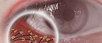
Radial keratotomy incisions (orange stripes)
Radial keratotomy is used as surgical treatment. Using special instruments, the surgeon makes incisions in the cornea. Intraocular pressure changes, and the cornea takes on a changed shape. As a result, its refractive function changes, and visual acuity returns to normal. Previously, the patient undergoes an examination, biomicroscopy and computer keratotopography are performed - assessment of the curvature of the cornea. Radial keratotomy is contraindicated for high myopia, inflammatory and oncological eye diseases. The operation is performed using local anesthesia. The patient should take anti-inflammatory drugs for 3-4 days after the intervention.
Read in a separate article: Eye astigmatism in adults: what it is, symptoms and treatment
Another acceptable method is photorefractive keratectomy. Using laser beams, the surgeon removes the surface epithelial layer. As a result, the curvature of the cornea changes. Photorefractive keratectomy is not recommended for patients under 18 years of age.
Advantages of treatment at MGK
You can receive adequate, high-quality treatment for this disease by contacting the Moscow Eye Clinic. Here you will have the opportunity to undergo a complete diagnosis of the organ of vision using the most modern medical equipment and receive comprehensive advice on further treatment strategies.
If necessary, you will be recommended to undergo laser vision correction surgery. Here, it is performed using LASIK technology using the latest excimer laser; the operation is performed by recognized Russian refractive surgeons.
Treatment of myopic eye astigmatism takes an average of 5-7 days, including diagnostic measures, surgery and postoperative observation. Come, and clear vision without annoying interference will once again become a daily reality for you!
You can make an appointment and ask clarifying questions to our specialists by calling 8 (800) 777-38-81 and 8 (499) 322-36-36 (daily from 9:00 to 21:00) or using the contact form connections on the site.
Molchanova Anna Alexandrovna
Eye exercises
Performing visual exercises will help reduce the manifestations of the disease with mild astigmatism of the 1st degree. Exercises also help relax the eye muscles and are a good prevention of ophthalmic diseases.
It is recommended to perform the following complex:
- go to the window, focus your gaze on an object located behind it, for example, a tree, after 30 seconds focus on the glass. Repeat 5 times;
- close your eyes for 3-4 seconds, open them wide and look from right to left and from top to bottom. Repeat 3-4 times;
- with closed eyelids, rotate your eyes clockwise and then counterclockwise;
- massage closed eyelids with your fingertips.
It is recommended to complete the complex by blinking frequently for 15-20 seconds.
Features of the course of the disease in pregnant women and children
In babies from the newborn period to 11-12 months, congenital astigmatism is often detected. As long as its indicators do not exceed the limits of 0.5-0.75 diopters, it can be considered a physiological norm, in which case correction is not required. A child with increasing astigmatism after school may complain of eye fatigue and distorted vision of surrounding objects. When reading, such children often confuse letters that are similar to each other.
Complex myopic astigmatism in pregnant women may be an indirect indication for surgical delivery. It does not pose a threat throughout pregnancy, but during childbirth it can lead to complete or partial retinal detachment, which threatens the woman with loss of vision. To find out whether natural childbirth is possible, it is necessary to conduct a fundus examination.
Prevention and prognosis
To prevent the occurrence of astigmatism, it is worth learning how to correctly distribute high load on the eyes. This means regularly practicing eye exercises and taking breaks when working for long hours at the computer. You also need to protect your eyes from possible mechanical injuries, and always consult a doctor if you have symptoms of infectious ophthalmological diseases: burning, itching in the eyes, tearing, redness and swelling of the eyelids. Young children should have routine eye exams with an ophthalmologist on a scheduled basis. It is on them that the disease is most often detected in the initial stage.
Astigmatism, in the absence of treatment measures, leads to a noticeable decrease in visual acuity or strabismus.
Author of the article: Elizaveta Viktorovna Klokova, specialist for the website glazalik.ru Share your experience and opinion in the comments.