Causes of angle-closure glaucoma
Anatomical and functional changes lead to the appearance of pathology. Anatomical factors - causes of glaucoma - include:
- short anterior-posterior axis of the organ of vision (small size);
- farsighted refraction;
- shallow anterior chamber of the eye and its narrow angle;
- large lens, including due to swelling due to cataracts;
- increase in the volume of the vitreous body.
Angle-closure glaucoma also occurs for functional reasons: due to pupil dilation, increased secretion of aqueous humor, or intraocular vessels overflowing with blood.
Treatment of an acute attack of glaucoma
Therapy should be aimed at restoring the normal position of the iridolenticular diaphragm and, accordingly, the natural dimensions of the anterior chamber. Prescribe hot foot baths, mustard plasters on calves, laxatives (according to indications). Hirudotherapy - leeches on the temple - is effective, as well as other methods of normalizing blood pressure in the vascular system of the eye and removing excess fluid from the vitreous body (diacarb is usually prescribed according to an individual regimen.
Forms of angle-closure glaucoma
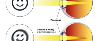
All clinical forms are characterized by closure of the anterior chamber angle (ACA) by the iris root. They differ in the blockade mechanism and prognosis for the treatment of angle-closure glaucoma.
In 80% of all patients with ocular closed-angle glaucoma, the disease occurs with pupillary block. The course of this form is wavy, attacks are replaced by calm, asymptomatic intervals. As a result of each attack, adhesions remain in the drainage system, subsequently leading to chronically high intraocular pressure and changes in the visual fields characteristic of glaucoma.
Angle-closure glaucoma with flat iris accounts for 10%. The attack occurs under the influence of pupil dilation (physiological - for example, in the dark, during stress or medicinal - when mydriatics are instilled).
Even less common (7%) is the “creeping” (with shortening of the anterior chamber angle) form. With it, a gradual fusion of the root of the iris with the wall of the UPC occurs for unknown reasons. At the beginning, the disease is asymptomatic, invisible and becomes an incidental finding during gonioscopy. As the PC angle “overgrows,” subacute attacks of ocular hypertension appear and a typical clinical picture develops.
The rarest form (1%) is angle-closure glaucoma with vitreolenticular block (“malignant”). Its appearance is caused by the accumulation of aqueous humor in the posterior part of the organ of vision predisposed to glaucoma.
| Treatment of malignant angle-closure glaucoma with miotics is contraindicated - these drugs, by constricting the pupil, relax the lens ligaments and aggravate the vitreolenticular block. |
Diagnostics
Taking an anamnesis is the first phase of the diagnostic process for primary angle-closure glaucoma. It is very important, since the disease has a wave-like course, without complaints in the intervals between attacks. Symptoms appear only in subacute and acute attacks of the pathology. The disease is bilateral, although the attack rarely occurs in both eyes at once.
As a rule, medical history indicates a stressful situation as the main cause of the attack. Also, it often develops with overexcitation, prolonged work with a bowed head, drinking a large amount of liquid, drug-induced mydriasis, and hypothermia. Additional complaints include: headache, intestinal disorders, nausea, vomiting.
Physical tests ordered include:
- External examination to assess the general condition of the eyes
- Visometry and perimetry to study visual function
- Biomicroscopy, which reveals the condition of internal structures (mucous membrane, cornea, iris, pupil and its reaction to light). In addition, it is used to examine the depth of the anterior chamber, the volume of intraocular fluid, the transparency of the lens and vitreous body
Instrumental studies are also required, which include gonioscopy, ophthalmoscopy, ultrasound biometric studies and biomicroscopy.
Characteristic features of the diagnosis of various forms of angle-closure glaucoma:
With pupillary block
There are no symptoms, only after performing biomicroscopy, some shallowing of the anterior chamber is revealed, which is caused by an anterior displacement of the iridolenticular diaphragm.
In the latent course of glaucoma, the diagnosis is retrospective, which is based on the condition of the paired eye that suffered an attack. In the absence of an acute attack, the situation is interpreted as an anatomical anomaly of the organ of vision.
A subacute attack of this form of glaucoma is diagnosed based on very limited data: slight blurred vision, multi-colored circles around the light source, an unpleasant sensation in the eye. In rare cases, such symptoms are accompanied by a headache or pain in the eyebrow. Examination reveals a small injection, slight swelling of the cornea, and slight dilation of the pupil.
At the same time, gonioscopy reveals varying degrees of blockade of the anterior chamber angle. Tonometry - a significant increase in IOP due to fluid retention in the chamber. A subacute attack usually leaves no visible consequences.
An acute attack, as a rule, does not cause difficulties in diagnosis. The patient indicates pain in the eye and in the eyebrow on the corresponding side. He notes a sharp, often significant decrease in vision.
Ophthalmological examination reveals congestive injection, sometimes with conjunctival edema. The cornea is opaque and edematous, occasionally with bullous lesions and decreased sensitivity. The anterior chamber has lost its depth and is sometimes found only in the pupillary area. The intraocular fluid often changes transparency, as the protein content in it increases sharply. The iris is hyperemic with a shaded pattern. The pupil is irregularly shaped and paralytically dilated.
With angle shortening (“creeping”)
This form of glaucoma does not have a systemic course. Features of gonioscopic examination are expressed in the replacement of the apex of the angle from the ciliary body to the root of the iris. The path of the light focal line hits the iris without shifting.
With a flat iris
This form occurs when the angle of the anterior chamber is closed and the typical attachment of the iris is flat. This structure of the iris is the reason for the shallowing of the anterior chamber at the periphery, while its center is relatively deep.
With vitreolens block
This is a malignant form of glaucoma, which is very difficult and important to diagnose. When it occurs spontaneously, a closed angle, a small chamber, high intraocular pressure and a relative block of the pupil are always detected. As a rule, the disease is detected accidentally.
After antiglaucoma operations
The disease is malignant and is accompanied by:
- Mixed injection of different severity.
- Lack of external filtration along the created outflow pathways.
- Small anterior chamber (slit-like or preserved only in the pupillary region).
The purpose of gonioscopy reveals the processes of the ciliary body in contact with the lens equator, directed anteriorly in the lumen of the basal coloboma. Very high or significantly increased intraocular pressure. If the cornea and lens are transparent, biomicroscopy reveals free zones in the vitreous body. Ultrasound scanning adds to the information obtained the anterior position of the iridolenticular diaphragm and ciliary body, fluid in the vitreous chambers, changes in the depth and shape of the posterior chamber.
Differential diagnosis of primary angle-closure glaucoma during an acute attack is carried out with iridocyclitis, migraine, hypertensive crisis, acute abdomen, and gastrointestinal infection.
Malignant postoperative glaucoma must be differentiated from small anterior chamber syndrome.
Symptoms of angle-closure glaucoma
This disease occurs in waves and is characterized by attacks: acute or subacute. Outside of an attack, there are no symptoms of angle-closure glaucoma.
During the interictal period at the onset of the disease, intraocular pressure is normal; as the process progresses and becomes chronic, ophthalmotonus steadily increases.
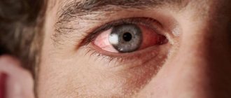
Symptoms of an acute attack of angle-closure glaucoma:
- severe dull pain in the eye, eyebrow, temporal region;
- Possible nausea and vomiting that does not bring relief;
- iridescent areolas around the light source;
- “milky” fog in the field of view;
- severe redness around the cornea;
- pupil dilation.
On palpation (feeling), the diseased organ is hard, rocky-dense. At the height of the attack, IOP can increase to 40-60 mm Hg. Art.
If the angle of the anterior chamber is not completely or not completely blocked, a subacute attack develops. Its symptoms in angle-closure glaucoma are smoothed out; most often, patients complain of rainbow circles around light sources and blurred vision. Soreness and redness are mild.
Diagnostics
An attack of angle-closure glaucoma occurs with characteristic symptoms; the diagnosis is confirmed by gonioscopy, examination of the anterior segment of the eyeball at the slit lamp and tonometry.
During the interictal period, the diagnosis of closed-angle glaucoma is based on examination of the PC angle, optical coherence tomography (OCT of the PC and optic nerve head), examination of the anterior and posterior segments. Perimetry is required - examination of visual fields.
In doubtful cases, stress tests are performed: IOP is measured before and after instillation of drops that dilate the pupil (mydriatics).
For patients with farsightedness over the age of 40 years, due to the anatomy of the organ of vision, an examination by an ophthalmologist is required once a year.
Signs of an acute attack
Diagnostic criteria are:
- redness, hyperemia, “overcrowding” of the eye with blood due to pathological vasodilatation and impaired outflow.
- corneal turbidity, surface unevenness, swelling, decreased sensitivity;
- anterior shift of the iridolenticular diaphragm (which should be eliminated first), reduction in the size of the anterior chamber;
- mydriasis (pupil dilation);
- “stone hardness” of the eye upon palpation (it is easy to compare tactile sensations by touching your own eye or the patient’s healthier eye).
The full complex or any combination of such symptoms indicates an acute attack of glaucoma and requires immediate specialized intervention.
Acute attacks predominantly develop with angle-closure glaucoma. In practice, however, such patients are thoroughly informed and instructed by the attending ophthalmologist, so they are able to recognize the development of an attack in time and have the necessary medications with them to prevent it.
Treatment of angle-closure glaucoma
Measures for the treatment of angle-closure glaucoma differ during an attack and in the inter-attack interval.
| Treatment of acute and subacute attacks of glaucoma requires emergency care from an ophthalmologist. |
During the first hour, 1-2% pilocarpine is instilled every 15 minutes, then every hour, then every 4 hours. At the same time, a beta blocker (timolol 0.5%) is used once and a diuretic is given. In the absence or low effectiveness of drug treatment for angle-closure glaucoma during an attack within 24 hours, surgical intervention is indicated - iridectomy. If the attack is stopped, glaucoma surgery is performed without fail, but at a later date. After the incident, visual functions may not be restored.
Conservative treatment of angle-closure glaucoma is carried out with drops that narrow the pupil. These are drugs from the group of cholinomimetics (pilocarpine). Combination medications can be used (Fotil - pilocarpine 1% + timolol 0.5%, Fotil forte - pilocarpine 2% + timolol 0.5%). If the effect is insufficient, treatment of angle-closure glaucoma is supplemented with carbonic anhydrase inhibitors (Azopt, Dorzopt, Trusopt) and osmotic diuretics according to indications.
Conservative antihypertensive therapy is justified in the interictal period as preparation for surgery, as well as in the event of an attack of angle-closure glaucoma and the impossibility of surgical intervention.
Surgical or laser treatment of angle-closure graucoma involves the formation of artificial holes at the root of the iris - iridectomy. This opens the angle of the PC and equalizes the pressure between the chambers of the eye. In some cases, surgical iridocycloretraction is performed - widening the PC angle. Laser treatment of glaucoma is preferable, as it is low-traumatic, bloodless and outpatient.
Preventive measures
Prevention of this type of glaucoma includes performing peripheral laser iridectomy (PLIE) to prevent the development of an acute attack.
It is recommended for patients with angle-closure glaucoma:
- intellectual or light physical labor;
- limiting heavy physical activity, especially with prolonged bending of the head and torso;
- work in hot shops, visiting baths and saunas is prohibited;
- avoid prolonged nervous tension;
- Clothes with tight collars, ties, etc. are contraindicated;
- Simultaneous intake of large amounts of liquid is unacceptable.
Persons predisposed to the development of angle-closure glaucoma are not recommended to spend long periods of time in dark or poorly lit rooms (watching TV in a dark room, visiting a cinema, etc.).
First aid
It is important that in the first minutes after the onset of the attack the patient takes 1-2 tablets of Fonurite or Diacarb. An analgesic is also administered, possibly a narcotic (for example, promedol subcutaneously 1 ml of a 2% solution). Medicine is instilled into the eyes three times to reduce pressure: pilocarpine (1% solution), phosphacol (1:5000 solution), armin (1:20000 solution). To reduce pressure, you can also use techniques to deposit blood in the veins of the lower extremities (a hot bath for the legs with mustard) or other methods of reducing the volume of intravascular fluid (leeches on the temporal region). It is important to understand that the earlier treatment is started, the greater the chance of maintaining vision. If it is not possible to start therapy at home, then the patient should be taken to the hospital as soon as possible, without delaying the intermediate stages. An acute attack of glaucoma is one of those few ophthalmological diseases where hours and minutes count.
Cost of surgical treatment of glaucoma
| № | Service name | Price in rubles | Make an appointment |
| 2010025 | Set of disposable consumables for antiglaucomatous surgery | 36000 | Sign up |
| 2010024 | Molteno valve implantation | 54000 | Sign up |
| 2010023 | Implantation of the EX-Pres shunt valve | 54000 | Sign up |
| 2010022 | Ahmed valve implantation | 54000 | Sign up |
| 2010021 | Summing collagen or silicone drainage | 9000 | Sign up |
| 2010020 | Pupilloplasty | 30000 | Sign up |
| 2010019 | Iridoplasty | 22200 | Sign up |
| 2010018 | Basal iridotomy | 10800 | Sign up |
| 2010001 | Sinutrabeculectomy (STE) | 42000 | Sign up |
| 2010002 | Non-penetrating deep sclerectomy (NPDS) | 46200 | Sign up |
| 2010004 | Antiglaucomatous surgery for primary glaucoma of the first category of complexity | 23400 | Sign up |
| 2010005 | Antiglaucomatous surgery for primary glaucoma of the second category of complexity | 30600 | Sign up |
| 2010006 | Antiglaucomatous surgery for primary glaucoma of the third category of complexity | 37200 | Sign up |
| 2010007 | Antiglaucomatous surgery for secondary or refractory glaucoma of the first category of complexity | 29500 | Sign up |
| 2010008 | Antiglaucomatous surgery for secondary or refractory glaucoma of the second category of complexity | 42000 | Sign up |
| 2010009 | Antiglaucomatous surgery for secondary or refractory glaucoma of the third category of complexity | 48000 | Sign up |
| 2010010 | Antiglaucomatous surgery with drainage of the anterior chamber angle for primary glaucoma of the first category of complexity | 28200 | Sign up |
| 2010011 | Antiglaucomatous surgery with drainage of the anterior chamber angle for primary glaucoma of the second category of complexity | 39300 | Sign up |
| 2010012 | Antiglaucomatous surgery with drainage of the anterior chamber angle for primary glaucoma of the third category of complexity | 45600 | Sign up |
| 2010013 | Antiglaucomatous surgery with drainage of the anterior chamber angle for secondary or refractory glaucoma of the first category of complexity | 33360 | Sign up |
| 2010014 | Antiglaucomatous surgery with drainage of the anterior chamber angle for secondary or refractory glaucoma of the second category of complexity | 43800 | Sign up |
| 2010015 | Antiglaucomatous surgery with drainage of the anterior chamber angle for secondary or refractory glaucoma of the third category of complexity | 54000 | Sign up |
| 2010017 | Reconstruction of the anterior chamber angle in secondary glaucoma | 27000 | Sign up |
Making an appointment Today registered: 19
Prevention and prognosis
In order not to aggravate the course of the disease and prevent the development of an acute attack, people with angle-closure glaucoma must adhere to certain recommendations:
- Don't stay in the dark for a long time with your eyes open
- Do not engage in activities with the head and torso tilted
- Avoid contrasting temperature changes
- Avoid hypothermia
- Avoid steam rooms and saunas
- Avoid heavy physical work and sports
It is also worth eliminating or limiting smoking and alcohol; do not drink large amounts of liquid, try to avoid emotional stress.
Early diagnosis and adequate treatment of the disease makes the prognosis for vision relatively favorable, especially in chronic and latent glaucoma. If attacks occur, the prognosis is less favorable due to the rapid progression of the process with each attack. With malignant glaucoma, the prognosis is unfavorable.
Causes of glaucoma development
A healthy eye normally produces a special fluid every day that fills it. Approximately 4 milliliters of fluid are produced per day. In the corner of the anterior chamber (between the cornea and iris) of the eye there is a drainage system through which fluid flows out of the eye. Normal eye pressure ranges from 16 to 25 mm. mercury column. Increased pressure leads to intense compression of the optic nerve fibers and blood vessels, followed by gradual death of the nerve and blindness.
Glaucoma with functional pupillary block
Cause:
pronounced anterior position of the lens, and a tight fit of the iris to it.
Development mechanism:
due to the tight fit of the iris to the lens, the flow of aqueous humor through the pupil (from the posterior eye chamber to the anterior one) is disrupted. Pressure increases in the posterior chamber of the eye, and the peripheral part of the iris protrudes anteriorly, closing the APC. This contributes to the development of an acute glaucomatous attack.
Characteristic:
this form of glaucoma is more common in older women. The disease manifests itself in acute attacks, often with a transition to a chronic course (due to the formation of adhesions, goniosynechia, between the wall of the iridocorneal angle and the iris).