Structure
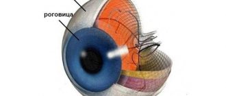
The sclera occupies 5/6 of the fibrous membrane of the eye. The thickness varies from 0.3 to 1 mm in different places. The thinnest part is located in the area of the equator of the eye, in the area of the optic nerve exit, near the cribriform plate, where the axons of ganglion cells extend beyond the eyeball. Staphylomas or bumps can form in these areas when intraocular pressure increases.
Sometimes excavation of the optic disc occurs. This condition is characteristic of glaucoma. The scleral membrane along with the attachment of the extraocular muscles is damaged after injury.
The sclera consists of the following elements:
- the episclera is a loose layer through which many veins and arteries pass in the superficial and deep parts of the vascular network;
- the substance of the sclera, consisting of collagen and elastic fibers;
- the dark scleral plate, formed from connective tissue and pigment cells, is located between the vascular network and the sclera.
In the back of the eyeball, the sclera is formed in the form of a narrow lattice plate, allowing the optic nerve and blood vessels to supply the retina with useful substances. The protective layer of the optic nerve is formed 1/3 from the cribriform plate and 2/3 from scleral tissue.
The cornea, sclera and conjunctiva are connected at the limbus. This structural feature affects the development of polymorphic diseases, inflammation, allergic reactions, tumors and genetic abnormalities.
A dense nerve plexus is located on the circumference of the limbus, formed from the ciliary nerves, and the fibers responsible for the sensitivity of the cornea extend from it. The scleral tissue has few nerve endings; this feature affects the development of diseases characteristic of collagenosis.
Three pairs of extraocular muscles attach to the surface of the sclera. Nerves and arteries pass to the choroid through special channels or emissaries. A circular groove up to 0.75 mm thick is located on the inner surface of the anterior part of the sclera. The ciliary body is attached to dense tissue in the form of a ring through which blood vessels pass.
The anterior part of the groove is located near Descemet's membrane. At its bottom, near the posterior edge, there is a venous sinus of the sclera, which is called Schlemm’s canal. The trabecular meshwork occupies the remainder of the scleral recess.
Indications for retinotomy and retinectomy
Today, there are many more indications for retinotomy and retinectomy; their implementation is an integral, routine surgical procedure in the work of vitreoretinal surgeons.
Currently, excision and dissection of retinal tissue are widely used to provide access under the retina to remove subretinal hemorrhages, subfoveal neovascular membranes trapped inside parasites, liquid PFOS and SM, for biopsy, in the case of tumors of the choroid. In addition, such interventions are necessary to localize small retinal tears during rhegmatogenous detachment, since a vital dye is injected through a pre-made retinotomy hole.
The technology of retinotomy, performed to access photoreceptors if surgical intervention is necessary on them and the retinal pigment epithelium (for installation of subretinal electrodes in case of photoreceptor death), is under research.
Excision and dissection of the retina can be not only full-layer, but also layer-by-layer (lamellar). For example, with retinoschisis, drainage of the cavity fluid is achieved by retinotomy of only its inner wall. Retinectomy is also used - complete excision of the inner wall in order to eliminate tension (traction) from the vitreous body. In addition, there is a technique for retinotomy (puncture) of the inner wall of macular cysts. This achieves their drainage in the case of chronic macular edema that is not amenable to drug, laser or other surgical treatment.
Functions
The main functions of the sclera:
- 2/3 of its thickness is the protective sheath of nerve fibers;
- in the area of the equator of the eye, 4-6 vortex veins pass through the sclera;
- the sclera is the attachment point for the eye muscles, which ensure good mobility of the organ of vision in all directions;
- The sclera passes sensory nerve fibers to the eyeball, sympathetic innervation is directed from the superior cervical ganglion.
The arteries pass through the sclera to the back of the eye.
Manifestation of the disease
Patients complain that they feel as if a foreign body has entered the eye. In this case, there is often increased tearing, redness of the eye and painful discomfort.
Not the whole eye turns red, but only a small area (sector). May go away without treatment in 10 days.
Nodular episcleritis is characterized by limited, hyperemic, clearly defined nodules that do not connect directly to the conjunctiva. At the same time, the sclera does not swell. Palpation of the eye is painless. The disease lasts a long time and is recurrent. During relapse of the disease, the nodules migrate and appear in various places around the limbus. Most often, both eyes are affected. The process lasts about a month and a half.
Migratory episcleritis is characterized by the sudden appearance of hyperemic lesions that cause pain and discomfort. This process lasts two to three days. Nodules can be found in the episclera in rosacea keratitis. Often, nodules form on the sclera at the same time that rosacea appears on the skin on the face. Rarely, migrating episcleritis can become protracted and recurrent.
Pathologies of the sclera
Congenital diseases:
- Melanosis occurs due to poor carbohydrate metabolism with an increased amount of melanin in the scleral tissues. The tunica albuginea turns yellow in this disorder.
- Aniridia is a rare disorder involving the absence of the iris. The pathology manifests itself due to changes in the gene responsible for the normal development of the visual organs. Sometimes acquired aniridia occurs after injury or inflammation of the iris.
- Blue sclera syndrome, in which the whites turn bright blue. The disorder is accompanied by deterioration of vision, hearing and a decrease in the amount of iron in the body. Sometimes the syndrome develops as a symptom of hereditary pathologies.
Acquired diseases:
- Episcleritis is an inflammation of fibrous tissue that becomes covered with dense nodules. The disorder is diagnosed more often in women over 40, and is rarely detected in children. The pathology affects both eyes and becomes chronic without timely treatment.
- Scleritis is an inflammation of all layers of the sclera. The disorder is accompanied by pain, swelling, and blurred vision. Destruction of the tunica albuginea occurs without timely therapy. The disorder affects only one eye and is more common in women.
Staphylomas occur after loosening of collagen tissues in complex forms of myopia. Patients' vision decreases, fatigue increases, and a feeling of heaviness arises.
Diseases
The organ is susceptible to inflammatory processes caused by bacteria.
Pathologies that develop in the eyes at the level of the sclera are most often of an inflammatory nature, provoked by infections. However, primary sources are not always located directly in the organ. Painful manifestations in the eye shell can only act as symptoms of the main processes. First of all, the ophthalmologist looks for the main diseases of the sclera, these include the following:
- Scleritis. An inflammatory pathology in which the inner layers of the membrane are affected.
- Staphyloma. The disease is caused by destructive processes, as a result of which the membrane is depleted.
- Episcleritis. Damage to the upper layer, accompanied by the formation of nodules.
How is scleral condition diagnosed?
An examination by a doctor is carried out after identifying a violation of the sclera. Symptoms associated with scleral tissue disorder:
- eyes hurt when muscle tissue contracts;
- sensation of a foreign object in the eye;
- uncontrollable tearfulness;
- the color of the sclera changes;
- changes in the structure of the eye, deformation of certain areas of the surface, dilation of the arteries.
Diagnostic methods:
- listing the number of Placido rings;
- scanning through an optical slit;
- examination using a Schleimpflug camera;
- study of raster images;
- laser interferometry.
Deformation or clouding of the cornea is a complication after which a person becomes partially or completely blind.
Diagnostics
Based on the results of a blood test, the doctor determines the presence of an inflammatory process.
If you notice dangerous symptoms, you should consult a doctor. First of all, the ophthalmologist does a blood test and examines the eyes and determines the presence of obvious signs of inflammation in them. Further diagnostics are aimed at studying the source and stage of the pathology. First of all, the shell is examined using a microscope. Additionally, ultrasound is prescribed to determine structural changes. Further diagnosis depends on the root cause of the process.
Useful video
More useful information about the functions of the white membrane of the eye can be obtained by watching the video.
Author's rating
Author of the article
Alexandrova O.M.
Articles written
2029
about the author
Was the article helpful?
Rate the material on a five-point scale!
( 1 ratings, average: 5.00 out of 5)
If you have any questions or want to share your opinion or experience, write a comment below.
Chimpanzee, don't hide your eyes!
One of the unique features of humans is the light sclera - the outer layer of the eyeball. In most mammals, and in particular in monkeys, the sclera is dark and brown. In addition, the shape of the eyes is usually such that the sclera is almost invisible. For a long time, it was believed that humans are the only species with white sclera. In addition, due to the elongated shape of the eyes, humans have the largest portion of the sclera open among all mammals. The analysis showed that the eyes are more elongated horizontally in terrestrial primate species, which more often “scan” the surrounding space, turning their heads from side to side. At the same time, the sclera is more open in large species, probably to increase visibility. It is easier for great apes to move their eyes than to move their heavy, massive heads. People turned out to be record holders for horizontal eye elongation.
Why do primates have dark sclera? Scientists have proposed this explanation: the pupil is more difficult to notice against a brown background. Try to guess where the monkey is looking! Perhaps this is how animals hide their gaze from predators and from their fellow tribesmen - competitors for food. At least this has been a popular hypothesis for a number of years. Humans were considered the only species in which the border of the eyes and the position of the iris were clearly visible, especially in those with dark skin. It is interesting in this regard that already human babies begin to closely monitor the views of the people around them. Judging by observations, monkeys cope with this task much worse. Understanding where another person is looking is necessary for planning your own behavior. Perhaps among our ancestors, “reading” the gaze became important for cooperation, for example, in hunting.
Chimpanzee eye.
However, in 2015, after studying the eyes of 85 gorillas, researchers found that although the eyes of humans are indeed more elongated, the visible area of the sclera in gorillas is similar to that of humans. In addition, 10 gorillas had human-like white sclera. The researchers also reasonably concluded that one should not draw a conclusion about “gaze masking” solely on the basis of the color of the sclera. After all, the point is primarily in the contrast of the sclera and the iris. A light circle on a dark background is no worse visible than a dark circle on a light one. In a new study published in PNAS, primatologists compared the eyes of humans (52 individuals), bonobos (51) and chimpanzees (50). Chimpanzees turned out to be the owners of the darkest sclera, bonobos took an intermediate place between chimpanzees and humans. And if in bonobos and humans the iris is darker than the sclera, in chimpanzees the opposite is true: the iris is light against the background of the dark sclera. It became clear why in experiments bonobos follow the direction of human gaze, but chimpanzees do not.
After assessing the iris/sclera contrast in all three species, the study authors concluded that the relative difference in iris and sclera hues in humans, bonobos, and chimpanzees is approximately the same. It turns out that none of these species is going to hide their eyes. Depigmentation (lightening) of the sclera is one of the evolutionary ways of emphasizing the gaze. Lightening the iris against the background of dark sclera (or darkening the sclera against the background of the iris) is obviously another option with the same result. By the way, researchers noticed that chimpanzees' sclera darkens with age. This way the contrast is enhanced.
Scientists conclude that the ability to follow the gaze of fellow tribesmen must have been possessed by the common ancestor of great apes (including gorillas and orangutans, since the latter also exhibit lightening of the sclera).
The study's authors ask why, unlike the common chimpanzee, bonobos and humans followed the path of scleral depigmentation. According to one hypothesis, this is a side effect of selection to reduce aggression. A similar effect—lightening of the sclera—is observed in domesticated animals that have been selected for friendliness. By the way, it has been noted that lowland gorillas have lighter sclera than mountain gorillas, and Sumatran orangutans have lighter sclera than Borneo orangutans. Is there any connection here with the level of aggressiveness?
Comparison of eye contrast of chimpanzees, bonobos and humans.
In any case, we now know that chimpanzees, although they have dark sclera, can quite easily follow the direction of another chimpanzee's gaze. It is worth adding that other factors, such as complexion, can also affect the contrast of the eyes. In any case, the popular idea of “gaze masking” in monkeys should now be tested more carefully.
Methods of treatment and prevention of scleral diseases
Drops with glucocorticoid and antibacterial properties should be installed up to eight times every 24 hours. With increased intraocular pressure, antihypertensive drugs that have a local effect are prescribed. For example, timolol maleate, dorzolamide, betaxolol and others. It is also recommended to take Diclofenac, Ibuprofen, Meloxicam, etc. orally. If there is an individual intolerance to these drugs, then prescribe 75 mg of prednisol to be taken orally per day.
If there is no necrosis of the sclera, then doctors prescribe subconjunctival injections of glucocorticoids, antibiotics, long-acting glucocorticoids - triamcinolone. If a pathological process is detected that caused scleritis. Let's say tuberculosis or syphilis. Then you need to first or comprehensively treat the underlying disease.
For necrosis of the sclera, combination therapy must be performed. To do this, prescribe prednisol 100-120 mg per day for three days. It is combined with immunosuppressants (Azathioprine, Cyclosporine, Cyclophosphamide).
To treat scleritis, measures are taken against allergies, against inflammation, as well as desensitizing treatment.
Purulent scleritis is treated with general antibiotic therapy. Antibacterial drugs are also administered subconjunctivally, in close proximity to the lesion. Electrophoresis with antibacterial drugs is also used.
Doctors often recommend taking fluoroquinolones, aminoglycosides, and semisynthetic penicillins orally.
Sometimes it happens that a purulent focus is opened in order to prevent it from breaking through into the membrane of the eye.