Definition of the concept
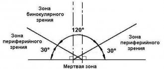
The human field of vision covers about 120 degrees. But visual perception is not the same throughout this field. We see objects in the center of our gaze more clearly than those located on the sides, above and below. The central is responsible for the first, and the peripheral is responsible for the second.
Photography, or more precisely, the photoeffect, will help to clearly demonstrate what peripheral vision is. Think of an image in which the subject in the center of the frame is clearly visible, but objects to the side and the background are blurry. If photographers artificially achieve such an effect, then for a person this state of affairs is the norm.
Peripheral vision is the ability to see objects outside the focus of attention. Central, or direct, vision has a small scope, but distinguishes the smallest details of an object. Peripheral vision sees everything else, but blurry.
Panoramic vision
This year marks the 105th anniversary of the “Battle of Borodino” panorama, the main exhibit of the capital’s museum of the same name. The development of multimedia technologies has not affected interest in historical painting - the museum’s halls are always full of visitors. One of the largest paintings in the world is very impressive. It takes the viewer to the center of the battle, allowing you to feel the enormous scale of military action.
The author of the panorama, Franz Roubaud, was born in 1856 in Odessa and learned the basics of his professional skills there.
Subsequently, while living abroad, he invariably considered himself a Russian artist. It was he who became the founder of the national school of panoramic painting. His fame was brought to him by paintings about the Russian-Persian War and military events in the Caucasus, panoramas “Assault on the village of Akhulgo”, “Defense of Sevastopol”. For the 100th anniversary of the Patriotic War of 1812, by order of Emperor Nicholas II, the artist painted a panorama of the Battle of Borodino, which until 1918 was exhibited in Moscow in the pavilion on Chistye Prudy.
Later, the painting was dismantled, stored rolled up for a long time and was badly damaged. By 1962, it was restored and exhibited in a specially built building on Kutuzovsky Prospekt - next to the restored “Kutuzovskaya Izba”, which once hosted the famous military council. Alas, the misfortunes did not end there: in 1967, there was a fire in the museum that destroyed a significant part of the canvas. Masters from the Studio of Military Artists named after. M.B. Grekov restored Roubaud's creation.
The length of the canvas in a circle is 115 m, the height is 15 m. It depicts more than two thousand figures. The Borodino field appears before the viewer - as it was on September 7, 1812. The front line runs along the Semenovsky ravine. The Russians are on the eastern bank, the enemy is on the western bank. A fierce battle is in full swing. In the foreground are our cavalry, infantry, artillery, Cossacks, and militias. One of the compositional centers of the work is a cavalry battle in the rye, another key episode is the guards infantry repelling the onslaught of the French heavy cavalry. The command posts of Kutuzov and Napoleon are visible in the distance. And all around is a romantic landscape of the Moscow region: fields of ripe rye, green grass, a forest touched with crimson, gray-blue distances. The subject plan, creating the illusion of volume, demonstrates the burnt buildings of the village and military weapons.
Interactive kiosks in the panoramic hall will help you study in detail everything depicted in the picture, find out the exact location of the troops, and find specific participants in the battle.
The stands will additionally tell about all the main events of the Patriotic War of 1812. The halls display authentic weapons and equipment, awards, household items, high-quality reconstructions of military uniforms of both armies, as well as paintings and graphics.
The personal belongings of Major Pavel Nikitin are kept under glass: a camp chest, a candlestick, a snuff box, a wallet, scissors and documents accidentally discovered in the attic of an old Moscow mansion. The pride of the museum is presumably the last lifetime portrait of Mikhail Kutuzov, painted by the artist Roman Volkov in 1813. Nearby is a display case telling the story of the commander-in-chief’s life and several items from his home in St. Petersburg. Copies of portraits of Peter Bagration, Mikhail Barclay de Tolly, Matvey Platov, Nikolai Raevsky and other outstanding military leaders were made especially for the museum. In addition, here you can see images of Emperor Alexander I, militia heroes, participants in the partisan movement and their commanders - Denis Davydov, Alexander Seslavin, Alexander Figner, leaders of peasant detachments Vasilisa Kozhina, Gerasim Kurin, Yegor Stulov.
A special section is dedicated to the heroism of Muscovites in the fight against the invaders, the fire in the Mother See and Napoleon's retreat. All subsequent events of the war are shown, up to the expulsion of the French from the country and the signing of the Manifesto by the Russian Tsar on December 25, 1812 (January 7, 1813), as well as the foreign campaign of 1813–1814, which ended with the triumphant entry of the Russian army into Paris.
The museum has a good tradition. Every year on January 7, the Manifesto holiday is celebrated: representatives of military-historical reconstruction clubs read out the text of the highest document, lay flowers at the monument to Kutuzov by Nikolai Tomsky, and military music is played.
In the upper room of the Kutuzov Izba there is a multimedia exhibition dedicated to the military council in Fili. Another department of this museum-historical complex - “Museum of Heroes of the Soviet Union and Russia” - is located on Bolshaya Cheryomushkinskaya. To celebrate the 105th anniversary of the panorama, an exhibition of portraits has been launched there under the general title “This is how I was in the past summers...” - more than a hundred works by Russian and European artists of the late 18th - 21st centuries.
I asked its director Vladimir Presnov to tell us about the present day and the immediate plans of the panorama museum “Svoy”:
— We receive our main support from the founder - the Moscow Department of Culture. Moreover, its leader, Alexander Kibovsky, once worked for us. As an expert in the field of portrait attribution, he recently gave a very insightful lecture here. For our part, we try to make our events as interesting and useful as possible. We organize exhibitions in other cities. We participate in many programs of the Moscow government. We pay special attention, of course, to working with children and adolescents. For example, one of the museum’s educational programs is called “The Thunderstorm of the Twelfth Year.” We conduct thematic excursions: “Tell me, uncle” based on the well-known poem by Lermontov, as well as “Through the pages of the novel by L.N. Tolstoy "War and Peace". We are implementing a project called “The Year of National Glory” together with sixteen metropolitan libraries. They display books about the Patriotic War of 1812 from the museum’s collection that were published at the beginning of the 20th century. Master classes, lectures, and interactive classes are also held there. We also have Internet projects. For example, since last year, “MARCHRouteRubo” has existed in the virtual world - about the life and work of a wonderful artist.
Since June 2017, the online project “Voices of 1812” has been operating: on Thursdays, video recordings are posted on the museum’s Facebook page in which artists and cultural figures read out historical documents, reports, and memories of that war. Thanks to this, the story becomes somewhat closer to viewers and listeners; before their eyes, one might say, the emotional atmosphere of a distant era is recreated.
In the fall, a catalog of exhibits from the funds dedicated to the fire of Moscow in 1812 will be published. It will be illustrated with original comics by young artists. Recently, the excursion “Behind the Scenes of the Panorama” has been operating, allowing you to see the picture behind the subject plan, at arm’s length, and also learn amazing facts from its history. A large-scale exhibition of exhibits will open in November this year in the Manege Central Exhibition Hall. It will operate for a year, representing the museum while the main building is closed for major renovations. Then an open restoration of the panorama is planned, which will take place right in front of visitors. This will not only not disrupt the work schedule, but will also make those who come participate in fate, in the cause of preserving the “Battle of Borodino” panorama.
Peculiarities
Unlike central vision, peripheral vision does not allow you to see clearly. This is explained by the structure of the eyeball. The retina consists of special nerve cells - rods and cones. There are more cones concentrated in the center of the eye. They provide a clear, high-resolution picture and convey the color and shape of the object well. There are more rods in the periphery of the eye, which do not provide such high-quality vision. But they help you see in low light. Therefore, we see clearly with the central parts of the pupils, and blurry with the peripheral parts.
Side view has features:
- Perceives moving or illuminated objects better than static and dark ones;
- Easier to catch white objects;
- Developed through training.
The lateral view is more developed in women, while the central view is more developed in men. Scientists explain this by the way of life of ancient people. The main occupation of the stronger sex was hunting. To track down prey and hit it, good focus was required. It is provided by a central vision.
Diagnostic methods
A variety of procedures are used to make a diagnosis. In this case, the condition of each eye is analyzed separately. The doctor asks the patient to look at one point, noting the appearance of an object in different areas. The eyes that are not being checked are covered with a special shield or palm.
Main diagnostic methods:
- Test. It makes it possible to evaluate optical fields, takes a minimum of time and does not require specialized equipment. The main condition is that the doctor has no deviations in the visual field. During the examination, the patient notes where fingers or other objects appear.
- Kinetic. It is carried out using a manual perimeter. The patient's chin is fixed on a stand. As soon as you see a glowing object on the screen in your peripheral vision, tell your doctor about it.
- Static. Conducted using automatic perimeter. The computer shows the illuminated object in different areas, increasing its brightness until the patient notices it and presses a button.
- With double frequency. When diagnosing, a person examines vertical stripes of white and black. They flicker at different frequencies, which creates a visual doubling effect. If the patient does not see them at a certain frequency, this indicates a pathology of the optic nerve or retina.
Return to contents
The value of lateral vision
Thanks to peripheral vision, we move freely and orient ourselves in space. For humans, this is a common phenomenon, and we do not think about how it works. Yes, peripheral vision is not as sharp as central vision. But with its help, a person catches a large number of things. It could be a stone on the road that you vaguely saw, walked around and didn’t stumble.
For drivers, lateral vision is necessary to notice other cars, pedestrians and various obstacles.
Central and peripheral vision are responsible for different areas of activity. Direct vision is used during concentrated work - to search, look, read. For animals, this activity is hunting. This explains why predators have eyes in the front, while herbivores have eyes on the sides. In humans, the front position of the eyes is associated with active mental work and creativity.
Some people have better peripheral vision than others. The explanation lies in their field of activity. Some professions promote the development of lateral vision.
Scientists from the University of East Anglia conducted a series of studies and found that peripheral vision is better at recognizing the positive emotions of the interlocutor. To determine the partner’s state, the brain analyzes parameters: facial expression, posture, gestures. If these stimuli are perceived from the side, then a person will easily notice the good mood of the interlocutor. In the findings, the scientists report that central vision is better at recognizing fear, while peripheral vision is better at recognizing joy and surprise.
Extreme peripheral vision
Side view of the human eye at approximately 90° temporal view, illustrating how the iris and pupil appear to be turned toward the observer due to the optical properties of the cornea and aqueous humor.
When viewed at high angles, the iris and pupil appear to be turned toward the viewer due to optical refraction in the cornea. As a result, the pupil may still be visible at angles greater than 90°.[39][40][41]
Cone-shaped edge of the retina
The retinal rim contains a large concentration of cones. The retina extends furthest in the superonasal 45° quadrant (from the pupil to the bridge of the nose) with the greatest extent of the visual field in the opposite direction, the inferior temporal 45° quadrant (from the pupil of either eye to the bottom of the nearest ear). Vision in this extreme portion of the visual field is thought to be related to threat detection, measurement of optic flow, color constancy, or circadian rhythm.[42][43][44]
Disorders and diseases
What happens if a person loses peripheral vision but retains central vision? He will not be able to move without assistance, i.e. he will become disabled. Such a person will not be able to notice objects around him while moving. It will also not be possible to take in large objects with your gaze. The pathology is called tunnel vision. A person sees the surrounding space as if through a narrow pipe.
Impaired peripheral vision is also manifested by the appearance of a blind area in the visual field. This area is called a scotoma. There may be several of them. In these places, photosensitivity is completely or partially absent. Non-functioning areas come in different sizes: spots, half or a quarter of the visual field. Pathology occurs in one or both eyes.
The main causes of peripheral vision impairment:
- Diseases associated with increased pressure inside the eyes (glaucoma). The optic nerves are damaged, which leads to the appearance of areas of the retina with impaired functionality. The early stage is characterized by a small number of such areas. The development of the disease leads to a reduction in the boundaries of peripheral vision. In some cases, the disease also affects central vision, then blindness occurs;
- Retinal damage. The reasons are different: detachment, problems in the blood supply to the arteries of the eye, inflammation, degeneration of nerve cells;
- Brain injuries;
- If a person has pathologies of the vascular system, this can cause problems in the functioning of the entire visual mechanism, including peripheral vision;
- Defects in the functioning of the optic nerve. One of the main reasons is increased intracranial pressure;
- Suffered a stroke.
Peripheral vision impairment
Peripheral vision impairments are quite often temporary; for example, the field of vision is narrowed during severe alcohol intoxication. It is restored when the person returns to normal.
With severe blood loss, injuries, shock, stress, nitrogen poisoning - all this leads to short-term impairment of peripheral vision.
There is organic damage to the retina, when the problem is practically insoluble, and the course of the disease can only be slowed down, it cannot be cured, for example, as with glaucoma.
- There is a lack of peripheral vision when there is only central vision. In this case, a person sees all objects as if through a pipe. This disorder is called tunnel vision. If this condition is caused by glaucoma or retinal degeneration, treatment may be prescribed. The same condition often occurs in people in extreme situations, when there is overload of the optic nerve - in astronauts, military pilots, divers, climbers at high altitudes, and in other cases of oxygen starvation. But in this case, tunnel vision does not last long and the eyes quickly return to normal without treatment. They just need to give it a rest.
- The opposite also happens - peripheral vision is present, but central vision is not. This condition is called central scotoma. There are several types of them, often scotoma is caused by depression of the cerebral cortex. Then a person in the central part of the eye sees a flicker, while in the periphery the image is clear.
In both cases, vision functions are impaired.
Ischemic optic neuropathy
This is damage to the optic nerve that occurs when there is a sudden deterioration in its blood supply. Then the field of vision and visual acuity suddenly and sharply narrow, and peripheral vision suffers. Mostly men over 40 years of age are susceptible to it, and it is not an independent eye disease - it is concomitant with other systemic diseases. This is a very serious condition that, if left untreated, most often leads to complete irreversible blindness.
Most often, the attack occurs in only one eye, but a third of patients also have bilateral disorders. Usually the second eye is attacked after a few days, but it happens that it takes from two to five years. The attack develops suddenly and rapidly - after sleep, physical stress, sauna, hot bath, stress. Immediately, vision deterioration occurs, down to tenths. There may be a complete loss of light perception, total blindness. Moreover, the disease can develop within a few minutes, so when visiting a doctor, the patient will indicate the time of the onset of the attack with an accuracy of a few minutes. So-called warning symptoms often occur - short-term blurred vision, pain behind the eye, severe headaches. If such signs occur, you should not delay consulting a doctor.
At the first symptoms, treatment for peripheral neuropathy is immediately started - decongestants, anticoagulants, vitamins are immediately prescribed, thrombolytic, antispasmodic therapy, magnetic therapy, electrical and laser stimulation of the optic nerve are carried out.
The prognosis is most often unfavorable, as rapid atrophy of the optic nerve occurs. In rare cases, it is possible to increase vision by 0.1 unit.
To prevent this disease, general vascular therapy and treatment of other systemic diseases of the body are carried out. Patients who have had this disease in one eye are registered with an ophthalmologist, they are under lifelong dispensary registration, and they are prescribed appropriate preventive therapy.
Research methods
To diagnose a disease of the optic nerves and retina, the patient consults a neurologist or ophthalmologist. If a violation in the functioning of the organ is confirmed, the doctor will issue a referral to an ophthalmologist in order to make an accurate diagnosis and determine the method of treatment. The study of peripheral vision includes three methods; we will analyze them in more detail.
Control
With this method, the indicators of the subject and the doctor are compared. The second one is considered to have normal vision. The patient sits opposite the doctor, facing the light at a distance of 1 m. They have one eye closed - if the patient’s left is closed, then the doctor’s right is closed and vice versa. The doctor begins to move the object, most often a finger, from the edge to the center. The patient reports when an object enters his field of vision. The views of the doctor and the subject are fixed in front and do not move. If the doctor begins to see an object before the patient, it means that there are deviations in the work of lateral vision.
This method is indicative, since it only records the presence of impairment of peripheral vision. It does not provide data on the degree of narrowing of the boundaries of the color perception field. The use is advisable when it is not possible to undergo examination with special devices or if the patient is bedridden.
Campimetry
This method determines the location of scotomas (blind spots) and size. It is carried out using a campimeter - a matte black field, in the center of which there is a white dot. The subject sits with his back to the light at a distance of 1 m from the campimeter. The chin is placed on a stand, which is installed opposite the white point.
Objects from 1 to 10 mm in diameter begin to move across the field from the center to the periphery. They move up, down, right, left and diagonally at different angles. Places where points disappear from the patient's field of vision are recorded.
When performing campimetry, the following conditions are observed:
- The chin is placed on the stand so that the eye being examined is in the center of the screen;
- The other eye is covered with a tight bandage;
- The examinee must fix a point in the center of the screen and not move his gaze while the examination is underway;
- The patient is given 5-10 minutes to get used to the examination conditions.
Perimetry
There are two types of perimetry:
- Kinetic;
- Static.
For kinetic perimetry, a device is used that represents a 180-degree arc of matte black color. The patient's chin is placed on a stand, one eye is covered with a bandage - it does not participate in the examination. The open eye is located in the center of the hemisphere. The patient, without looking away, looks at the white dot in the center of the screen.
The doctor moves a white or colored object from the edges to the center, and the examinee reports its visibility. Then the screen is rotated and the actions with the mark are repeated. The data is plotted on a map, and a schematic representation of the human color perception field is obtained.
Automatic static perimetry is now more common. When examining the field of view, stationary objects are used; their size and brightness may change. The visibility of the appearing marks is fixed by pressing a button. The data is reflected on the computer. This is how the light sensitivity of different parts of the retina is determined. The procedure is painless and takes 10-15 minutes.
Reasons for violations
The nature of the loss of optical fields directly depends on the cause that provoked the development of the pathology. Most often these are anomalies affecting the light-receiving apparatus of the organ of vision.
If the loss of the field is accompanied by the formation of a curtain on one side, the reason lies in the detachment of the retina or the development of pathology of the conductive paths of the optical system. Also, with such a defect, the size of the drop-down fields changes depending on the time of day.
| In some cases, patients say that they look as if through a wall of water, the picture seems to “float”. Retinal detachment can be caused by trauma or a high degree of myopia. If the outer halves of the field (from the temple) fall out in both eyes, then the pituitary gland can be suspected. |
If the pathology is accompanied by the formation of a dense curtain on the side of the nose, there is a high risk of developing glaucoma. At the same time, patients complain of blurred vision, the appearance of rainbow circles when looking at a luminous object.
Loss of optical fields in the form of a translucent curtain on either side can provoke pterygium, cataracts, and vitreous opacification. If “blind spots” occur in the center, the cause should be sought in macular degeneration or partial atrophy of the optic nerve.
Concentric narrowing of the fields in most cases becomes a consequence of pigmentary degeneration of the retina. At the same time, high visual acuity is maintained for a long time. Late-stage glaucoma can also lead to tube vision. In everyday life, deviation manifests itself as follows. A man approaches the door and for a long time tries to find the keyhole to insert the key. Return to contents
Treatment and prevention
Peripheral vision disorder is a consequence of other diseases. It is necessary to determine the cause and try to eliminate it. To treat glaucoma in the initial stages, the doctor prescribes eye drops. When the disease progresses, surgery is required.
As with any other disease, the time factor is of great importance. It is important to take care of your health and not ignore the manifestations of vision problems:
- Difficulty in orientation in space;
- Poor visibility in semi-darkness;
- Interference in the field of view.
If you have alarming symptoms, you should consult a doctor promptly.
What is visual field loss
Loss of visual fields is accompanied by different symptoms for each person. Sometimes a translucent film may appear before the eyes. The cause may lie in retinal detachment or optic nerve disorders. When the retina is detached, the shape of objects can become distorted. Floating areas appear in the fallout area.
Many factors can cause violations. This may be due not only to the organs of vision, but also to disorders in the brain. The main reasons include:
- eye injuries;
- glaucoma and increased intraocular pressure;
- cataract;
- development of pathological processes;
- retinal detachment;
- neuralgic diseases;
- hypertension;
- atherosclerosis;
- diabetes.
The true cause can only be determined after diagnosis and examination by an ophthalmologist. For prevention, you need to visit a doctor 1-2 times a year.
How to develop?
Lateral vision improves if you perform special exercises. Developing peripheral vision is necessary for people in certain professions (drivers, athletes, police officers, rescuers), as well as for those who want to improve their speed reading skills.
Here are simple but effective exercises:
- Sit down and fix your gaze in front of you. Without moving your pupils, try to recognize all the objects that are in the periphery. Perform the exercise daily for 5-10 minutes. When this becomes easy, go outside and do the same. This training can be done at any convenient time: on the road, while standing in line, during a break at work;
- This exercise will require large numbers and letters, printed or handwritten. Place the cards on the side and try to see them with your peripheral vision. Complicate the task by moving the inscriptions away from the center of focus and making the font smaller;
- Use Schulte tables. These are cards with rows of numbers or letters. The more rows, the more difficult the task. Start with a small amount. There is a red dot in the center of the card. You need to focus your gaze on the point and read all the numbers and letters;
- Pick up two identical objects, for example, pencils. Extend your arms in front of you and fix your gaze on objects. Extend your arms out to the sides until they disappear from view. At first, the distance by which you can spread the pencils will be very small. It will increase as you train;
- While walking, direct your gaze straight ahead, and with your peripheral vision try to see as many details around as possible. This exercise also trains far and near focusing, which is especially useful for car enthusiasts.
Do exercises regularly, but no more than 15 minutes a day. At first, you may experience discomfort, pain in the eyes and head. This suggests that previously unused eye muscles and areas of the brain were involved in the work. Over time, the symptoms will go away.
Peripheral vision training
It is useful to develop such vision by performing special exercises. They are designed to prevent disorders and strengthen the visual organs. Such exercises will be useful for brain function. They contribute to the development of its functionality and support the activity of thinking for a long time.
It is definitely recommended to perform for patients who have the following professions:
- truck drivers;
- professional athletes;
- military;
- teachers and educators;
- policemen.
It is also useful to practice for people whose work activities involve computers. The exercises are very simple and do not require much time. But it is important to take into account that in order to achieve an effective result, training must be carried out constantly.
Exercises:
- Initially, you need to fix your gaze on the object that should lie in front of the person. Then you need to try to look at objects on the sides and not move your pupils.
- You can select a specific object that is located on the wall. It should be at a distance of about 3 m. Then you need to take two pencils or felt-tip pens, stretch them out in front of your eyes and begin to slowly move them apart. When the pencils move apart, you need to focus your gaze on the wall object.
- For this exercise you will need pictures with large letters or numbers. It doesn't matter what symbols are shown in the pictures. A person should sit on a chair and place such images in front of him. They must be in side view so that they can be distinguished. Slowly you need to move them to the sides so that the viewing circle increases.
- The workout is suitable for office workers, teachers, doctors and other professionals. To carry it out, a person must go to the window. Then select a specific object and look at it. Without moving the pupils, you should indicate objects that are located next to the selected object. This workout can be done at home or at work.
- This exercise will require a book, newspaper or magazine. A person must choose a specific word and focus his gaze on it. Then you need to voice the words that are located next to this word. The pupils should not move.
Such training is beneficial for every person. They do not require money or a lot of time. This is a significant advantage. In order for the lateral view to perform its function without disruption, such exercises should be performed every day. This will help strengthen your peripheral vision. Preventive measures will help reduce the risk of injuries, pathological processes, and severe complications.