The normal functioning of blood vessels in the eyes depends entirely on blood circulation. It is the blood that saturates the organs with oxygen and nutrients. First of all, this is necessary for the nervous tissues of the body, which includes the retina.
The slightest disturbance in the blood flow can affect vision functions, cause vasospasm or other diseases. The circulatory system of the eye has a rather complex structure; it consists of many large arteries, veins and small capillaries.
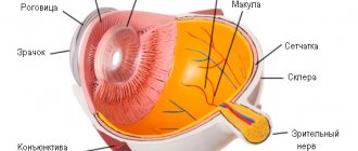
The main vessels of the eyes supply the retina and optic nerve. Blood flows to the fundus through the main ophthalmic artery, which branches off from the carotid artery.
The network of capillaries also ensures the movement of blood to the tissues of the eye. The venous system removes harmful substances that enter the blood from food. It completely replicates the arterial network, but does not have valves that provide reverse outflow of blood. Therefore, any purulent process can enter the brain and pose a threat to life.
Causes
Most often, the inflammatory process on the choroid is observed due to the influence of the following factors:
- mechanical damage to the eyeball, which can be minor or extensive;
- the entry of a foreign object into the eyes, which can vary in size, not only damages the mucous membrane, but also contributes to the spread of bacterial infection;
- infection with bacteria (staphylococci, streptococci, pneumococci, gonococci, chlamydia, tuberculosis);
- spread of viruses to the choroid of the eyes, for example, adenovirus, herpes, influenza, parainfluenza;
- spread of helminths and other parasites to the visual area;
- the presence of seasonal or periodic allergies, which are formed due to animal hair, flowering plants, bird feathers, house dust, chemicals;
- the impact of excessively low temperatures on the organs of vision, especially in strong winds;
- exposure to a large amount of ultraviolet radiation on the organs of vision, which affects not only the retina, but also the outer structure of the eyes.
Each cause of the condition has its own treatment methods, so it is necessary to conduct diagnostic tests, only then use medications.
Pathologies of the vascular tract
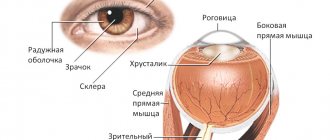
The middle layer of the eye is the vascular tract of the eye (uvea) , which embryogenetically corresponds to the pia mater and consists of three parts: the choroid itself (choroid), the ciliary body (corpus ciliare) and the iris (iris).
The vascular tract is separated from the sclera by the suprachoroidal space and is adjacent to it, but not along its entire length. It consists of branching vessels of various calibers, forming tissue whose structure resembles cavernous tissue.
All intraocular veins do not have valves.
Classification
- Inflammatory diseases It is known that the blood supply to the choroid itself comes from the posterior short ciliary arteries, and the iris and ciliary body from the anterior and posterior long ciliary arteries, therefore there may be separate damage to the anterior and posterior parts of the vascular tract. The lesion of the anterior section is called anterior uveitis or iridocyclitis, the lesion of the posterior section is called choroiditis. Damage to all parts of the vascular tract is called panuveitis or generalized uveitis. The choroid is anatomically and functionally intimately connected with the outer layers of the retina, which in choroiditis is always involved in the process, and infectious inflammatory diseases of the retina are usually complicated by damage to the choroid. Iritis
- Iridocyclitis
- Choroiditis
- Panuveitis
- Fuchs syndrome. Fuchs' heterochromia is accompanied by the presence of precipitates, vitreous opacities and often cataracts, as well as heterochromic glaucoma. Congenital simple heterochromia has no other pathology or anomalies and the functions of the organ of vision are not impaired.
Essential mesodermal progressive dystrophy of the iris - changes in iris color and deformation of the pupil, increased vascular pattern of the limbus, petechial hemorrhages. Small grayish or weakly pigmented deposits and swelling of the corneal endothelium appear on the posterior surface of the cornea. Subsequently, the process moves to the posterior, ectodermal layer. The pupil is pulled to one side, acquiring a pear-shaped and then an ellipsoid shape. The appearance of peripheral synechiae and the proliferation of fibrous tissue in the corner of the anterior chamber on the side of the iris defects leads to difficulty in the outflow of intraocular fluid.
- Aniridia (complete and partial) is the absence of the iris, often bilateral. Even with side lighting, the contours of the lens and zonular fibers are visible. Sometimes the remainder of the iris root and ciliary processes are identified, which are clearly visible during biomicroscopy. Aniridia is often accompanied by photophobia and nystagmus. It can be combined with congenital posterior embryotoxon, blockade of the anterior chamber angle, mesenchymal - embryonic tissue, anterior synechiae, Schlemm's canal aplasia with the development of secondary glaucoma. Frequent symptoms of aniridia are subluxation, rarely luxation of the lens, microphakia, colobomas, and cataracts. As a rule, there is hypoplasia of the central fovea of the retina, leading to a sharp decrease in central vision. Inherited in a dominant manner. Treatment of secondary glaucoma, cataracts, subluxations, dislocations of the lens - surgical, correction of refractive errors, cosmetic contact lens.
- Coloboma is a slit-like defect located downward, downward inward, less often downward outward. It can be one or two-sided, complete or partial. The pupil is pear-shaped with a pigment border, which distinguishes it from an artificial coloboma. Coloboma can be combined with coloboma of the choroid itself and other anomalies of eye development. In cases where there is no vascular coloboma or other anomalies, vision is not altered. It has a family-hereditary, often dominant character.
- Polycoria is a congenital anomaly in which each iris has 2-3 pupillary openings, each with its own sphincter that reacts to light. Treatment is sometimes plastic surgery.
- Congenital microcoria can be combined with other eye development defects: microphthalmos, microcornea, congenital cataracts, arachnodactyly. Congenital miosis is differentiated from sympathetic nerve palsy and spinal miosis.
- Congenital iris cysts develop as a result of defects in the embryogenesis of the eyeball. Often, congenital cysts of the iris are accompanied by other anomalies of eye development, such as remnants of the pupillary membrane, dispersion of pigment on the lens capsule, congenital cataracts, and colobomas. Congenital cysts do not change for a long time, and then grow quickly and can reach significant sizes. Treating cysts is challenging. Treatment methods proposed include diathermocoagulation, puncture, electrolysis, radiation therapy, laser coagulation and surgical treatment of cysts.
- Corectopia is usually a bilateral, symmetrical displacement of the pupil. The pupil can be round or slit-like, sometimes barely noticeable, the size of a small pin head. A sharp decrease in vision, the development of amblyopia and strabismus are possible. Recessive or dominant inheritance is noted.
The pupillary membrane is the remnants of the anterior section of the vascular bag of the lens. The remnants of the pupillary membrane look like threads or rough strands running from the small arterial circle of the iris to the small arterial circle on the opposite side or the anterior capsule of the lens. Rarely, the pupillary membrane can cover the entire pupil area and then surgery is required. Sometimes the remnants of the film take the form of pigmented stellate cells accumulated on the anterior capsule of the lens, which usually do not interfere with vision.
Such patients often have pronounced nystagmus and very low vision. Wearing smoky glasses is shown. Sometimes surgical treatment of nystagmus. Anomalies of the vascular tract can be combined with other malformations, such as microphthalmos, microcornea, cleft lip, cleft palate, etc.
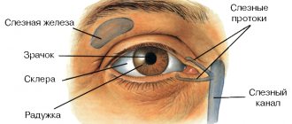
Symptoms
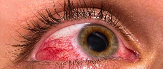
Inflammation of the choroid of the eyes is accompanied by the following characteristic clinical symptoms:
- severe redness of the eyeball;
- discomfort: itching, burning, pain, sensation of a foreign body under the eyelid that is not actually there;
- increased production of tear fluid, which can later be replaced by dry eyes, due to which the cornea is damaged;
- complications of bacterial infection, as opportunistic microflora begins to actively multiply due to the influence of negative factors;
- photophobia;
- spasm of the eyelids, due to which the patient is unable to open his eyes.
The patient may have all or only part of the above symptoms. For example, if a foreign object gets into the patient’s eye, he will have photophobia, eyelid spasm, and increased lacrimation. If the cause is a bacterial infection, pus will form and the eyes will become red.
Structure and functions of the vascular system of the eye
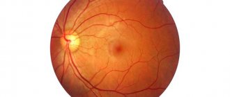
The ophthalmic artery, the main branch of the internal carotid artery, which supplies the organs of vision, is responsible for a significant portion of the blood flow in the eyeball. Tissue nutrition is provided by a network of capillaries. The most important of these are those vessels that carry blood to the retina of the eye, as well as to the optic nerve - the central retinal artery and the posterior short ciliary arteries. If blood flow in these vessels is disrupted, serious vision loss and even blindness can occur.
Harmful products also enter the bloodstream from the cells, but they are removed through the veins. The network of veins is similar to the structure of arteries. The only peculiarity is the absence of valves to limit the reverse flow of blood. The veins of the orbit are connected to the venous network of the face and brain.
Treatment
The method of therapy depends on the cause that caused the patient's condition. The most commonly used means are:
- antibacterial therapy with topical agents;
- antiviral drugs that are used systemically and locally;
- non-steroidal anti-inflammatory drugs based on diclofenac, which can be used topically;
- steroidal anti-inflammatory drugs based on dexamethasone, hydrocortisone and other hormones;
- removal of a foreign body from the eyes with further treatment with an antibacterial agent;
- antihistamines, which can be used systemically or locally in the form of drops and ointments;
- moisturizing eye drops.
All medications must be used strictly according to the instructions; self-medication is not allowed.
The patient should remember the course of therapy. If antibiotics are used, they can be used for more than 7 days. After this, the bacteria develop resistance to the active substance. Steroids are used for no more than 10-14 days. After this time, atrophy of the mucous membranes and skin and changes in pigmentation occur. Other drugs can be used in a prolonged course, but only with the permission of a doctor.
Preventive measures
Diseases of the retina and choroid need to be treated in a timely manner.
Knowledge of ways to prevent diseases of the retina and visual apparatus will be useful for the reader, so we will describe the main methods of prevention.
Basic preventive measures:
- Make regular appointments with your ophthalmologist, especially if you have a family history of eye disease. It is recommended to conduct an examination at least once a year.
- Add to your diet as many foods as possible that contain vitamins A and C. Wild berries are beneficial.
- Monitor intraocular pressure. If episodes of increased blood pressure are detected, you should discuss ways to prevent glaucoma with your doctor.
- Do not overwork your visual apparatus by working with text for a long time. Use moisturizing drops as needed.
It must be remembered that early preventive measures help preserve vision.
ethnoscience
As a traditional medicine, you can use washing or instilling eyes with herbal decoctions.
For these purposes, chamomile, calendula, string, coltsfoot are suitable. They can be brewed separately or together. The finished liquid is carefully infused and filtered so that foreign particles do not spread to the organs of vision.
The liquid can be instilled into the eyes, applied to the eyelids as a lotion, and washed out the mucous membrane.
Oak bark has a good analgesic effect. It is pre-brewed and filtered. After this, apply to cotton pads and apply to closed eyes. Through the gap between the eyelids, the active substance will penetrate to the mucous membrane of the eyes.
Classification of inflammatory diseases of the choroid
1. Endogenous, i.e. primary (when infection enters the blood, when the body and eyes are sensitized); exogenous, i.e. secondary (with perforated wounds of the eye, after operations, purulent processes in the cornea), forms.
2. Focal and diffuse uveitis.
3. According to the morphological picture - granulomatous (metastatic hematogenous) and non-granulomatous (toxic-allergic) inflammation.
4. According to the clinical course, uveitis is divided into acute and chronic.
5. Pathomorphological features of uveitis: acute inflammation corresponds to an exudative-infiltrative process, and chronic inflammation corresponds to an infiltrative-productive process.
6. Based on localization, inflammation of the anterior part of the vascular tract (iritis and iridocyclitis or anterior uveitis), the posterior part (choroiditis or posterior uveitis) and the entire vascular tract (iridocyclochoroiditis or panuveitis) are distinguished.
Herpetic uveitis
| Herpetic uveitis accounts for up to 25% of all inflammatory diseases of the iris and ciliary body and most often occurs as keratouveitis (see.
|
Only an ophthalmologist can make a correct diagnosis. In our clinic, you will undergo all the necessary examinations using modern high-precision equipment and will prescribe the necessary treatment.
Diagnostics
The appearance of uveitis symptoms is a reason to immediately consult a doctor. Delaying the visit is fraught with serious consequences, including blindness.
The doctor conducts an external examination, determines visual acuity and fields, and measures eye pressure.
The reaction of the pupils to light is studied in the light of a slit lamp, retinitis is visible when examining the fundus. Additionally, ultrasound, angiography and MRI are used.
Prevention measures
This disease can be prevented. To do this, you must follow the following recommendations:
- promptly treat infectious diseases;
- Wear safety glasses when performing work that is hazardous to the eyes;
- exclude injuries;
- prevent eye burns;
- visit an ophthalmologist periodically;
- monitor hormonal levels;
- do not contact with allergens;
- lead a healthy lifestyle.
The most common causes of uveitis are infection, trauma, and systemic disease. They need to be prevented or treated in the early stages. Most often, uveitis is a complication of another pathology. Prevention should be carried out from a young age. To protect children from this pathology, it is necessary to prevent bacterial and viral infections.
Characteristics of the disease