Causes
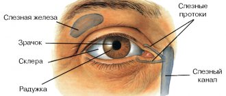
The front part of our eye - the sclera - is white when healthy.
Its shade can vary from whitish-blue to whitish-pink, but it should be uniform. A noticeable change in the shade of the sclera and the appearance of yellow spots on it indicates the development of pathology. Moreover, this can be either a disorder of the eye itself or a systemic disease of the body. If yellow spots appear on the white of the eye, we advise you to contact an ophthalmology clinic.
Often, in addition to yellow spots on the eyes, the patient also notices other symptoms
:
- Itching and pain in the eye area
- Deterioration of vision
- Increased sensitivity to bright light
- The appearance of discharge from the eyes.
Yellow spots can signal various eye diseases. As a rule, these diseases are not very dangerous and cannot lead to complete loss of vision, but you should definitely not ignore their symptoms. Among the most common reasons are the following
:
- A conjunctival cyst is a formation with fluid inside, which may have a yellowish tint and appear as a small spot on the white of the eye.
- Pterygium is an eye disease in which the conjunctival tissue begins to grow from the nasal corner of the eye to the center of the cornea. The yellow spot on the squirrel has a triangular shape and sometimes changes color to scarlet or pink
- A pinguecula is a yellow spot that is a sign of aging of the conjunctiva or a lack of vitamin A. Most often, a pinguecula is localized on the inside of the eye near the bridge of the nose, so Patients do not immediately notice it
- Nevus is a kind of “mole” on the white, which signals serious pathologies of the eye
- Horner-Trantas spots are spots that occur as a result of an allergic reaction. They look like grains and are localized mainly around the cornea. The reason for the appearance of this type of spots is keratitis (inflammation of the cornea) or allergic conjunctivitis (allergic reaction of the conjunctiva).
Physiological role of the macula of the eye
The main functions that the macula performs in the human optical system include the following:
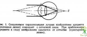
1. The fovea itself is responsible for the clearest central vision. Due to the large accumulation of cone receptors, the image of the external world is transmitted to the central structures, which recognize it. 2. Color perception and color perception are also provided by cones, which are connected to nerve pathways. The nerve impulse is quite quickly transmitted from the receptor apparatus to the central nervous system, where the visual image is formed.
Diseases associated with the appearance of yellow spots on the white of the eye
If the cause of the spots is a general disease of the body, then nausea, chills, increased fatigue, loss of appetite, etc. may be added to the listed symptoms.
Yellow spots may be evidence of the following disorders in the body:
A:
- Jaundice - occurs when there is a violation of the metabolism of bilirubin (bile pigment, one of the most important components of bile)
- Hepatitis (a viral disease caused by a pathogen with pronounced hepatotropic properties
- Liver cirrhosis (pathological condition of the liver, which is a consequence of impaired blood circulation in the hepatic vascular system and dysfunction of the bile ducts)
- Anemia or anemia (a pathological condition characterized by a decrease in the concentration of hemoglobin and, in the vast majority of cases, the number of red blood cells per unit volume of blood
- Liver cancer
- Gallstones are formed in the body under the influence of a number of unfavorable factors and can take on different shapes, structures, and sizes.
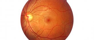
Structure of the macula of the eye
The macula coincides with the central zone of the retina. The diameter of the macula is about 5.5 mm. In the very center of the macula there is a depression (pit), the size of which is about 1.5 mm.
Due to the fact that in the macular zone there is a large amount of a special pigment, the color of which is yellow, during ophthalmoscopy this area is colored yellow. In this regard, the macula received its second name. The main pigments contained in the macula are lutein and zeaxanthin. The human body is not able to synthesize these substances on its own; therefore, all pigment reserves come from outside (with food, drugs). Green, orange and yellow foods contain a lot of lutein and zeaxanthin. The main role of pigments is to protect the photoreceptors of the retina from reactive oxygen species and other free radicals. The latter are formed as a result of oxidative reactions and negatively affect nerve cells and the eyeball.
Interestingly, there are no blood vessels in the macula area, so nutrition comes from the adjacent choroid (choroid).
Diagnosis and treatment of yellow spots on the white of the eye
Yellow spots on the white of the eye can be eliminated only after identifying the root cause of their appearance and promptly prescribing the correct treatment. In each case it will be individual.
For example, pinguecula and pterygium in the initial stages are treated with drops and gels that moisturize the conjunctiva, or with anti-inflammatory drugs. Surgical intervention is used only in cases where long-term therapy has not helped. Drug therapy is also used to treat Horner-Trantas spots.
But advanced cases of conjunctival cysts are treated only surgically. Modern technologies make it possible to do this using a laser painlessly for the Patient.
Doctors at Dr. Belikova’s Eye Clinic will conduct a comprehensive examination of the visual organs and, if necessary, select the most effective treatment or give recommendations for visiting a specialized specialist.
Diagnostic methods for macular pathology
Patients with suspected macular pathology should be comprehensively examined using the following methods:
- Ophthalmoscopy, which can use various types of lighting and magnifying devices.
- Optical coherence tomography, which allows you to evaluate the anatomical structure of all layers of the retina in the macula area.
- Fluorescein angiography is based on the use of a contrast agent that stains the retinal vessels. It helps in the diagnostic search for diseases of the macula.
- Computer perimetry is necessary to determine visual field loss (scotomy) in the central zones.
Preventive measures
To prevent yellow spots from appearing on your eyes, you should follow some preventive rules:
- adhere to a healthy lifestyle;
- get enough sleep during the day;
- promptly treat various pathologies of the visual organs;
- monitor normal hydration of the eye mucosa;
- use contact lenses correctly;
- using various means to protect the organs of vision from wind, dust, sun and computer rays.
Any formation on the organ of vision can pose a serious danger to human health. If a yellow spot appears on the eye, it is recommended to consult a doctor and not self-medicate. A timely diagnosis allows you to slow down the development of pathology and prevent the occurrence of dangerous complications.
It makes no sense to prevent the appearance of circles before the eyes - you should fight the diseases that cause such a symptom. Common pathologies that lead to visual impairment (arterial hypertension, diabetes mellitus) must be treated in a timely manner and stable compensation maintained.
To avoid the short-term appearance of spots before the eyes, you need to regularly perform eye exercises, observe the lighting regime, alternate visual stress with rest, and avoid looking directly at bright light sources without protective devices.
Noticed a mistake? Select it and press Ctrl Enter to let us know.
Symptoms
Diseases of such a part of the visual system as the macula have their own characteristic symptoms.
Since this area of the retina is primarily responsible for the central fragments of the visual image, the first signal of macular dysfunction may be the loss of the center from the overall picture when looking at an object. It may seem to the patient that the central part of the image is obscured by something.
Also, disorders of this kind may be indicated by a disorder associated with the perception of color, or rather its brightness and contrast.
Sometimes diseases of the macula are expressed in various curvatures of the visual image or erroneous judgment about the size of an object.
Any of the above symptoms can be the first signal of a serious visual disorder, so such incidents should never be ignored.
Treatment
Due to some diseases that appear as the human body ages in adulthood, the macula of the eye can gradually degenerate.
Dry and wet forms of this process are known. Moreover, wet-type degeneration is much more dangerous than its dry counterpart and can reduce visual acuity much faster.
Modern equipment and new treatment methods, which are used in specialized clinics, make it possible to successfully treat such diseases with a minimal degree of injury. By the way, in this case, both combination therapy and surgical intervention can be used. However, sometimes the time factor plays a decisive role in the issue of complete restoration of visual functions.
Diseases of the macula of the eye
Some pathologies that can lead to macular damage are presented below:
- Age-related degeneration of the macular zone, which develops with a decrease in the amount of pigments in this area. This condition is more common in older patients.
- Macular swelling.
- Epiretinal gliosis, which consists of the formation of special adhesions between the vitreous body and the central zone of the retina.
- Retinitis, which affects the macula area.
- Macular hole.
Prevention methods
Macular degeneration can be prevented if you lead a healthy lifestyle with sufficient physical activity, and also do not put excessive stress on the visual analyzer. It is necessary to avoid direct sunlight entering the eye, as ultraviolet radiation promotes atrophy of light-sensitive cells. It is necessary to consume sufficient amounts of vitamins and microelements; carotenes, which protect the retina from radiation damage, are especially useful.
Prevention of eye problems is considered proper nutrition, taking vitamins. The action of such active beneficial substances is aimed at improving the functioning of visual function. Taking vitamin A, blueberry extract, and lutein shows high effectiveness. The listed components protect the retina from degenerative changes that can lead to negative consequences.
Symptoms of damage to the macula of the eye
When the macula area is damaged, manifestations occur that mainly affect central vision. Among them are characteristic features:
- Reducing the brightness of surrounding colors;
- The appearance of a darkening in the central zone of the image, which can even cover part of the object being studied;
- Reducing the brightness of objects;
- Metamorphopsia, which manifests itself as distortion of shape;
- Changing the size of objects that are perceived larger or smaller than their true size. This is due to a change in the distance between the cone receptors.
Types of degeneration
Aging leads to such changes in this part of the organ.
This pathology occurs in the human retina more often during aging and is associated with a violation of the condition of rods and cones, as well as a decrease in their number. In this case, the patient’s central vision is impaired, but peripheral vision remains the same. This disease rarely occurs in young people and its development is caused by a lack of vitamins and minerals. Nutrients essential to the eye include antioxidants, vitamins A, E and C, and zinc.
What is important is the reduction in the macula zone of zeaxanthin and lutein, protective substances from exposure to ultraviolet rays of the sun. Exposure to certain viruses, such as cytomegalovirus and Epstein-Barr, can cause macular degeneration. When pathology appears, a person complains of blurred vision, the appearance of distortions when looking at straight lines, and the inability to see details. There may be a dark dot in the field of view.
What symptoms accompany
Often, when a yellow spot appears in the pupil area, a person does not pay attention to it and does not associate it with the appearance of other atypical signs of the visual system. Periodically, penguenticula may be accompanied by the following symptoms:
- rashes of various types on the eyelids;
- severe itching and burning;
- staining the mucous membrane red;
- increased eye fatigue and discomfort;
- sensation of the presence of a foreign object in the organ of vision;
- the formation of dark circles before the eyes and decreased vision.
Unpleasant symptoms can noticeably intensify when the child is in the wind or in direct sunlight, as well as when dust gets into the eyes.