Retina
The retina lines the inside of the eyeball; normally its thickness reaches 281 micromillimeters. Moreover, in the area of the macula, the membrane is several times thinner than at the periphery. The element extends from the optical disk to the jagged line. In the optic disc, the retina is attached very tightly; in other areas the connection is loose. This explains the easy development of retinal detachment.
The layers of the shell differ in structure and function, forming a complex structure. Thanks to the close interaction of different elements of the visual apparatus, a person is able to distinguish colors, sizes of objects, and estimate distance.
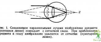
Penetrating into the eye, light fluxes pass through several refractive media. In the absence of deviations in refraction, people receive a reduced and inverted, but real image on the retina. Subsequently, the impulses are transformed and enter the brain, where the final processing of the image of the external world is carried out.
Content:
- 1 How the retina works 1.1 Structure of the retina
- 1.2 Visual activity 1.2.1 Distribution
Description
The retina, located at the back of the eyeball, contains specialized cells called photoreceptors that are sensitive to light in different areas of the spectrum. This allows us to see in both light and darkness.
The eye has evolved from a primitive collection of photosensitive cells into a complex organ that is extremely sensitive to light. However, the bulk of the eye tissue does not perceive light. The muscles surrounding the eyeball, the iris, cornea and lens work together to focus light onto the retina, a relatively small area located on the back surface of the eyeball that contains photoreceptors.
Structure of the retina
The retina, from a functional point of view, is divided into two components:
- Optical area. Occupies most of the retina (2/3 of all tissues), forms a light-sensitive structure (thin and transparent film).
- The blind part. The ciliary-iridal area occupies less space and makes up the outer pigment layer.
The visual area is characterized by uneven thickness:
- The densest area (0.4 millimeters) is located near the edge of the optic disc.
- The thinnest area (up to 0.075 mm) is part of the macula. It is distinguished by the best perception of optical stimuli.
- The area of average thickness (within 0.1 millimeters) is located not far from the jagged line.
Photoreceptor apparatus
Consists of cones and rods. The first contains the optical pigment iodopsin, the second rhodopsin. Cones are responsible for color and central vision and are six micromillimeters in diameter. Rods provide black-and-white, peripheral and crepuscular perception. The diameter of the elements reaches two micromillimeters.
| The retina consists of three types of cones, which differ in their visual pigment. They perceive light streams of different lengths, which ensures the uninterrupted functioning of the color perception mechanism. |
The main segments of photoreceptors:
- Outer. It contains a photosensitive substance.
- Interior. It contains cytoplasm with ornelles. A special role is assigned to mitochondria, which provide photoreceptor functions with a sufficient amount of energy.
- Core.
- Synaptic body. It is part of the cones and rods and connects to nerve cells, which are components of the optical pathway.
Histological structure of the retina
The structure of retin is very complex. All elements are closely interconnected and damage to any of them can lead to serious complications. The retina consists of ten layers. Four relate to the light-sensitive apparatus of the membrane, six represent brain tissue.
Layers of the retina:
- Pigment epithelium and Buch's membrane. Acts as a barrier, prevents the entry of light radiation and absorbs segments of rods and cones. With the development of certain pathologies, hard or soft spots of small size and a yellow tint (drusen) form here.
- Inner nuclear layer. Here are the bodies of Müllerian, amacrine and horizontal cells. The former are necessary for the maintenance of nervous matter. All the rest are engaged in processing the signals transmitted by photoreceptors.
- Nerve fibers. Send information to the optic nerve.
- Photosensory layer. This is where the cones and rods are located.
- External limiting membrane. Formed by terminal plates and flat adhesive contacts of photoreceptors. The processes of Müller cells are also located here. They perform a light-conducting function, i.e., they collect rays on the front surface of the retina and redirect them to cones and rods.
| Outer nuclear layer. Photoreceptors, their bodies and nuclei are located here. The outer processes (dendrites) are directed towards the pigment epithelium, the inner ones - towards the outer layer of the retina, where they contact bipolar cells. |
- Outer mesh layer. Formed by synapses between photoreceptors, associative neurons and bipolar cells.
- Inner mesh layer. It consists of axons of various nerve cells of the retina.
- Ganglion cells receive signals from photoreceptors through bipolar neurons and transmit them to the optic nerve. They are not covered with myelin, therefore they are absolutely transparent and easily transmit light.
- Internal limiting membrane. Acts as a barrier between the retina and the vitreous body.
Return to contents
Macular area
After the light fluxes pass through the optical structures of the visual apparatus and the vitreous body, they penetrate the retina from the inside. Before the impulses reach the rods and cones, they have to cross the ganglion cells, reticularis and nuclear layers.
In the area of the fovea, the inner layers move apart to reduce vision loss. One of the most important areas of the retina is the macular area. It consists of several parts:
- Fovea (the darkest area in the macula area). The diameter of the element is from 1.5 to 1.8 millimeters.
- Foveola (light spot in the center of the macula). The area size is 0.35-0.5 mm.
- A zone without vessels with a diameter of approximately 0.5 millimeters.
| Up to ten percent of photoreceptors are concentrated in the fovea. The sharpness of the eyes depends on its functionality. In infants, the macula has blurred boundaries. A clear outline appears by the end of the first year of life. |
Optic disc
The area where the optic nerve of the eye enters the brain structures. The area of the element is approximately three square millimeters, the diameter of the optic disc is 2 mm. The vessels are concentrated in the middle of the disc; they are represented by the retinal vein and the central artery. Their main purpose is to supply the retina with blood.
Blood supply to the retina
The process comes from two sources. The six inner layers deliver the “red fluid” from the branches of the central artery. The external ones receive nutrients from the choriocapillaris region of the choroid.
The central artery is very important in the blood supply. It is divided into two branches: upper and lower. They are also classified into nasal and temporal branches. The outflow of blood from the retina occurs through the venous system.
Macula (retinal spot)
The fundus of the eye has a specific formation in the center - the macula. It also contains a hole - a funnel on the inner surface of the retina. The size of the spot corresponds to the volume of the optic nerve head and is located opposite the pupil.
| The macula is the area of the optical analyzer where visual acuity is most pronounced. The spot is responsible for clarity and clarity of perception. |
↑ Structure of the retina
To a first approximation, the retina can be imagined as consisting of four layers:
* Along the back surface of the retina is the outer pigmented layer - the epithelial cells contained in it absorb light (but do not “see” it), thus preventing the reflection of light inside the eye.
* Next is a layer of photoreceptors that are capable of converting light energy into electrical energy.
* Electrical potentials produced by photoreceptors are transmitted to bipolar neurons.
* The bipolar neurons in turn communicate with the ganglion neurons, whose axons then connect and bend at right angles before leaving the eye as the optic nerve, which carries images to the brain.
Thus, light first passes through a layer of ganglion and bipolar neurons before reaching the photosensitive photoreceptors. However, such an “inverted” structure does not in the least prevent the photoreceptors from capturing it.
Retinal functions:
The main task of the retina is photoreception. This is a chain of biochemical reactions during which light impulses are converted into nerve signals. It occurs due to the breakdown of rhodopsin and iodopsin - visual pigments formed when there is a sufficient amount of vitamin A in the body.
The retina of the eye performs the following functions:
- Central vision. It allows a person to read and see objects at different distances. It is provided by the retina cones located in the macula area.
- Peripheral vision. Required for orientation in space. This is possible due to the location of rods in the retina, which are located on the periphery of the shell.
- Color vision. Allows you to distinguish shades. It is provided by three different types of cones, each of which perceives light fluxes that differ in length. As a result, a person recognizes the colors green, red and blue. Problems with the perception of shades lead to color blindness. Some people have a fourth cone or rod and are able to distinguish up to one hundred million colors.
- Night vision. Allows you to see in low light conditions. Only chopsticks provide it. Cones do not function in the dark.
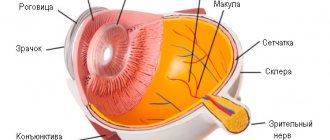
Structure of the human eye
The retina is only one of the layers of the eye. Several layers:
- Eye drops with vitamins Eyelight Vita Yellow contain useful components;
- Vitamins B1, B2 and PP to improve vision;
- Prevents age-related changes and the development of inflammatory diseases;
- Strengthen local immunity.
- The cornea is a transparent membrane that is located at the front of the eye, it contains blood vessels and borders the sclera.
- The anterior chamber is located between the iris and cornea, filled with intraocular fluid.
- The iris is the area where the opening for the pupil is located. It consists of muscles that relax and contract, changing the diameter of the pupil, regulating the flow of light. The color may vary depending on the amount of pigment. For example, brown eyes require a lot of it, and blue eyes require less.
- The pupil is a hole in the iris through which light enters the inner areas of the eye.
- The lens is a natural lens; it is elastic, can change shape, and is transparent. The lens changes its focus instantly so that you can see objects at different distances from a person.
- The vitreous body is a transparent, gel-like substance; it is this part that maintains the spherical shape of the eye and takes part in metabolism.
- The retina is responsible for vision and participates in metabolic processes.
- The sclera is the outer layer and extends into the cornea.
- Vascular part.
- The optic nerve is involved in transmitting signals from the eye to parts of the brain; nerve cells are formed by one of the parts of the retina, i.e. they are its continuation.
Symptoms of retinal pathology
A characteristic sign of retinal damage is a decrease in visual acuity and narrowing of optical fields. In some cases, the formation of absolute or relative scotomas located in different parts of the retina is observed. Damage to photoreceptors is indicated by the development of color blindness and night blindness.
A pronounced drop in central vision indicates damage to the macula area. If there are problems with peripheral vision, there is a high risk of developing fundus abnormalities in the periphery. The formation of scotomas indicates local damage to a certain area of the retina.
An increase in the volume of the blind spot, accompanied by a severe deterioration in visual acuity, may signal pathologies of the optic nerve. Occlusion of the central retinal artery results in sudden (within seconds) blindness in one eye. When the retina ruptures and detaches, flashes, lightning and spots appear in front of the organ of vision.
There is usually no pain in retinal pathologies, since nerve impulses are not transmitted due to the lack of sensitive innervation. Return to contents
Methods for diagnosing diseases
The standard examination program includes measuring intraocular pressure, checking visual acuity, determining the level of refraction, analysis of optical fields (perimetry), biomicroscopy and ophthalmoscopy.
Diagnostics may also include:
- Fluorescein angiography of the retina. It is carried out to assess the condition of the vascular system.
- Study of contrast sensitivity, color perception.
- Electrophysiological diagnostics (optical coherence tomography).
- Taking a fundus photograph. Required for follow-up and comparison of indicators.
Retinal diseases
Among all ophthalmological diseases, the share of anomalies affecting the retina accounts for less than one percent. They can be divided into several categories:
- Dystrophic pathologies. They are congenital or acquired.
- Inflammatory diseases.
- Damage to the retina due to trauma to the visual apparatus.
- Deviations associated with concomitant diseases. For example, disorders of the endocrine or cardiovascular system.
Vascular pathology
The most common anomaly in this category is angiopathy. It is characterized by damage to various vessels. Cause of the disease: diabetes, hypertension, vasculitis, etc.
| First, patients develop retinal vasospasm or dystonia, later it changes into fibrosis and thinning of the blood vessels. This leads to ischemia and the appearance of angioretinopathy. Hypertensive patients are diagnosed with arteriovenous decussation, in which the vessels resemble copper and silver wire. |
Angiodystonia is accompanied by a decrease in visual acuity and increased fatigue. Arteriospasm develops with high or low blood pressure and a number of neurological abnormalities.
A common vascular anomaly is occlusion of the central retinal artery. The disease is accompanied by blockage of a vessel or one of its branches, which leads to ischemia. Central artery embolism most often occurs in patients with atherosclerosis, hypertension and arrhythmia.
Dystrophies, injuries, developmental defects
The most common anomaly is coloboma (absence of part of the retina). Patients often experience macular, central and peripheral dystrophy. The latter is further subdivided into ethmoid, fine cystic, frost-like, and “snail track.” With the development of these pathologies, holes of different sizes appear in the fundus.
After blunt trauma and contusions, Prussian opacities may occur on the retina. Treatment of the disease involves the use of a complex of vitamins and antihypoxants. Sometimes hyperbaric oxygen therapy sessions are prescribed. Unfortunately, therapy does not always bring the expected effect.
Neoplasms
Retina tumors have recently become increasingly common among people visiting an ophthalmologist. It accounts for approximately 1/3 of all neoplasms. Patients are usually diagnosed with retinoblastoma. Nevus, angioma and other benign tumors are much less common.
Angiomatosis is usually combined with various developmental defects. Treatment is selected for each patient individually.
Vascular pathologies
Vascular diseases of the retina are associated with damage to the choriocapillaris and blockage of blood flow by thromboembolism.
Diabetic retinopathy
Expert opinion
Kim Oksana Alexandrovna
Head of the ophthalmology clinic. Ophthalmologist with more than 10 years of experience.
The pathology occurs in patients who have suffered from diabetes mellitus for a long time and do not correct the level of glucose in the body. Hyperglycemia in the blood plasma disrupts the permeability and density of the walls of the choriocapillaris. As a result, they can rupture, causing hemorrhages into the vitreous cavity. As a compensatory mechanism, the formation of new vessels that grow through the retina begins. But during this period, photosensitive cells become cut off from the supply of oxygen and nutrients. As a result, dystrophic changes develop in the nervous tissue.
There are 3 stages of development of the pathological process: