The human eye is a paired organ that provides the function of vision. The properties of the eye are divided into physiological and optical, and therefore are studied by physiological optics - a science located at the intersection of biology and physics.
The eye is shaped like a ball, which is why it is called the eyeball.
The skull has an orbit - the location of the eyeball. A significant part of its surface is protected there from damage.
The extraocular muscles provide the motor ability of the eyeball. Constant hydration of the eye, creating a thin protective film, is provided by the lacrimal glands.
Structure of the human eye - diagram
Structure and functions of the human eye
Our eyes are a complex apparatus that allows a person to receive information and see the world in all its glory.
The eyes have a very complex structure - they combine various tissue structures and vascular systems. Understanding what the eye consists of, how its components work and what functions they perform, helps you to carefully and knowledgeably treat your own vision, recognize “signals” of violations and, accordingly, make decisions to eliminate them.
Alternative medicine
One of the methods of alternative medicine based on the structure of the eye is iridology. The diagram of the iris helps the doctor make a diagnosis for various diseases in the body:
This analysis is based on the assumption that different organs and areas of the human body correspond to certain areas on the iris. If an organ is sick, this is reflected in the corresponding area. These changes can be used to determine the diagnosis.
The importance of vision in our lives cannot be overestimated. In order for it to continue to serve us, we need to help it: wear glasses to correct vision, if required, and sunglasses in bright sunshine. It is important to understand that age-related changes occur over time, which can only be delayed by prevention.
Related articles:
- Optic nerve: functions, diseases, treatment
- Binocular vision: definition, treatment, research
- Eye pressure: normal, symptoms of increase, treatment
- Retina of the eye: structure and functions, main pathologies
Eyeball
Due to its spherical or spherical shape, the human eye is called the “eyeball”. It is located in the orbit, a special bony structure of the skull that protects the eyeball from damage. The front surface of the eyeball is protected by the eyelids.
Six extrinsic muscles move the eyeball and create binocular vision—that is, seeing with two eyes, which creates a three-dimensional image (stereoscopic vision).
Tear fluid flows out through the lacrimal ducts. The lacrimal glands produce tears, which moisturize the surface of the eyeball and create a protective film on its surface.
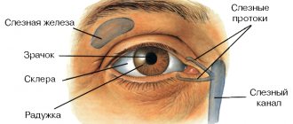
Lacrimal apparatus of the eye
The lacrimal apparatus includes the gland, canaliculi, lacrimal puncta and sac, as well as the nasolacrimal duct. The lacrimal gland is located in the area of the upper outer wall of the orbit. It secretes tear fluid, which penetrates through the channels into the eye area, and then into the lower conjunctival fornix.
After this, the tear, through the lacrimal puncta located in the area of the inner corner of the eye, enters the lacrimal sac through the lacrimal canals. The latter is located between the inner corner of the eyeball and the wing of the nose. From the bag, tears can flow through the nasolacrimal duct directly into the nasal cavity.
The tear itself is a rather salty, transparent liquid that has a slightly alkaline environment. A person produces about 1 ml of such liquid with a varied biochemical composition per day. The main functions of tears are protective, optical, and nutritional.
Eye shells
There are several membranes of the eye.
Conjunctiva
- This is the outer transparent membrane that covers the surface of the eye and the inside of the eyelids. The conjunctiva assists in the necessary gliding of the eyeballs.
Fibrous membrane
- This is a membrane of gas that consists of the sclera, limbus and cornea.
Sclera
- a white shell that performs protective and supporting functions. This is the densest part of the eyeball.
Limbo
- the transitional layer of the eye between the sclera and the cornea. The limbus contains stem cells that regenerate the outer layers of the cornea.
Cornea
- transparent part of the fibrous membrane. It is abundantly supplied with nerves, so it is characterized by high sensitivity. The transparency of the cornea allows light rays to penetrate into the eye.
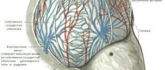
Choroid
- part of the eye that provides blood supply to the eye and trophism (nutrition) of its cells. The choroid consists of:
- The choroid is a structure that is in close contact with the retina and sclera, performing the functions of nutrition and shock absorption.
- Ciliary body - (providing vision at different distances) and producing (producing) aqueous humor (intraocular fluid)
- The iris, which determines the color of our eyes, and the pupil, the opening for light rays to enter
- The retina is the inner lining of the eye. The retina is a very thin, less than one-third of a millimeter (300 microns) thick, light-sensitive tissue. It provides the basic functions of the visual organs - central and peripheral (lateral) vision. This is where the fibers of the optic nerve begin, through which impulses pass to the brain, providing our visual perception.
the macula, is responsible for central vision in humans.
.
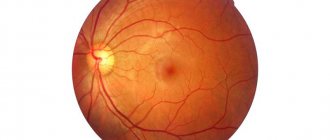
↑ Retina
Retina
- the inner membrane of the eyeball, adjacent to the choroid along its entire length up to the pupil.
The origin of the optic nerve of the retina is the optic disc, which is located 3-4 mm medially (towards the nose) from the posterior pole of the eye and has a diameter of about 1.6 mm. There are no light-sensitive elements in the area of the optic nerve head, so this place does not provide visual sensation and is called a blind spot.
Lateral (to the temporal side) from the posterior pole of the eye there is a spot (macula) - a yellow area of the retina that has an oval shape (diameter 2-4 mm). In the center of the macula there is a central fovea, which is formed as a result of thinning of the retina (diameter 1-2 mm). In the middle of the central fovea lies a dimple - a depression with a diameter of 0.2-0.4 mm; it is the place of greatest visual acuity, contains only cones (about 2500 cells), and there are no rods.
The retina is distinguished by a dentate line, which divides it into two sections: light-sensitive and non-light-sensitive.
The photosensitive section is located posterior to the dentate line and carries light-sensitive elements (the visual part of the retina). The part that does not perceive light is located anterior to the dentate line (blind part).
Structure of the blind part:
- The iris part of the retina covers the posterior surface of the iris, continues into the ciliary part and consists of a double-layered, highly pigmented epithelium.
- The ciliated portion of the retina consists of a double-layered cuboidal epithelium (ciliated epithelium) covering the posterior surface of the ciliary body.
In the retina, there is an outer layer of epithelium containing pigment cells - the pigment part of the retina and an inner layer, devoid of pigment, - the nervous part.
The nervous part (the retina itself) has three nuclear layers:
- outer - neuroepithelial layer consists of cones and rods (the cone apparatus provides color perception, the rod apparatus provides light perception), in which light quanta are transformed into nerve impulses;
- the middle - ganglion layer of the retina consists of the bodies of bipolar and amacrine neurons (nerve cells whose processes transmit signals from bipolar cells to ganglion cells);
- The inner ganglion layer of the optic nerve consists of multipolar cell bodies, unmyelinated axons, which form the optic nerve. The blood supply to the retina is provided by the central retinal artery (a branch of the ophthalmic artery).
At the optic disc, the central retinal artery divides into the superior and inferior papillary arteries. These arteries near the disc again divide dichotomously, and this division extends to the third-order arteries. All ordinal arteries anastomose with each other. The macula macula is surrounded by a thin vascular network in the form of a corolla. In the central fossa, as a rule, there are no capillaries. The blood flow is carried out by the central retinal vein, which leaves the eye in the central part of the optic nerve head next to the central retinal artery.
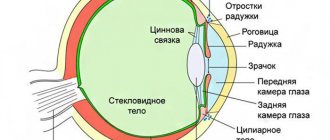
Cameras of the eye
Cameras of the eye
- these are closed and interconnected spaces. They contain intraocular fluid that nourishes the cornea and lens. There are two chambers in the eyeball - anterior and posterior.
Front
located between the cornea and iris. And between the peripheral part of the cornea and the iris, the angle of the anterior chamber is distinguished. In this place, the outflow of intraocular fluid occurs.
Rear camera
located between the iris and the posterior surface of the ciliary body.
The lens is located behind the iris
. It has the shape of a biconvex lens and is attached to the ciliary (ciliary) body with the help of a large number of thin ligaments.
Behind the lens, the cavity of the eyeball is filled with the vitreous humor.
, which is necessary to maintain the tense state of the cell membranes of the eye (turgor).
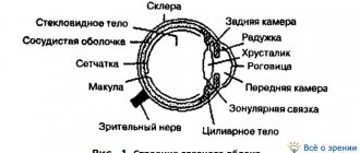
Structure of the eye
1 - vitreous body, 2 - serrated margin, 3 - ciliary muscle, 4 - ciliary girdle, 5 - Schlemm's canal, 6 - pupil, 7 - cornea, 8 - iris, 9 - lens nucleus, 10 - lens cortex, 11 - conjunctiva , 12 - ciliary process, 13 - medial rectus muscle, 14 - retinal arteries and veins, 15 - blind spot, 16 - dura mater, 17 - central retinal artery, 18 - central retinal vein, 19 - optic nerve, 20 - yellow spot, 21 - central fovea, 22 - sclera, 23 - choroid, 24 - retina, 25 - superior rectus muscle.
Optical system of the eye
Optical system of the eye
- this is our vision. The optical system allows a person to visually perceive all kinds of objects in the external world. The clarity of vision is determined by the state of the most important structures of the optical system - the cornea, lens and vitreous body.
Cornea
refracts light rays, which then pass through the pupil and form an image. The pupil is often compared to the aperture of a camera - it is responsible for the “clarity” of the resulting picture.
Through the lens
light rays strike the retina, the light-receiving structure of the eye.
Vitreous body
and the degree of transparency of the intraocular fluid in the eye chambers affect the quality of our visual acuity. So, sometimes blood clots or other floaters form in the aqueous humor, which can reduce vision.
After hitting the retina, light rays transmit special impulses to the brain along the fibers of the optic nerve. The brain analyzes the information and produces the final real image.
Our eye, like a separate organism, literally performs complex work every second, the clarity and success of which depends on the healthy state of each structure of the visual apparatus.
[~DESCRIPTION] =>
Structural parts of the eye
The information the eye receives is light reflected from objects. The final stage is information entering the brain, which actually “sees” the object. Between them is the eye - an incomprehensible miracle created by nature.
Photo with description
The first surface that light hits is the cornea . This is a “lens” that refracts the incident light. Parts of various optical instruments, such as cameras, are designed like this natural masterpiece. The cornea, which has a spherical surface, focuses all rays at one point.
But before the final stage, light rays have to go a long way:
- The light first passes through the anterior chamber containing a colorless liquid.
- The rays fall on the iris , which determines the color of the eyes.
- The rays then pass through the pupil of the eye , a hole located in the center of the iris. The lateral muscles are capable of dilating or constricting the pupil depending on external circumstances. Too bright light can harm the eye, so the pupil narrows. In the dark it expands. The diameter of the pupil reacts not only to the degree of illumination, but also to various emotions. For example, a person experiencing fear or pain will have larger pupils. This function is called adaptation.
- The posterior chamber contains the next miracle - the lens . This is a biological biconvex lens, the task of which is to focus the rays on the retina, which acts as a screen. But, if the glass lens has constant dimensions, then the radii of the lens can change with compression and relaxation of the surrounding muscles. This function is called accommodation. It consists in the ability to see sharply, both distant and close objects, changing the radii of the lens.
- The space between the lens and the retina is occupied by the vitreous . The rays pass through it calmly, thanks to its transparency. The vitreous helps maintain the shape of the eye.
- The image of the object is displayed on the retina , but upside down. It turns out this way due to the structure of the “optical scheme” for the passage of light rays. In the retina, this information is recoded into electromagnetic impulses, after which they are processed by the brain, which reverses the image.
This is the internal structure of the eye and the path of light flow inside it. Video:
Structure and functions of the human eye
Our eyes are a complex apparatus that allows a person to receive information and see the world in all its glory. The eyes have a very complex structure - they combine various tissue structures and vascular systems.
Understanding what the eye consists of, how its components work and what functions they perform, helps you to carefully and knowledgeably treat your own vision, recognize “signals” of violations and, accordingly, make decisions to eliminate them.
Eyeball
Due to its spherical or spherical shape, the human eye is called the “eyeball”. It is located in the orbit, a special bony structure of the skull that protects the eyeball from damage. The front surface of the eyeball is protected by the eyelids.
Six extrinsic muscles move the eyeball and create binocular vision—that is, seeing with two eyes, which creates a three-dimensional image (stereoscopic vision).
Tear fluid flows out through the lacrimal ducts. The lacrimal glands produce tears, which moisturize the surface of the eyeball and create a protective film on its surface.
Useful video
If you follow a daily routine, exercise, and a balanced diet, your eyes will acquire natural beauty, in which there is no need for an additional increase in the size characteristics of the eyeball.
Author's rating
Author of the article
Alexandrova O.M.
Articles written
2029
about the author
Was the article helpful?
Rate the material on a five-point scale!
( 1 ratings, average: 5.00 out of 5)
If you have any questions or want to share your opinion or experience, write a comment below.
Eye shells
There are several membranes of the eye.
Conjunctiva
- This is the outer transparent membrane that covers the surface of the eye and the inside of the eyelids. The conjunctiva assists in the necessary gliding of the eyeballs.
Fibrous membrane
- This is a membrane of gas that consists of the sclera, limbus and cornea.
Sclera
- a white shell that performs protective and supporting functions. This is the densest part of the eyeball.
Limbo
- the transitional layer of the eye between the sclera and the cornea. The limbus contains stem cells that regenerate the outer layers of the cornea.
Cornea
- transparent part of the fibrous membrane. It is abundantly supplied with nerves, so it is characterized by high sensitivity. The transparency of the cornea allows light rays to penetrate into the eye.
Choroid
- part of the eye that provides blood supply to the eye and trophism (nutrition) of its cells. The choroid consists of:
- The choroid is a structure that is in close contact with the retina and sclera, performing the functions of nutrition and shock absorption.
- Ciliary body - (providing vision at different distances) and producing (producing) aqueous humor (intraocular fluid)
- The iris, which determines the color of our eyes, and the pupil, the opening for light rays to enter
- The retina is the inner lining of the eye. The retina is a very thin, less than one-third of a millimeter (300 microns) thick, light-sensitive tissue. It provides the basic functions of the visual organs - central and peripheral (lateral) vision. This is where the fibers of the optic nerve begin, through which impulses pass to the brain, providing our visual perception.
the macula, is responsible for central vision in humans.
.
Eyelids
The entire eyeball is reliably protected from the effects of negative environmental factors and accidental injuries by special partitions - for centuries.
The eyelid itself consists of muscle tissue covered on top with a thin layer of skin. Thanks to the muscles, the eyelid can move; when the upper and lower protective septum are closed, the entire eyeball is evenly moistened, and foreign objects that accidentally enter the eye are removed.
The preservation of the shape and strength of the eyelid itself is ensured by cartilage, which is a dense formation of collagen, in the thickness of which there are special meibomian glands, designed to produce a fatty component that improves the closure of the eyelids and the contact of the eyeball with their surface. The mucous membrane, the conjunctiva, is attached to the cartilage on the inside, designed to produce a moisturizing fluid that improves the sliding of the eyelid relative to the eye.
The eyelids have a very extensive blood supply system, and all their work is completely controlled by the oculomotor, facial and trigeminal nerve endings.
Cameras of the eye
Cameras of the eye
- these are closed and interconnected spaces. They contain intraocular fluid that nourishes the cornea and lens. There are two chambers in the eyeball - anterior and posterior.
Front
located between the cornea and iris. And between the peripheral part of the cornea and the iris, the angle of the anterior chamber is distinguished. In this place, the outflow of intraocular fluid occurs.
Rear camera
located between the iris and the posterior surface of the ciliary body.
The lens is located behind the iris
. It has the shape of a biconvex lens and is attached to the ciliary (ciliary) body with the help of a large number of thin ligaments.
Behind the lens, the cavity of the eyeball is filled with the vitreous humor.
, which is necessary to maintain the tense state of the cell membranes of the eye (turgor).
Average eye size
The visual organ grows until about 25 years of age, during which it increases in size by almost 1.5 times. According to research, the size of the apple is identical in all people and is 24 mm in diameter. The volume of the organ of vision is 7.5 cm cubic.
The size of the eyeball depends on the age category of the person. In a newborn, the average weight of the organ is 2.3 g. In the adult population, the mass of an apple does not exceed 7.5 g. The size of the eyeball does not depend on gender.
Structure and functions of the human eye
Our eyes are a complex apparatus that allows a person to receive information and see the world in all its glory. The eyes have a very complex structure - they combine various tissue structures and vascular systems.
Understanding what the eye consists of, how its components work and what functions they perform, helps you to carefully and knowledgeably treat your own vision, recognize “signals” of violations and, accordingly, make decisions to eliminate them.
Eyeball
Due to its spherical or spherical shape, the human eye is called the “eyeball”. It is located in the orbit, a special bony structure of the skull that protects the eyeball from damage. The front surface of the eyeball is protected by the eyelids.
Six extrinsic muscles move the eyeball and create binocular vision—that is, seeing with two eyes, which creates a three-dimensional image (stereoscopic vision).
Tear fluid flows out through the lacrimal ducts. The lacrimal glands produce tears, which moisturize the surface of the eyeball and create a protective film on its surface.
Eye shells
There are several membranes of the eye.
Conjunctiva
- This is the outer transparent membrane that covers the surface of the eye and the inside of the eyelids. The conjunctiva assists in the necessary gliding of the eyeballs.
Fibrous membrane
- This is a membrane of gas that consists of the sclera, limbus and cornea.
Sclera
- a white shell that performs protective and supporting functions. This is the densest part of the eyeball.
Limbo
- the transitional layer of the eye between the sclera and the cornea. The limbus contains stem cells that regenerate the outer layers of the cornea.
Cornea
- transparent part of the fibrous membrane. It is abundantly supplied with nerves, so it is characterized by high sensitivity. The transparency of the cornea allows light rays to penetrate into the eye.
Choroid
- part of the eye that provides blood supply to the eye and trophism (nutrition) of its cells. The choroid consists of:
- The choroid is a structure that is in close contact with the retina and sclera, performing the functions of nutrition and shock absorption.
- Ciliary body - (providing vision at different distances) and producing (producing) aqueous humor (intraocular fluid)
- The iris, which determines the color of our eyes, and the pupil, the opening for light rays to enter
- The retina is the inner lining of the eye. The retina is a very thin, less than one-third of a millimeter (300 microns) thick, light-sensitive tissue. It provides the basic functions of the visual organs - central and peripheral (lateral) vision. This is where the fibers of the optic nerve begin, through which impulses pass to the brain, providing our visual perception.
the macula, is responsible for central vision in humans.
.
Iris
The iris is the front part of the eye choroid. It is shaped like a disk, with a hole in the center. Moreover, the color of this element of the eye depends on the density of the stroma and pigment.
If the amount of pigment is not large and the tissues are loose, then the iris may have a bluish tint. In the case when the tissues are loose, but there is enough pigment, the iris is colored green. And the density of tissues is characterized by a gray tint of this element when there is a small amount of pigment substance and brown when there is a sufficient amount of pigment.
The thickness of the iris is not large and ranges from two to four tenths of a millimeter, and the anterior surface is divided into two sections - the ciliary and pupillary zone, which are separated from each other by a small arterial circle, consisting of a plexus of thin arteries.
Cameras of the eye
Cameras of the eye
- these are closed and interconnected spaces. They contain intraocular fluid that nourishes the cornea and lens. There are two chambers in the eyeball - anterior and posterior.
Front
located between the cornea and iris. And between the peripheral part of the cornea and the iris, the angle of the anterior chamber is distinguished. In this place, the outflow of intraocular fluid occurs.
Rear camera
located between the iris and the posterior surface of the ciliary body.
The lens is located behind the iris
. It has the shape of a biconvex lens and is attached to the ciliary (ciliary) body with the help of a large number of thin ligaments.
Behind the lens, the cavity of the eyeball is filled with the vitreous humor.
, which is necessary to maintain the tense state of the cell membranes of the eye (turgor).
Optical system of the eye
Optical system of the eye
- this is our vision. The optical system allows a person to visually perceive all kinds of objects in the external world. The clarity of vision is determined by the state of the most important structures of the optical system - the cornea, lens and vitreous body.
Cornea
refracts light rays, which then pass through the pupil and form an image. The pupil is often compared to the aperture of a camera - it is responsible for the “clarity” of the resulting picture.
Through the lens
light rays strike the retina, the light-receiving structure of the eye.
Vitreous body
and the degree of transparency of the intraocular fluid in the eye chambers affect the quality of our visual acuity. So, sometimes blood clots or other floaters form in the aqueous humor, which can reduce vision.
After hitting the retina, light rays transmit special impulses to the brain along the fibers of the optic nerve. The brain analyzes the information and produces the final real image.
Our eye, like a separate organism, literally performs complex work every second, the clarity and success of which depends on the healthy state of each structure of the visual apparatus.
[SECTION_META_TITLE] => The structure of the human eye: anatomy, description [SECTION_META_DESCRIPTION] => Understanding what the eye consists of, how its components work and what functions they perform, helps you to be attentive and knowledgeable about your own vision, to recognize “signals” about violations and - accordingly - make decisions to eliminate them ) ) )
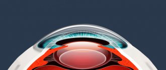
Apparatus for changing and processing light
Light refractive structure
This is a lens system. The first lens is the cornea. Thanks to it, the field of view is 190 degrees. The result of a malfunction of this lens is tunnel vision.
The final refraction of light occurs in the lens, which focuses light rays onto a small area of the retina. The lens is responsible for visual acuity; changes in its shape lead to the development of myopia or farsightedness.
Accommodative structure
The system regulates the intensity of incoming light and its focus. It consists of the iris, pupil, annular, radial and ciliary muscles; it can also include the lens. Focusing for seeing distant or close objects occurs by changing its curvature. The curvature of the lens is changed by the ciliary muscles.
Regulation of the light flux occurs due to changes in the diameter of the pupil, expansion or contraction of the iris. The circular muscles of the iris are responsible for constricting the pupil, and the radial muscles are responsible for its expansion.
Receptor structure
It is represented by the retina, consisting of photoreceptor cells and the endings of neurons corresponding to them. The anatomy of the retina is complex and heterogeneous. Has a blind spot and an area of increased sensitivity. It itself consists of 11 layers. Photoreceptor cells, divided by shape into rods and cones, are responsible for the main function of processing light information.