In the vision system, each element has a strict purpose, even the cameras of the eye, despite the fact that they represent only empty space, of a given volume are of great importance for the reliable operation of the visual apparatus.
After all, there is nothing superfluous in nature, and even cavities and voids in the structure of internal organs are not random oversights, but rather, on the contrary, a high flight of scientific thought.
Structure
Functions
Symptoms
Treatment
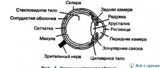
The structure of chamber formations
The anterior chamber of the eye is located directly behind the cornea. The peculiarity of the “element” is that it has different depths along its entire length. The greatest depth is in the area of the pupil, the indicator reaches three and a half millimeters, it decreases towards the periphery. Aberration in camera parameters can become a symptom of an eye disease. For example, the depth increases after removal of the lens or decreases when the mucous membrane is detached.
The posterior chamber of the eye is located directly behind the anterior chamber and contains many tiny zonules of Zinn, which act as a “connecting” element between the lens and the ciliary body. They are also responsible for the contraction of the ciliary muscles, whose task is to transform the shape of the lens. Thanks to this, a person sees well at any distance.
Both chambers are filled with a liquid identical in composition to blood plasma. Moisture contains a large amount of useful substances that are transferred to the organ of vision and ensure its smooth operation. The liquid also accepts metabolic products from the eye, after which it “redirects” them to the circulatory system. Moisture is produced through the ciliary processes of the ciliary body, and outflow occurs through the drainage system.
| The volume of the posterior and anterior chambers holds from 1.23 to 1.32 cubic centimeters of intraocular fluid. It is important to maintain a balance between the decrease and increase of moisture inside the eye. When the indicator shifts, the smooth operation of the visual apparatus is disrupted. |
If more fluid is produced than comes out, glaucoma develops. In the opposite situation, there is a high risk of eye subatrophy. Any slight imbalance has a negative impact on the functioning of the eyes and can even lead to blindness.
The volume of chamber formations in the visual apparatus must be stable; this is the only way to ensure uninterrupted production of intraocular moisture and its outflow.
Color perception
| This section is missing references to information sources. Information must be verifiable, otherwise it may be questioned and deleted. You may edit this article to include links to authoritative sources. This mark was set on April 3, 2013 . |
Blue eye
Multicolor is perceived due to the fact that the cones react to a certain spectrum of light in isolation. There are three types of cones. Type 1 cones respond predominantly to red, type 2 to green, and type 3 to blue. These colors are called primary. When exposed to waves of different lengths, each type of cone is excited differently. As a result, each wavelength is perceived as a special color. For example, when we look at a rainbow, the primary colors (red, green, blue) seem most noticeable to us.
By optical mixing of primary colors, other colors and shades can be obtained. If all three types of cones are excited simultaneously and equally, the sensation of white color occurs.
Some people, the so-called tetrachromats, are able to see radiation that goes beyond the spectrum visible to the eye of an ordinary person and distinguish colors that are perceived as identical by an ordinary person.
Some people (approximately 8% of men[6] and 0.4% of women[ source not specified 843 days
]) have a color perception feature called color blindness.
Colorblind people perceive color in their own way, confusing some shades that are contrasting for the majority and distinguishing their own colors, which seem the same for the rest of the majority of people [ source not specified 843 days
].
It is believed that incorrect color discrimination is associated with an insufficient number of one or more types of cones in the retina[6]. There is also acquired color blindness due to disease or age-related changes. Colorblind people may not feel their vision until they are faced with the need to choose between two similar shades, which are perceived as different colors by a person with normal vision. Due to the possibility of color perception errors, some professions impose restrictions on allowing colorblind people to work. It is interesting that the reverse side of color blindness is increased sensitivity to some shades that are not available to others, which have not yet been well studied and are rarely used in households [ source not specified 843 days
].
Physiological role of eye cameras
Their main purpose is to “monitor” the circulation of intraocular fluid. The production of moisture occurs in the ciliary processes by filtering the capillary blood flow. First it appears in the posterior chamber (secreting), then moves to the anterior one. Afterwards, due to low blood pressure, moisture leaves through the UPC, which is responsible for the outflow of fluid.
Also, chamber formations have a number of additional functions:
- Responsible for the conductivity of light rays;
- Form “good relationships” between all structures of the eye and intraocular tissues;
- On “their shoulders” lies light refraction. Thanks to this, the rays are focused on the retina, i.e. cameras act as conductors of sorts.
Literature
| Eye at Wiktionary |
| Eye on Wikiquote |
| Eye on Wikisource |
| Eye on Wikimedia Commons |
| Eye on Wikinews |
- Eye // Encyclopedic Dictionary of Brockhaus and Efron: in 86 volumes (82 volumes and 4 additional). - St. Petersburg, 1890-1907.
- G. E. Kreidlin. Eye gestures and visual communicative behavior // Proceedings on cultural anthropology M.: 2002. pp. 236-251
- Bykov V.L.
Private human histology. - St. Petersburg: Sotis, 2001. - 304 p. — ISBN 5-85503-116-0.
Angle of the anterior chamber - general structure
The UPC is a peripheral plane where the cornea smoothly flows into the sclera, and the iris into the ciliary body. The main value of the anterior chamber angle is the drainage system, which is responsible for the outflow of intraocular moisture into the circulatory structure.
It includes:
- Venous sinus, located in the white of the eye;
- Trabecular diaphragm, which is a network with a porous-layered structure. It decreases in size closer to the outside, this has a positive effect on the outflow of intraocular fluid;
- Collecting tubules.
First, the moisture released in the eyes enters the trabecular diaphragm, then it is “directed” into the lumen of Schlemm’s canal (located near the limbus in the sclera of the eyeball).
In some cases, the outflow of intraocular fluid occurs in a different way, through the uveoscleral pathway. Thus, approximately fifteen percent of the total volume of moisture penetrates into the bloodstream. In this case, it first enters the ciliary body, then moves in the direction of the muscle fibers and penetrates the suprachoroidal space. From here, the fluid passes through the veins into Schlemm's canal or the white membrane of the eye.
Collecting tubules in the sclera remove moisture in three directions:
- In the episcleral veins;
- In the vessels of the ciliary body;
- In the venous plexus located on the surface of the white membrane of the eyes.
You will learn more about the structure of the anterior chamber and its functions from the video
Return to contents
Perception of the location of objects in space
Correct assessment of the location of objects in space and the distance to them is achieved by using the eye. It can be improved, like any property. The eye meter is especially important for pilots and drivers. Improved perception of objects is achieved through characteristics such as field of view, angular velocity, binocular vision and convergence.
The field of view is the space that can be covered by the eye when the eyeball is in a fixed state. The field of view can cover a significant number of objects, their location at a certain distance. However, the image of objects that are in the field of view, but located closer, partially overlaps with the images of those behind them. As objects move away from the eye, their size, the relief of their shape, the difference in shadows on the surface, the saturation of colors, etc. decrease, until the object disappears from the field of view.
There are many objects moving in space, and we can perceive not only their movement, but also the speed of movement. The speed of movement of objects is determined based on the speed at which they move across the retina, the so-called angular velocity. The angular speed of closely located objects is higher, for example, the carriages of a moving train rush past the observer at high speed, and an airplane in the sky disappears from view slowly, although its speed is much greater than the speed of the train. This is because the train is much closer relative to the observer than the plane. Thus, nearby objects disappear from the field of view earlier than distant ones, since their angular velocity is greater. However, the movement of objects that move extremely quickly or too slowly is not perceived by the eye.
Binocular vision also contributes to an accurate assessment of the spatial arrangement of objects and their movement. This allows not only to perceive a three-dimensional image of an object, since both the left and right parts of the object are simultaneously covered, but also to determine the location in space and the distance to it. This can be explained by the fact that when the cerebral cortex combines sensations from images of objects in the left and right eyes, it evaluates the sequence of objects’ arrangement and their shape.
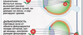
If the refraction in the left and right eyes is not the same, this leads to impaired binocular vision (seeing with two eyes) - strabismus. Then a sharp image appears on the retina from one eye and a blurry image from the other. Strabismus is caused by a violation of the innervation of the muscles of the eye, a congenital or acquired decrease in visual acuity in one eye, etc.
Another mechanism of spatial perception is eye elevation (convergence). The axes of the right and left eyes, with the help of the oculomotor muscle, converge on the object being examined. The closer the object is, the more the internal rectus muscles are contracted and the external rectus muscles are stretched. This allows you to determine the distance of objects.
Posterior chamber of the eye
It is protected in front by the iris and on the back by the vitreous body. On the outside it has a boundary, the role of which is played by the ciliary body; on the inside, a part of the lens acts as a limiter. The entire space of the chamber is filled with connecting threads, which are responsible for the weakening and tension of the ciliary muscles.
Thanks to this function, a person can equally well distinguish between objects located at close and far distances.
You will learn a lot of interesting things about the structure of the rear camera and its functions by watching the video
Notes
- Volkova I. P.
The role of vision in human life and the consequences of its impairment in mental and personal development. koleso.mostinfo.ru (May 20, 2008). Retrieved April 3, 2013. - Bykov, 2001, p. 220-221.
- article “Jellyfish and flies have confirmed the common origin of the eyes” on the website membrana.ru (July 30, 2010). Retrieved August 7, 2010. Archived February 1, 2013.
- Zhimulev I.F.//General and molecular genetics (course of lectures for 3rd year students) - online publication. Chapter 14.1 “Developmental Genetics”, p. 14/17 (inaccessible link - history
). Retrieved August 22, 2009. Archived April 19, 2009. - Gehring W.J.
The genetic control of eye development and its implications for the evolution of the various eye-types (English) // Int J Dev Biol.. - 2002. - No. 46(1). - P. 65-73. — PMID 11902689. Archived April 3, 2013. - ↑ 1 2 D. Hubel.
Eye, brain, vision / ed. A. L. Byzova. - M.: Mir, 1990. - 172 p. - ↑ 1 2
Land, M. F. (1992).
"The evolution of eyes". Annual Review of Neuroscience 15
: 1–29. DOI:10.1146/annurev.ne.15.030192.000245. PMID 1575438. - (2008) "Assembly of the cnidarian camera-type eye from vertebrate-like components" (PDF). Proceedings of the National Academy of Sciences 105
(26):8989–8993. DOI:10.1073/pnas.. PMID 18577593. Bibcode: 2008PNAS..105.8989K. - Fernald, Russell D. (September 2006). "Casting a Genetic Light on the Evolution of Eyes." Science 313
(5795):1914–1918. DOI:10.1126/science.1127889. PMID 17008522. Bibcode: 2006Sci…313.1914F.
Diseases affecting chamber formations
Ailments that affect the back or front of the eye are classified into congenital and acquired. The first category includes:
- Lack of angle in the “facade” chamber;
- Abnormal attachment to the iris from the anterior edge;
- Blocking of the UPC by rudiments of embryonic matter.
The group of acquired pathologies includes:
- Failure in the outflow of intraocular fluid due to blocking of the anterior angle by a pigment spot or enlarged iris;
- Accumulation of purulent discharge;
- Uneven change in the depth of the “facade” chamber. The cause of the deviation may be previous damage to the visual apparatus, displacement of the lens or asthenopia of the zonules;
- Blood condensation (hyphema);
- Formation of connecting cords;
- CCP cracking;
- A hard deposit forms on the endothelial layer of the cornea;
- Glaucoma occurs due to a disruption in the circulation of moisture produced by the eyes;
- Rupture of the anterior part of the ciliary body;
- Injury to the lens or damping of the ligaments responsible for its support ultimately changes the depth of the anterior chamber;
- Reduction in the size of the anterior chamber, the cause of the pathology most often becomes a closed pupil.
To maintain visual acuity, do not ignore visits to the ophthalmologist. Only a professional doctor, with the help of special tests and examinations, will be able to identify the pathology and select the optimal therapy to prevent its progression. As a preventative measure to prevent eye diseases, visit an ophthalmologist once every twelve months.
| If eye problems begin suddenly, pain appears, or you notice the appearance of blood clots on the whites, visit the doctor unscheduled. |
Symptoms of damage to the chambers of the eye
If there are deviations in the functioning of the “elements” of the organ of vision, the following signs are observed:
- Cloudiness of the cornea;
- Painful sensations;
- The appearance of new growths or spots on the eyes;
- Fear of bright light;
- The resulting image is unclear and has blurry contours;
- Problems with visual acuity;
- With hemorrhage in the anterior chamber, a change in the color of the iris is observed.
If dangerous symptoms appear, consult a doctor immediately to identify the disease at an early stage. Return to contents
Diagnosis of eye chamber pathologies
If the development of a particular disease is suspected, the ophthalmologist sends the patient for several examinations:
- Biomicroscopy. It is carried out using a slit lamp;
- Microscopy of the anterior chamber. Helps detect glaucoma;
- Analysis of intraocular moisture, study of its circulation process;
- Coherence optical tomography;
- Ultrasonography;
- Pachymetry. Used to measure the depth of the anterior chamber;
- Automated tonometry. It is used to determine the level of pressure exerted by intraocular moisture.
| Modern equipment and innovative technologies will help detect any abnormality at an early stage, and the doctor will be able to select treatment to block its progression. |