The human eye is a very complex optical system, consisting of a variety of elements, each of which is responsible for its own tasks. In general, the eye apparatus helps to perceive an external image, process it and transmit information in an already prepared form to the brain. Without its functions, the organs of the human body could not interact as fully. Although the organ of vision is complex, every person should understand at least a basic description of the principle of its functioning.
General operating principle
Having understood what the eye is and understanding its description, let’s consider the principle of its operation. The eye works by perceiving light reflected from surrounding objects. This light hits the cornea, a special lens that allows the incoming rays to be focused. After the cornea, the rays pass through the chamber of the eye (which is filled with a colorless liquid), and then fall on the iris, which has a pupil in its center. The pupil has a hole (palpebral fissure) through which only the central rays pass, that is, part of the rays located at the edges of the light stream are eliminated.
https://www.youtube.com/watch?v=DBMNKvUayt4
The pupil helps adapt to different levels of light. It (more precisely, its palpebral fissure) filters out only those rays that do not affect the quality of the image, but regulates their flow. As a result, what is left goes to the lens, which, like the cornea, is a lens, but only intended for something else - for more accurate, “finish” focusing of light. The lens and cornea are the optical media of the eye.
Next, the light passes through a special vitreous body, which enters the optical apparatus of the eye, onto the retina, where the image is projected like on a movie screen, but only upside down. In the center of the retina is the macula, the area that responds to visual acuity, which includes the object we look at directly.
During the final stages of image acquisition, retinal cells process what is on them, translating everything into electromagnetic impulses, which are then sent to the brain. A digital camera functions in a similar way.
Of all the elements of the eye, only the sclera, a special opaque membrane that covers the outside of the eyeball, does not participate in signal processing. It surrounds it almost entirely, approximately 80%; in the anterior part it smoothly passes into the cornea. People usually call its outer part protein, although this is not entirely correct.
Report on the topic “The structure of the human eye”
Underneath it is the choroid, which passes from the front into the iris, in the center of which there is a hole - the pupil, which can contract and expand under the influence of muscles. The choroid contains the ciliary muscle, which regulates the curvature of the lens. The inner layer of the eye, the retina, contains light-sensitive receptors called rods and cones. In them, light energy is converted into an excitation process, which is transmitted along the optic nerve to the brain. Cones are located in the center of the retina, opposite the pupil - in the corpus luteum and provide daytime vision, perceiving colors, shapes and details of objects. On the periphery of the retina there are only rods that are irritated by weak twilight light, but they are not sensitive to color.
The inner nucleus of the eyeball forms (together with the cornea) the optical system of the eye and consists of the lens, vitreous humor and aqueous humor of the chambers of the eye. The transparent and elastic lens, located behind the pupil, has the shape of a biconvex lens. It, together with the cornea and intraocular fluids, refracts light rays entering the eye and focuses them on the retina.
Attention!
If you need help with academic work, we recommend turning to professionals. More than 70,000 experts are ready to help you right now.
Cost calculation Guarantees Reviews
So, the reflex mechanism by which light rays emanating from an object are focused on the retina is called accommodation. It includes two processes - a reflex change in the diameter of the pupil and refraction (refraction) of light.
Reflex change in pupil diameter.
In bright light, the circular muscles of the iris contract, and the radial muscles relax; As a result, the pupil narrows and the amount of light entering the retina decreases, which prevents damage. In dim light, on the contrary, the radial muscles contract and the circular muscles relax. An additional benefit of constricting the pupil is that the depth of field is increased, so that differences in the distance from the subject to the eye have less impact on the image.
Light refraction.
From an object more than 6 cm away, almost parallel rays of light enter the eye, while rays coming from closer objects diverge noticeably. In both cases, in order for light to be focused on the retina, it must be refracted, and for close objects the refraction must be stronger. The normal eye is able to accurately focus light from objects at a distance of 25 cm to infinity. The shape of the cornea cannot change, so refraction here depends only on the angle of incidence of light on the cornea, which in turn depends on the distance of the object. The strongest refraction of light occurs in the cornea, and the function of the lens is the final “focusing”. When the ciliary muscle contracts, the lens changes its curvature, taking a shape for distant or near vision. Refracted rays of light from the object in question, falling on the retina, form a reduced inverse image of the object on it.
However, we see objects in direct form thanks to the daily training of the visual analyzer, which is achieved by the formation of conditioned reflexes, readings from other analyzers, their interactions, constant checking of visual sensations, and everyday practice.
The motor system of each eye consists of 6 muscles, the contractions of which allow you to change the direction of gaze.
In people with normal vision, a clear image of objects appears on the retina, because it is focused on the center of the retina.
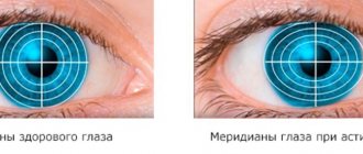
Visual impairment is often associated with abnormal eyeball length. Myopia develops as the longitudinal axis of the eye increases. Parallel rays coming from distant objects are concentrated (focused) in front of the retina, which receives diverging rays, resulting in a blurry image. For myopia, glasses with biconcave lenses are prescribed, which reduce the refraction of rays so much that the image of objects appears on the retina.
Order work from 200 rubles
If you need help with academic work, we recommend turning to professionals. More than 70,000 experts are ready to help you right now.
Cost calculation Guarantees Reviews
When the axis of the eyeball is shortened, farsightedness occurs. The image is focused behind the retina. Correction requires biconvex glass. Senile farsightedness usually develops after 40 years, when the lens loses elasticity, hardens and loses the ability to change curvature, which makes it difficult to see clearly at close range. The eye loses the ability to clearly see objects at different distances.
The Guinness Book of Records contains a record for human visual acuity. It was directed by Dennis M. Levy from Texas, USA. In April 1984, he repeatedly determined the relative position of the thin, bright green line to within 0.85 arcseconds. This is equivalent to a displacement of 6 mm observed from a distance of 1.6 km.
Number of distinguishable colors
The human organ of vision perceives images in color, and the number of shades of colors that it can distinguish is very large. How many different colors the eye can distinguish (more precisely, how many shades) can vary depending on the individual characteristics of the person, as well as the level of his training and the type of his professional activity. The eye “works” with so-called visible radiation, which is electromagnetic waves with a wavelength from 380 to 740 nm, that is, with light.
If we take average indicators, then a person can distinguish about 150 thousand color tones and shades in total.
However, there is an ambiguity here, which lies in the relative subjectivity of color perception. Therefore, some scientists agree on another figure for how many shades of colors a person usually sees/distinguishes – from seven to ten million. In any case, the figure is impressive. All these shades are obtained by varying the seven primary colors located in different parts of the rainbow spectrum. It is believed that professional artists and designers have a higher number of perceived shades, and sometimes a person is born with a mutation that allows him to see many times more colors and shades. How many different colors such people see is an open question.
Internal structure of the eye
The main internal structures of the eye include:
- The iris is the colored part of our eye.
- The pupil, a black round hole in the center of the cornea.
- The lens of the eye behind the iris, allowing the gaze to focus on near and distant objects.
- The retina is a very thin layer of millions of photoreceptors called "cones and cones"
Every day the set penetrates the eye through the cornea and pupil. If you turn on the light in a dark room, your pupil will constrict to reduce the amount of light. The opposite happens if you move from a sunny room to a dark room. Your pupil will dilate to see better in your new environment.
After passing through the pupil and lens of the eye, the light is focused on the retina. This is the most amazing stage of the visual process when the image is upside down on the back wall of the eye. Yes that's right.
After reaching the back wall, the light travels along the nerve endings. These images enter the brain through the optic nerves. As the brain processes this information, the images rotate, and we do not see them upside down. Without this we would live in an unusual world!
Our visual system is truly efficiently designed.
While this may look strange, it is the most efficient and fastest way to process information. This begs the question: “What happens when the function of any organ of vision is impaired”?
Eye diseases
Like any other system of the human body, the organ of vision is susceptible to various diseases and pathologies. Conventionally, they can be divided into infectious and non-infectious. Common types of diseases that are caused by bacteria, viruses or microorganisms are conjunctivitis, stye and blepharitis.
If the disease is non-infectious, then it usually occurs due to serious eye strain, due to a hereditary predisposition, or simply due to changes that occur in the human body with age. Less commonly, the problem may be that a general pathology of the body has arisen, for example, hypertension or diabetes has developed. As a result, glaucoma, cataracts or dry eye syndrome may occur, and the person ultimately sees or distinguishes objects worse.
In medical practice, all diseases are divided into the following categories:
- diseases of individual elements of the eye, for example, the lens, conjunctiva, and so on;
- pathologies of the optic nerves/pathways;
- muscle pathologies, due to which the friendly movement of the apples is disrupted;
- diseases associated with blindness and various visual disorders, impaired vision;
- glaucoma.
To avoid problems and pathologies, the eyes must be protected, not kept pointed at one point for a long time, and optimal lighting maintained when reading or working. Then the power of vision will not decrease.
How do we see?
Processing impulses entering the brain from both eyes produces a three-dimensional image. Primary signals from the retinas of both eyes are transmitted along the optic nerves, which form a partial chiasm. The nerve fibers that initially come from each eye separately are redistributed in such a way that the right hemisphere of the cerebral cortex receives information from the right side of the retina of both eyes, and the left hemisphere receives information from the left side. After crossing, the nerve impulse enters the subcortical centers of the visual analyzer, where visual stimuli are analyzed, their color characteristics, spatial contrast and average illumination in different parts of the visual field are assessed. Next, the neurons of the subcortical layer transmit the converted signals through axons to the projection area of the visual cortex, where the image is formed.
Why do you need to have your eyes checked?
The eye in this complex system is just a “receiver” that converts the image into millions of nerve impulses. The slightest malfunction in the most complex mechanism is fraught with serious consequences, including complete blindness. Diagnostics using the latest generation devices allows you to identify any problem at an early stage and take measures to eliminate it.
External structure of the eye
The human eye has not only an internal structure, but also an external one, which is represented by centuries. These are special partitions that protect the eyes from injury and negative environmental factors. They mainly consist of muscle tissue, which is covered on the outside with thin and delicate skin. In ophthalmology, it is generally accepted that the eyelids are one of the most important elements, which can cause problems if problems arise.
Although the eyelid is soft, its strength and consistency of shape are ensured by cartilage, which is essentially a collagen formation. The movement of the eyelids is carried out thanks to the muscle layer. When the eyelids close, this has a functional role - the eyeball is moistened, and small foreign particles, no matter how many there are on the surface of the eye, are removed. In addition, due to the wetting of the eyeball, the eyelid is able to slide freely relative to its surface.
An important component of the eyelids is also an extensive blood supply system and many nerve endings that help the eyelids carry out their functions.
How the human eye works, an explanation for children
Vision is a person’s ability to perceive light, shape and color of surrounding objects or, in other words, the ability to see them.
This happens thanks to special light-sensitive cells in our body, which are collected in special organs - the eyes. How does the human eye work? There are two types of light-sensitive cells, called rods and cones. Rods perceive only dark and light, while cones perceive color. The cones and rods are located on the thin inner layer of the eyeball, called the retina. The retina is penetrated by many blood vessels. The eyeball itself consists of dense, multi-layered connective tissue that gives it its shape. The front part of the eyeball is the transparent cornea, through which light penetrates into the eyeball. Then the light is captured by a kind of “diaphragm” of the eye - its iris. The iris, thanks to the pigment cells it contains, determines the color of the eyes. If there are many of them, then the person’s eyes are brown; if there are few or none at all, then they are light green or blue. Through the iris of the eye, light enters through an opening called the pupil. The pupil is equipped with two muscles, one of which makes it larger in the dark, and the other narrows it in bright light. Having passed the hole of the pupil, the light enters the spherical lens. This is the name of an elastic organ that is enclosed in a ring of muscles. By stretching, they reduce the convexity of the lens and change the curvature of its surface. The lens, like a lens, refracts rays and directs them to light-sensitive cells located on the retina. This is how we see it.
If a person examines objects that are close, the lens becomes more convex and refracts light rays more strongly. If we look at objects located far away, the lens becomes flatter and refracts the rays less. Over the years, the lens loses its elasticity and has to be “helped” with glasses. By the way, thanks to the lens, all objects are reflected on the retina upside down, but our brain corrects such a distorted picture. You can draw a parallel between how the human eye works and the camera. The cornea is the window of the lens, the iris and pupil are the diaphragm, the crystalline lens is the adjustable lens, and the photosensitive layer of the retina is the photographic film. However, a person has two eyes, our brain constantly “compares” what they see and, thanks to this, we have spatial vision.
Structure of the human ear
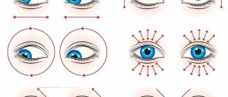
Eye movement
Human eyes move with the help of special muscles that ensure normal, constant functioning of the eyes. The visual apparatus moves with the help of the coordinated work of dozens of muscles, the main of which are four straight and two oblique muscle processes. The rectus muscles surround the optic nerve on different sides and help rotate the eyeball around different axes. Each group allows you to turn a person’s eye in its own direction.
The muscles also help lift and lower the eyelids. When all the muscles work harmoniously, this not only allows you to control the eyes individually, but also to carry out their harmonious work and coordination of their direction.