We see all the bright shades of the surrounding world that delight us at any time of the day only through the retina of the eye, or rather special photoreceptors. These are rods and cones. Rods and cones belong to photographic receptors, and their structure provides the maximum degree of sensitivity. Thanks to this quality, the cones and rods of the retina transform light signals coming from outside into special impulses, which can then be perceived by the human nervous system.
The special structure of each type of photoreceptor allows them to perform specific functions. During daylight hours, the cones of the eye experience greater stress. When the light flux decreases, that is, at dusk, the rods of the retina begin to do their work.
The structure of rods and cones is different due to the fact that these photoreceptors have different principles of operation and are involved in light perception in different ways.
Content
- 1 The structure of photoreceptors - cones 1.1 Human cones
- 1.2 Morphology
- 1.3 Cones of reptiles and birds
Cones are sensitive to light over a wide range. In the dark, when the illumination is insufficient for the cones to work, only the rod receptors work in humans. At night, people become “color blind” - they perceive the world as monochrome.
The photosensitivity of the receptors is associated with the presence of a specific pigment in them - iodopsin; with the cis-trans transition of retinal and other mechanisms. In turn, iodopsin consists of several visual pigments. Today, two pigments are well known and studied: chlorolab (sensitive to the yellow-green part of the spectrum) and erythrolab (sensitive to the yellow-red part of the spectrum).
There are about 6 million in the retina of an adult human eye[1] cones. Their dimensions are as follows: length about 50 microns, diameter - from 1 to 4 microns.
Cones are about 100 times less sensitive to light than rods (another type of retinal cell), but are much better at detecting rapid movements.
The retina is a complex, layered structure with several layers of neurons connected by synapses. Single neurons that are directly photosensitive are cone and rod photoreceptor cells.
Symptoms of retinal diseases
Retinal diseases can be either congenital or acquired.
Acquired pathologies include retinal detachment and retinitis (inflammatory process).
Any damage to the retina is an insidious process: the disease can be asymptomatic for a long time. One of the main signs of their development is a decrease in visual acuity.
If the lesion is localized in the central zone, then in the absence of the necessary treatment, the Patient may experience complete loss of vision.
Disorders of the peripheral parts can occur without deterioration of vision, which is why it is so important to undergo an eye examination every six months to a year. As a rule, extensive damage to the peripheral region is still accompanied by pronounced symptoms:
- Loss of a portion of the visual field
- Changes in color perception
- Reduced orientation in low light conditions.
With detachment, flashes, black spots and lightning may appear before the eyes.
[edit] The structure of photoreceptors - cones
Cones in different species of animals have a diverse structure; in individual species, different structures of cones can be found.
[edit] Human cones
[edit] Morphology
Structure of the cone (retina)
Cones and rods are similar in structure and consist of four sections.
- 1 - OUTER SEGMENT (contains membrane disks with iodopsin),
- 2 — LINKING DEPARTMENT (waisting),
- 3 - INNER SEGMENT (contains mitochondria),
- 4 - SYNAPTIC AREA
The outer segment of the cone is filled with membranous half-discs formed by the plasma membrane, separated from it. They are folds of the plasma membrane. There are significantly fewer membrane half-discs in cones than discs in a rod, and their number is approximately several hundred. Each disk is formed by two membranes connected at the edges with a thickness of about 50 - 75 Angstroms, separated by a gap of about 50 Angstroms.[2].[3].
In the area of the connecting section (constriction), the outer segment is almost completely separated from the inner segment by an invagination of the outer membrane. The connection between the two segments is carried out through the cytoplasm and a pair of cilia passing from one segment to another. Cilia contain only 9 pairs of double filaments (fibrils). They extend in the connecting cilium from one of the two centrioles (basal body), which lie next to each other perpendicular to each other. The filaments connecting the cilia extend from the inner segment to the tip of the outer segment.[4].
The inner segment contains a cluster of radially oriented and densely packed mitochondria. When illuminated, the cone mitochondria swell and, probably, the activity of oxidative enzymes in them increases. This is an area of active metabolism. Mitochondria and polyribosomes supply energy to support light perception processes, while proteins involved in the formation of membrane discs and visual pigment are synthesized. The core is located in the same area.[5].[6].
In the synaptic area with nerve endings, the cones approach and protrude into the dendrites of the bipolar and horizontal retinal cells. In addition, contacts between the receptors (rods and cones) of the retina are described. A large number of synaptic vesicles (vesicles) containing transmitter were found in presynaptic terminals. The number and size of these bubbles appear to change with changes in illumination.[7].[8].[9].
Diffuse bipolar cells
can form synapses with several rods. This phenomenon is called synaptic convergence.
Monosynaptic bipolar cells
connect one cone to one ganglion cell, which provides greater visual acuity compared to rods.
Horizontal
and
amacrylic
cells bind together a number of rods and cones. Thanks to these cells, visual information undergoes certain processing even before leaving the retina; these cells are particularly involved in lateral inhibition.[10],[11]
[edit] Cones of reptiles and birds
The cones in the retina of birds, amphibians and other vertebrates differ in their structure from the cones found in the retina of primates.
In particular, in birds, fish, and turtles, “oil droplets” are present in the structure of the cones. In addition, their retinas contain both “regular” cones and so-called “double” cones.
Sticks
One of the photoreceptors of the retina. They look like small cellular processes. These elements got their name because of their special shape - cylindrical. In total, the retina is filled with about one hundred and twenty million rods. They are extremely small in size. Their diameter does not exceed 0.002 mm, and their length is about 0.06 mm. They are the ones who convert light stimulation into nervous stimulation. In simple words, they are the very element of the eye due to which it reacts to lighting.
Structure
The rods consist of an outer segment, which includes membranous disks, a connecting section, also called a cilium because of its shape, and an inner section with mitochondria. The nerve endings are located at the base of the rod.
The pigment rhodopsin, present in the rods, is responsible for sensitivity to light. When exposed to light rays, the pigment discolors.
The distribution of rods throughout the body of the retina is uneven. There can be from twenty to two hundred thousand rods per square millimeter. In the peripheral areas their density is less than in the central ones. This determines the possibility of night and peripheral vision. There are almost no rods in the macula.
[edit] Human color vision
Curves of absorption spectra of pigments contained in cones and rods of the human retina.
Spectra of short (S), medium (M) and long wavelength (L) pigments and the spectrum of rod pigment in low (twilight) lighting (R). NB: the wavelength axis in this graph is non-linear. Spectral sensitivity curves of cone receivers (photopigments) of normal trichromat, determined by the colorimetric method (A), and absorption spectra measured in the outer segments of single macaque cones (B). (After Marks et al., 1964). Solid curves in A represent the result of calculating the spectral sensitivity curves using the addition curves of normal trichromate (Bongard and Smirnov, 1955); circles—results of experiments with dichromates [12]. According to the currently prevailing three-component theory of vision, the three absorption peaks found in the visible region of the spectrum by retinal tissue are caused by the presence of three types of visual pigments and, accordingly, there are three types of cones that are sensitive to different wavelengths of light (colors). These are S-type cones, sensitive in blue (S from English Short - short-wave spectrum), M-type - in green (M from English Medium - medium wavelength), and L-type - red (L from English Long - long-wavelength ) parts of the spectrum. The assumption is that each type of cone contains only one of the three pigments. [13]
It is now known that the light-sensitive pigment iodopsin, found in all cones of the eye, includes pigments such as chlorolab and erythrolab. Both of these pigments are sensitive to the entire region of the visible spectrum, but the first of them has an absorption maximum corresponding to the green (absorption maximum about 540 nm), and the second - to the red (absorption maximum about 570 nm) parts of the spectrum. The third pigment, sensitive to the violet-blue region of the spectrum, is called cyanolab.
There is criticism of this concept: followers of the nonlinear theory of vision deny the existence of cyanolabe with reference to the work of 1964 [14], and they claim that they failed to find any difference between the cones in the retina (see also Cones in the nonlinear theory of vision).
Rice. K. Passage of waves of blue, green, red colors in the outer membrane of the cone.[15]
Taking into account modern views of biophysics and biochemistry, the foundations of the previous established process of color vision have been revised from different points of view:
- From a biological point of view, in the field of color vision from 1966 to 2009 (Works of Dr. R.E. Mark and his laboratory) [16] with basic experimental data from living cell studies, the work of cones (LMS (color vision)) was established on retinal sections |S,M,L) in the RGB and sticks block. It has been established that in daylight conditions (color vision) cones work, and during twilight and night lighting (not color vision) rods work in a mode isolated from cones. The work of photoreceptors is associated with modified varieties of photopigments based on opsin proteins (See Retinomotor reaction of retinal photoreceptors) - this is conopsin in cones, rhodopsin in rods.
- From a purely physical point of view, based on the work (2011) of the scientist physicist [17] Gerald K. Hut, it was proposed to consider the interactions of light with the outer lobes of the membranes of the photoreceptors of the retina on the basis of the primary interaction with light (receptor) at the level of “nano-antennas”. According to Doctor of Physics John Medeiros [18], the work of the outer lobes of the membrane of cones and rods is considered similar to the work of conical and cylindrical waveguides in the medium of the transparent body of the eye (liquid medium).
As a result, physicists K. Hut and John Medeiros came to the generally accepted principle of trichromatism. (See Revisiting traditional views on the visual process by physicist K. Huta, The work of the outer membranes of the cones and rods of the retina as a waveguide). At the same time, considering the cone as a biological waveguide of a conical shape, data on the passage of rays in the waveguide, associated with the value of the cross section of the electromagnetic oscillation, were obtained. For example, blue color is carried by waves with a cross section of ≈2-4 nm, green - ≈4-6 nm, red - ≈6-7 nm or more. In this case, each of the waves stops in its own section of the conical waveguide - the outer membrane of the cone. (See Figure K). From where we can say that each cone, perceiving the oppositely selected rays incident on it in the appropriate section, produces the opsin photopigment of the desired color.
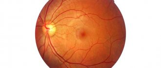
Diagnosis of diseases and treatment of the retina
To obtain a complete picture of the functioning of the retina and the functional state of its structure, various methods are used. The main one is ophthalmoscopy, as well as OCT (OCT) optical coherence tomography.
Treatment of retinal diseases is selected individually, depending on the specific case. This can be either drug treatment or the use of laser coagulation of the retina, and in difficult cases, surgical intervention.
Doctors at Dr. Belikova's Eye Clinic have extensive experience in diagnosing and treating diseases of the retina. Timely contact with ophthalmologists and preventive eye examinations, once every 6-12 months, will help avoid the development of serious pathological changes and preserve vision.