↑ Eyeball
The organ of vision includes:
- eyeball;
- protective apparatus (eye socket, eyelids);
- appendages of the eye (tear and muscular apparatus);
- conducting nerve pathways and centers of vision.
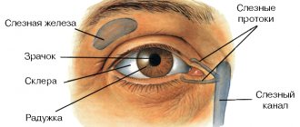
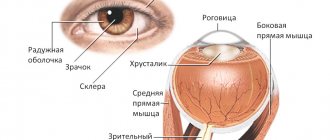
The eyeball has a spherical shape and is located in the orbit. The eyeball is separated from the walls of the orbit by a dense fibrous sheath (Tenon's capsule), behind which there is fatty tissue. Eye mobility is ensured by the activity of the extraocular muscles (four straight and two oblique). The front of the eye is protected by eyelids. The inner surface of the eyelids and the front part of the eyeball, with the exception of the cornea, are covered with a mucous membrane - the conjunctiva. At the upper outer edge of each orbit there is a lacrimal gland, which produces fluid that washes the eye (Fig. 1).
Eyeball parameters
Two poles:
- anterior pole - corresponds to the center of the cornea;
- posterior pole - located opposite the anterior pole (lateral to the exit point of the optic nerve).
Axles
The external (optical) axis or sagittal (front - back) size of the eyeball is a line connecting the anterior and posterior poles.
The internal axis of the eyeball is part of the optical axis, located between the posterior surface of the cornea and the internal surface of the retina.
The visual axis runs from the object under consideration through the central points of the cornea and lens and intersects with the retina. A plane perpendicular to the optical axis (ocular equator) divides the eyeball into anterior and posterior halves. The horizontal diameter of the equator (23.5 mm) is shorter than the external ocular axis (24 mm). The eyeball reaches its final size by the age of 25.
The membranes of the eyeball wall
- The outer one is a fibrous membrane (sclera), has two sections: the anterior transparent (cornea); posterior opaque (sclera). Functions: protective (determines the constancy of the shape and tone of the eye); place of attachment of the extraocular muscles; Vessels and nerves (including the optic nerve) pass through it. At the border between the cornea and the sclera there is a translucent shallow groove (width 1 - 1.5 mm) - the limbus, under which is located the circular venous sinus of the sclera - Schlemm's canal
- The middle is the choroid.
- The inner layer is the retina (retina). Inside the eyeball there are transparent light-refracting media - the lens, the vitreous body, and the intraocular fluid.
↑ Cornea
The cornea, or cornea, is convex in front and concave in back, a transparent, avascular plate of the eyeball, which is a direct continuation of the sclera.
Function.
The cornea is the optical structure of the eye; its refractive power is, on average, 45D (diopters) in children in the first year of life, and by the age of 7, as in adults, it is about 40D. The refractive power of the cornea in the vertical meridian is slightly greater than in the horizontal one (physiological astigmatism).
Dimensions:
- The horizontal diameter in adults is 11 mm (in newborns - 9 mm).
- Vertical diameter - 10 mm, in newborns - 8 mm.
- The thickness in the center is 0.4-0.6 mm, in the peripheral part - 0.8-1.2 mm.
- The radius of curvature of the anterior surface of the cornea in adults is 7.5 mm, in newborns - 7 mm.
The cornea grows by thinning and stretching the tissue.
Composition of the cornea.
The cornea contains water, collagen of mesenchymal origin, mucopolysaccharides, proteins (albumin, globulin), lipids, and vitamins. The transparency of the cornea depends on the correct location of the structural elements and the same refractive indices, as well as the water content in it (normally up to 75%; an increase in water above 86% leads to clouding of the cornea).
Changes in the cornea in old age.
The amount of moisture and vitamins decreases, globulin fractions of proteins predominate over albumin, calcium salts and lipids are deposited. In this regard, the area of transition of the cornea into the sclera, the limbus, changes first of all: the surface layers of the sclera seem to move onto the cornea, and the internal ones lag behind somewhat; the cornea becomes like glass inserted into a watch bezel. Due to metabolic disorders, a so-called senile arch is formed, and the sensitivity of the cornea decreases.
Structure of the cornea
- The surface layer of the cornea is squamous stratified epithelium, which is a continuation of the connective membrane of the eye (conjunctiva). The thickness of the epithelium is 0.04 mm. This layer regenerates well and quickly when damaged, without leaving cloudiness. The epithelium performs a protective function and is a regulator of water content in the cornea. The corneal epithelium, in turn, is protected from the external environment by the so-called liquid, or basal, layer.
- The anterior limiting plate - Bowman's membrane is loosely connected to the epithelium, so in case of pathology the epithelium can easily be torn off. It is structureless, inelastic, homogeneous, has a low level of metabolism, and is not capable of regeneration, so when it is damaged, opacities remain. The thickness in the center is 0.02 mm, and less at the periphery.
- The cornea's own substance (stroma) is the most basic and massive layer, up to 0.5 mm thick, which does not have blood vessels. Composition: a) thin connective tissue, regularly located plates containing collagen fibrils; b) mucoprotein - a transparent binding substance located in the spaces between the connective tissue plates; c) corneal cells - fibrocytes; d) single wandering cells - fibroblasts and lymphoid elements that perform a protective function.
- Posterior limiting elastic lamina - Descemet's membrane is located under the stroma and is not connected to it. High elasticity is due to a large amount of elastic fiber protein - elastane. It is durable, homogeneous, and regenerates well. The thickness of the shell is up to 0.05 mm, thickens to the periphery to 0.1 mm, in the area of the limbus it becomes fiberless and takes part in the formation of the framework of the trabeculae of the iridocorneal angle.
- The endothelium is the inner part of the cornea, facing the anterior chamber of the eye and washed by the intraocular fluid. It consists of single-layer squamous epithelium. Functions: protects the stroma from direct exposure to aqueous humor, simultaneously ensuring metabolic processes between it and the cornea; has a pronounced barrier function (regenerates well and quickly); participates in the formation of the trabecular apparatus of the iridocorneal angle. Layer thickness - up to 0.05 mm.
Physiology of the cornea.
The temperature of the cornea is approximately 10°C lower than body temperature, which is due to direct contact of the moist surface of the cornea with the external environment, as well as the absence of blood vessels in it. Since there are no lymphatic and blood vessels, nutrition and metabolism in the cornea occurs by osmosis and diffusion (due to tear fluid, anterior chamber moisture and pericorneal blood vessels).
The sensory innervation of the cornea is carried out by the trigeminal nerve. There are a lot of sensitive nerve endings in the superficial layers of the cornea, which determines its high sensitivity. The fewest nerve endings are in the posterior layers. Trophic innervation of the cornea is provided by trophic nerves that are part of the trigeminal and facial nerves. Sympathetic innervation comes from the superior cervical ganglion.
Types of eyes
Compounded eyes of a dragonfly
Photoreceptive ability has been found in some protozoan creatures. Invertebrates, many worms, as well as bivalves have eyes of the simplest structure - without a lens. Among mollusks, only cephalopods have compound eyes similar to those of vertebrates.
An insect's eye is a composite eye - made up of many individual facets, each of which collects light and directs it to a receptor to create a visual image. There are ten different types of structural organization of light-receiving organs. Moreover, all optical image capture schemes that are used by humans - with the exception of the zoom lens and the Fresnel lens - can be found in nature. The structure of the eye can be categorized as follows: “simple eye” - with one concave light-receiving surface and “compound eye” - consisting of several individual lenses located on a common convex surface [7]. It is worth noting that the word “simple” does not refer to to a lesser level of complexity or perceptual acuity. In fact, both types of eye structure can be adapted to almost any environment or type of behavior. The only limitation inherent in this diagram of the eye structure is resolution. The structural organization of compound eyes does not allow them to achieve resolution better than 1°. Also, superposition eyes can achieve higher sensitivity than apposition eyes. That is why superposition eyes are more suitable for residents of environments with low levels of illumination (ocean bottom) or almost complete absence of light (underground reservoirs, caves) [7]. Eyes are also naturally divided into two groups based on the structure of the photoreceptor cells: photoreceptors can be ciliary (as in vertebrates) or rhabdomeric. These two groups are not monophyly. For example, cnidarians also have ciliary cells as “eyes”[8], and some annelids have both types of photoreceptor cells[9].
↑ Sclera
The sclera is the posterior part of the fibrous membrane that is whitish in color. It is opaque because it consists of randomly arranged collagen fibers. The sclera is poor in blood vessels, but its superficial, looser layer - the episclera - is rich in them.
The structure of the sclera
- Episclera is a superficial, looser layer, rich in blood vessels. In the episclera, superficial and deep vasculature are distinguished.
- The sclera's own substance contains predominantly collagen and a small amount of elastic fibers.
- The dark scleral plate is a layer of loose connective tissue between the sclera and the choroid itself, containing pigment cells.
In the posterior part, the sclera is represented by a thin lattice plate through which the optic nerve and retinal vessels pass. Two-thirds of the thickness of the sclera passes into the optic nerve sheath, and only one-third (inner) forms the lamina cribrosa. The plate is a weak point of the eye capsule and, under the influence of increased ophthalmotonus or impaired trophism, can stretch, putting pressure on the optic nerve and blood vessels, leading to disruption of the function and nutrition of the eye.
The eyeball occupies the anterior part of the orbit and is separated from the rest of it by a fascial plate - the eyeball sheath, which connects to the muscle fascia and the optic nerve sheath. The vagina is connected to the sclera by a number of bridges and, together with its surface, limits the episcleral space.
Changes in the sclera with age.
In a newborn, the sclera is relatively thin (0.4 mm), but more elastic than in adults; the pigmented inner membrane shines through it, and therefore the color of the sclera is bluish. With age, it thickens, becomes opaque and rigid. In older people, the sclera becomes even more rigid and, due to lipid deposition, acquires a yellowish tint.
Functions of the sclera.
The sclera is the attachment point for the eye muscles, which allow the eyeballs to move freely in different directions.
Blood vessels penetrate through the sclera into the back of the eyeball - the short and long posterior ethmoidal arteries. 4-6 vortex (whirlpool) veins emerge from the eye in the equator region through the sclera, through which venous blood flows from the vascular tract.
Sensory nerves from the ophthalmic nerve (the first branch of the trigeminal nerve) reach the eyeball through the sclera. Sympathetic innervation to the eyeball is directed from the superior cervical ganglion. Two-thirds of the thickness of the sclera passes into the optic nerve sheath.
Specimen No. 89 The back wall of the dog’s eye is the iris.
Staining: hematoxylin-eosin.
The posterior wall of the eye includes the following layers. The sclera is a layer of connective
nitive tissue with bundles of collagen fibers, between which lie compact
polished fibroblasts. Choroid. Here lie the vessels and large
pigmented cells - chromatophores. Below lies the retina, consisting of
10 layers.
1 - layer of pigment epithelium;
2 - layer of rods and cones. Under a microscope it looks like a vertical
traced stripe;
3 - thin glial plate;
4 - outer nuclear layer. The nuclei of photoreceptors—rods and flasks—are visible.
check;
5 - outer mesh layer. Consists of fibers of adjacent nerve cells
layers;
6 - inner nuclear layer. The nuclei of bipolar, horizontal and
amacrine neurons;
7 - inner mesh layer. Consists of fibers of nerve cells adjacent
layers forming synapses;
8 - ganglion layer. Here lie cells similar to ganglion cells.
Their unmyelinated axons form the next layer. Lack of mye-
Lina and Schwann shells contribute to their transparency;
9 - layer of nerve fibers - axons of ganglion cells run parallel to
the surface of the posterior wall of the eye to the exit point of the optic nerve and form the
nerve;
10 - internal glial membrane - it consists of glial processes
cells and their basement membrane.
Preparation No. 44 Section of the cornea of the eye
Staining: hematoxylin-eosin.
The specimen is examined at low and high magnifications of the microscope.
In front, the cornea is covered with multilayered squamous non-keratinizing epithelium.
eat. Beneath it lies a thin Bowman's membrane. It is transparent homogeneous
a layer consisting of randomly arranged collagen fibrils. Below
the cornea's own substance is located. It contains flattened
fibroblasts and plates of collagen fibers running parallel to the surface
curl. Behind the corneal substance lies a second homogeneous non-
cell layer - Descemet's membrane. It represents a highly developed
thick basement membrane of single-layer squamous epithelium lining the internal
the inner side of the cornea.
Preparation No. 87 Organ of Corti
Staining: hematoxylin-eosin.
This is the receptor part of the mammalian auditory system, transforming
energy of sound vibrations into nervous excitement. received the organ of Corti
name after the Italian anatomist Alfonso Corti, who described it.
The specimen is a cross section of the cochlea of the inner ear. At
At low magnification of the microscope we find the bone labyrinth - the outer wall
snail curl. It contains a membranous labyrinth with three
coal form. It is enclosed between the scala vestibule and the tympanic le-
stnitsa. In the cochlear membranous labyrinth we find three limiting
its walls: the outer one, consisting of the spiral ligament - thickening of the periosteum -
tsy and vascular stria - multirow epithelium with vessels; drum
wall with basilar plate and organ of Corti; vestibule wall,
separating the cochlear canal from the scala vestibuli.
At high magnification, in the center of the Organ of Corti we find a triangular
the opening is the tunnel of Corti, formed by the cells of the pillars. On the sides of them
supporting cells are located, and on the latter are hair cells.
Above the hair cells is the integumentary (tectoral) membrane - gelatinous
new spiral formation. We also note the spiral bone plate, and
in its thickness there is a spiral ganglion with the bodies of neurons, the processes of which form
there is an auditory nerve.
Membranous canal and spiral organ. The cochlear canal is divided into the scala tympani and scala vestibular and the membranous canal, which houses the organ of Corti. The membranous canal is separated from the scala tympani by a basilar membrane. It contains peripheral processes of neurons of the spiral ganglion, forming synaptic contacts with outer and inner hair cells. [8]
Spiral organ. In the canal of the cochlea there is a scala tympani (scala tympani)
(7), the vestibular staircase
(scala vestibuli)
(6) and the membranous canal of the cochlea
(scala media,
middle staircase, cochlear duct). In the membranous canal of the cochlea, on the basilar membrane (5), the receptor apparatus of the cochlea is located - the spiral organ. In its composition, mechanoreceptor hair cells form synaptic contacts with the peripheral processes of sensory neurons of the spiral ganglion. The inner and outer rows of hair cells (1) and supporting cells (2) are separated by a tunnel (4). The integumentary membrane (3) comes into contact with the stereocilia of hair cells. Hematoxylin and eosin staining.
↑ Sections of the choroid of the eyeball.
Iris
Sections of the choroid of the eyeball:
- Iris;
- ciliary, or ciliary, body;
- the choroid itself (choroid). The iris is a round diaphragm with a hole (pupil) in the center, which regulates the flow of light into the eye depending on the conditions. Due to this, the pupil contracts in strong light and dilates in weak light.
Pupil width.
Optimal conditions for high visual acuity are provided with a pupil width of 3 mm (the maximum width can reach 8 mm, minimizing radially in the posterior layers of the iris, has sympathetic innervation.
Innervation of the iris:
sensitive - from the trigeminal nerve, parasympathetic - from the oculomotor nerve and sympathetic - from the cervical sympathetic trunk.
↑ Ciliary body
Ciliary body
- the anterior thickened part of the choroid, has the appearance of a closed ring 6-8 mm wide, 0.5 mm thick.
Functions
: production of intraocular fluid, partial outflow of intraocular fluid, accommodation.
Structure
. The anterior section - the stroma of the ciliary body, contains a large number of pigment cells - chromatophores, and is covered with an elastic glassy plate. The posterior portion of the ciliary body is covered by ciliary epithelium, pigment epithelium and an internal vitreous membrane. The zonular fibers are attached to the vitreous membrane, on which the lens is fixed. The posterior border of the ciliary body is the dentate line.
Muscles
. The ciliary (accommodative) muscle consists of smooth muscle fibers and is connected through the ciliary band (ligament of Zinn) to the lens, regulating its curvature.
Innervation
. Autonomic innervation: the meridian and radial parts of the muscle are innervated by the sympathetic cervical nerve, and the circular part by the parasympathetic fibers of the oculomotor nerve. Sensory innervation comes from the first branch of the trigeminal nerve.
Blood supply
: posterior long arteries and anastomoses with the vasculature of the iris and choroid.
↑ Choroid
The choroid proper (choroid) is the largest posterior part of the choroid. It is located under the sclera. Between the choroid and the sclera there is a perichoroidal space filled with flowing intraocular fluid.
Functions
: nutrition of avascular structures of the eye, participates in maintaining normal ophthalmotonus.
Structure
. The choroid consists mainly of blood vessels of different calibers (originating from the posterior short ciliary arteries). The outer layer is formed by large vessels. Between the vessels of this layer there is loose connective tissue with cells - chromatophores. Next comes the layer of middle vessels, where there is less connective tissue and chromatophores and veins predominate over arteries. Behind the middle vascular layer is a layer of small vessels, from which branches extend into the innermost layer - the choriocapillary layer (the most powerful layer in terms of the number of capillaries per unit area). The upper wall of the capillaries, i.e. The inner layer of the choroid is the vitreous plate.
The innervation of the choroid is mainly trophic (sympathetic).
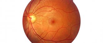
↑ Retina
Retina
- the inner membrane of the eyeball, adjacent to the choroid along its entire length up to the pupil.
The origin of the optic nerve of the retina is the optic disc, which is located 3-4 mm medially (towards the nose) from the posterior pole of the eye and has a diameter of about 1.6 mm. There are no light-sensitive elements in the area of the optic nerve head, so this place does not provide visual sensation and is called a blind spot.
Lateral (to the temporal side) from the posterior pole of the eye there is a spot (macula) - a yellow area of the retina that has an oval shape (diameter 2-4 mm). In the center of the macula there is a central fovea, which is formed as a result of thinning of the retina (diameter 1-2 mm). In the middle of the central fovea lies a dimple - a depression with a diameter of 0.2-0.4 mm; it is the place of greatest visual acuity, contains only cones (about 2500 cells), and there are no rods.
The retina is distinguished by a dentate line, which divides it into two sections: light-sensitive and non-light-sensitive.
The photosensitive section is located posterior to the dentate line and carries light-sensitive elements (the visual part of the retina). The part that does not perceive light is located anterior to the dentate line (blind part).
Structure of the blind part:
- The iris part of the retina covers the posterior surface of the iris, continues into the ciliary part and consists of a double-layered, highly pigmented epithelium.
- The ciliated portion of the retina consists of a double-layered cuboidal epithelium (ciliated epithelium) covering the posterior surface of the ciliary body.
In the retina, there is an outer layer of epithelium containing pigment cells - the pigment part of the retina and an inner layer, devoid of pigment, - the nervous part.
The nervous part (the retina itself) has three nuclear layers:
- outer - neuroepithelial layer consists of cones and rods (the cone apparatus provides color perception, the rod apparatus provides light perception), in which light quanta are transformed into nerve impulses;
- the middle - ganglion layer of the retina consists of the bodies of bipolar and amacrine neurons (nerve cells whose processes transmit signals from bipolar cells to ganglion cells);
- The inner ganglion layer of the optic nerve consists of multipolar cell bodies, unmyelinated axons, which form the optic nerve. The blood supply to the retina is provided by the central retinal artery (a branch of the ophthalmic artery).
At the optic disc, the central retinal artery divides into the superior and inferior papillary arteries. These arteries near the disc again divide dichotomously, and this division extends to the third-order arteries. All ordinal arteries anastomose with each other. The macula macula is surrounded by a thin vascular network in the form of a corolla. In the central fossa, as a rule, there are no capillaries. The blood flow is carried out by the central retinal vein, which leaves the eye in the central part of the optic nerve head next to the central retinal artery.
Perception of light
| This section is missing references to information sources. Information must be verifiable, otherwise it may be questioned and deleted. You may edit this article to include links to authoritative sources. This mark was set on April 3, 2013 . |
We perceive light due to the fact that its rays pass through the optical system of the eye. There, the excitation is processed and transmitted to the central parts of the visual system. The retina is a complex layer of the eye containing several layers of cells that vary in shape and function.
The first (outer) layer is the pigment layer, consisting of densely located epithelial cells containing the black pigment fuscin. It absorbs light rays, contributing to a clearer image of objects. The second layer is the receptor layer, formed by light-sensitive cells - visual receptors - photoreceptors: cones and rods. They perceive light and convert its energy into nerve impulses.
The human retina contains about 130 million rods and 7 million cones. They are located unevenly: in the center of the retina there are predominantly cones, further from the center there are cones and rods, and in the periphery rods predominate.
Cones provide perception of the shape and color of an object. They are insensitive to light and are excited only in bright light. More cones around the fovea. This area of cone accumulation is called the macula macula. The macula, especially its central fovea, is considered the place of best vision. Normally, the image is always focused by the optical system of the eye on the macula. At the same time, objects that are perceived by peripheral vision are distinguished worse.
The rods have an elongated shape, do not distinguish color, but are very sensitive to light and therefore are excited even in low, so-called twilight, lighting. Therefore, we can see even in a poorly lit room or at dusk, when the outlines of objects are barely different. Due to the fact that the rods predominate on the periphery of the retina, we are able to see “out of the corner of the eye” what is happening around us.
So, photoreceptors perceive light and convert it into the energy of a nerve impulse, which continues its path in the retina and passes through the third layer of cells formed by the connection of photoreceptors with nerve cells that have two processes (they are called bipolar). Next, information is transmitted along the optic nerves through the midbrain and diencephalon to the visual areas of the cerebral cortex. On the lower surface of the brain, the optic nerves partially overlap, so some information from the right eye enters the left hemisphere and vice versa.
The place where the optic nerve exits the retina is called the blind spot. It lacks photoreceptors. Objects whose images fall on this area are not visible. The area of the blind spot of the human retina (normally) ranges from 2.5 to 6 mm².
↑ Optic nerve
The processes of visual perception occurring in the eye are an integral part of brain activity. Light rays from the objects in question, passing through the cornea, aqueous humor of the anterior chamber, pupil, posterior chamber, lens, vitreous body, enter the retina, causing excitation of its nerve elements. The nerve elements of the retina form a chain of three neurons:
- 1st neuron - light-sensitive cells (rods and cones) that make up the receptor of the visual analyzer;
- 2nd neuron - bipolar neurocytes;
- The 3rd neuron is ganglion neurocytes, the processes of which continue into the nerve fibers of the optic nerve.
The optic nerves from the right and left eyes, leaving the sockets through the eye openings, approach the lower surface of the brain, where in the area of the sella turcica they merge with each other, forming a partial decussation - chiasmus. Only the parts of the nerve coming from the medial halves of the retina are crossed; the lateral parts of the nerve are not crossed.
After partial decussation of the optic nerves in the area of the chiasm, the right and left optic tracts are formed. The right optic tract contains uncrossed fibers from the right (temporal) half of the retina of the right eye and crossed fibers from the right (nasal) half of the left eye. The left optic tract contains uncrossed fibers from the left (temporal) half of the retina of the left eye and crossed fibers from the left (nasal) half of the right eye.
Both visual tracts are directed to the subcortical visual centers (superior colliculus, geniculate bodies, cushion of the visual thalamus, hypothalamus), where the peripheral part of the visual pathway ends. The central part of the visual analyzer begins from the cells of the subcortical visual centers, the axons of which pass through the posterior third of the posterior limb of the internal capsule to the cortical center of vision, located in the occipital lobes of the brain, where light perception occurs, as well as the formation of visual images.
↑ Lens
The lens, together with the cornea, aqueous humor and vitreous body, constitutes the optical (refractive) system of the eye.
Appearance.
The lens has the form of a biconvex lens with a diameter of 9-10 mm and a thickness of 4 mm; its anterior, less convex surface is adjacent to the iris, and its posterior, more convex surface is adjacent to the vitreous body. The central points of the front and rear surfaces are called the anterior and posterior poles, respectively. The peripheral edge where both surfaces meet each other is called the equator. Both poles are connected by the axis of the lens.
Structure
. The lens is enclosed in a thin capsule, the anterior part of which is lined with single-layer cuboidal epithelium. The posterior part of the capsule is thinner than the anterior one and has no epithelium. The lens is held in its position by the zonular ligament, which consists of many smooth and strong muscle fibers running from the lens capsule to the ciliary body, where these fibers lie between the ciliary processes. Between the fibers of the ligament there are fluid-filled spaces that communicate with the chambers of the eye.
The substance of the lens consists of a denser core located in the central part, which, without a sharp boundary, continues into the softer part - the cortex.
Functions
. The lens can automatically change its shape and adapt the eye to clearly see objects located at different distances, i.e. accommodate or participate in changing the refractive power of the eye. When the fibers of the ciliary muscle, innervated by the oculomotor and sympathetic nerves, contract, the zonular fibers relax. At the same time, the tension of the lens capsule decreases and, due to its elastic properties, it becomes more convex, creating conditions for viewing close objects. Relaxation of the ciliary muscle leads to flattening of the lens, creating the eye's ability to see well into the distance.
Lens composition:
water - 65%, proteins - 30%, inorganic compounds (potassium, calcium, phosphorus), vitamins, enzymes, lipids. The lens of young people contains mostly soluble proteins, in the redox processes of which cysteine is involved. Insoluble proteins - albuminoids do not contain cysteine; they contain insoluble amino acids (leucine, glycine, tyrosine and cystine).
Changes in the lens with age:
- cholesterol accumulates, the content of vitamins C and group B decreases, and the amount of water decreases;
- the permeability of the lens bag for nutrients worsens (nutrition is impaired);
- the regulatory role of the central nervous system in maintaining the quantitative ratios of mediators - adrenaline and acetylcholine, ensuring a stable level of nutrient permeability is weakened;
- The protein composition of the lens changes towards an increase in its insoluble fractions - albuminoids and a decrease in crystallins.
As a result of metabolic disorders in the lens, a dense nucleus forms in old age and its clouding occurs - cataracts. With the loss of the elastic properties of the lens, the ability to accommodate decreases, and senile farsightedness, or presbyopia, develops.
The lens does not have nerves or blood vessels, so it has no sensitivity and does not develop inflammatory processes. The lens is nourished by osmosis.
Perception of images of objects
| This section is missing references to information sources. Information must be verifiable, otherwise it may be questioned and deleted. You may edit this article to include links to authoritative sources. This mark was set on April 3, 2013 . |
A clear image of objects on the retina is provided by the complex unique optical system of the eye, consisting of the cornea, fluids of the anterior and posterior chambers, lens and vitreous body. Light rays pass through the listed media of the optical system of the eye and are refracted in them according to the laws of optics. The lens is of primary importance for the refraction of light in the eye.
For a clear perception of objects, it is necessary that their image is always focused in the center of the retina. Functionally, the eye is adapted for viewing distant objects. However, people can clearly distinguish objects located at different distances from the eye, thanks to the ability of the lens to change its curvature, and, accordingly, the refractive power of the eye. The ability of the eye to adapt to clearly seeing objects located at different distances is called accommodation. Violation of the accommodative ability of the lens leads to impaired visual acuity and the occurrence of myopia or farsightedness.
One of the reasons for the development of myopia is overstrain of the ciliary muscles of the lens when working with very small objects, reading for a long time in poor lighting, or reading in transport. When reading, writing or other work, the object should be placed at a distance of 30-35 cm from the eye. Too bright lighting greatly irritates the photoreceptors of the retina. It also harms your eyesight. The light should be soft and not blind the eyes.
When writing, drawing, drawing with the right hand, the light source is placed on the left so that the shadow from the hand does not darken the work area. It is important to have overhead lighting. If you have prolonged eye strain, you should take 10-minute breaks every hour. You should protect your eyes from injury, dust, and infection.
Visual impairment associated with uneven refraction of light by the cornea or lens is called astigmatism. With astigmatism, visual acuity usually decreases, the image becomes unclear and distorted. Astigmatism is eliminated using glasses with special (cylindrical) lenses.
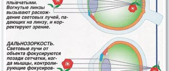
Myopia is a deviation from the normal ability of the optical system of the eye to refract rays, which consists in the fact that the image of objects located far from the eyes appears in front of the retina. Myopia can be congenital or acquired. With natural myopia, the eyeball has an elongated shape, so rays from objects are focused in front of the retina. Objects located at a close distance are clearly visible, but the image of distant objects is fuzzy and blurry. Acquired myopia develops when the curvature of the lens increases due to metabolic disorders or non-compliance with visual hygiene rules. There is a hereditary predisposition to the development of myopia. The main causes of acquired myopia are increased visual load, poor lighting, lack of vitamins in food, and physical inactivity. To correct myopia, glasses with biconcave lenses are worn.
Farsightedness is a deviation from the normal ability of the optical system of the eye to refract light rays. With congenital farsightedness, the eyeball is shortened. Therefore, images of objects located close to the eyes appear behind the retina. Mostly, farsightedness occurs with age (acquired farsightedness) due to a decrease in the elasticity of the lens. If you are farsighted, you need glasses with biconvex lenses.
↑ Vitreous body
The vitreous body is transparent, colorless, elastic, jelly-like. Located behind the lens.
Compound
: about 98% is water, and 2% comes from proteins (proteins - vitrosin and mucin - provide viscosity), mineral salts, glucose, vitamin C, hyaluronic acid, which is associated with mucoproteins and maintains eye turgor. The colloidal substance of the vitreous body has a high surface tension and is similar in composition to the intraocular fluid.
The structure of the vitreous body appears in the form of various shapes and sizes of soft gray ribbons, threads, in which whitish club-shaped and dotted formations seem to be interspersed. These structures, which sway when the eye moves, move along with the transparent areas of the vitreous body.
Structure
. On the anterior surface of the vitreous body there is a depression - the vitreous fossa, corresponding to the lens. The vitreous body is fixed in the region of the posterior pole of the lens, in the flat part of the ciliary body and near the optic nerve head. Throughout the rest of its length it is only adjacent to the internal limiting membrane of the retina. Between the optic disc and the center of the posterior surface of the lens there passes a narrow, downwardly curved vitreous canal, the walls of which are formed by a layer of compacted fibers. In embryos, the vitreous artery passes through this canal.
Functions:
- Supportive function (support for other eye structures).
- Transmission of light rays to the retina.
- Passively participates in accommodation.
- Creates favorable conditions for constant intraocular pressure and a stable shape of the eyeball.
- Protective function - protects the inner membranes of the eye (retina, ciliary body, lens) from displacement due to injury.
There are no vessels and nerves in the vitreous body, therefore its vital activity and constancy of the environment are ensured by osmosis and diffusion of nutrients from the intraocular fluid through the vitreous membrane.
The structure of the eye and its functioning
Our eyes are very complex, and the analogy of a camera is often used to explain how they function. Indeed, both the eyes and the camera (the comparison with a modern digital camera is even closer than with a film camera) use light rays emanating from objects around us to create their images. (You can brush up on the basic concepts of optics by reading the section “Light Rays and Lens Optics”).Light rays enter the eye through the cornea and lens, the role of which in the camera is performed by the lens of the photographic lens. The cornea and lens collect and refract light rays so that they are collected precisely on the retina of the eye and form a clear image on it (similar to the way a camera lens collects rays on a light-sensitive matrix). The amount of light entering the eye (camera) is limited by the size of the pupil, which contracts when there is too much light or, conversely, expands when there is not enough light. In a camera, this function is performed by a diaphragm, which similarly regulates the light flux entering inside. Finally, the image formed on the retina (photosensitive matrix of the camera) enters the brain (camera computer) to create our visual sensations (the image visible on the camera screen).
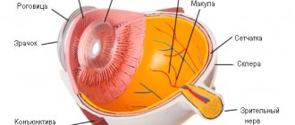
Basic optical structures of the eye
The cornea is the transparent layer of the eye that covers the iris and pupil. Light rays pass through the cornea and are refracted to form an image on the retina. The cornea in the optical sense is a collective lens with an optical power of about +40 diopters (a diopter is a unit of measurement of the optical power of a lens, abbreviated as D).
In addition to its optical function, the cornea performs a protective function, preventing germs, dust and other foreign objects from entering the human eye.
The pupil is a dark, round hole in the center of the eye that contracts or dilates to regulate the amount of light reaching the retina.
The lens is a transparent body of almost spherical shape, capable of changing the curvature of its surface, depending on the distance to the object of observation, to obtain a clear image on the retina. A change in the curvature of the surface of the lens leads to a change in its optical power. In a relaxed state (minimum curvature), the optical power of the lens is about +20.0 D. For reading (or to perform other visual work at close range), the curvature of the lens must increase to obtain a clear image, and its optical power must increase by several diopters. This mechanism for automatically changing the optical power of the lens depending on the distance to the observed object is called accommodation .
The retina is a light-sensitive membrane lining the back surface of the eye. On it, like on the matrix of a digital camera, the eye creates an image of the observed object. The light-sensitive elements of the retina (rods and cones) send signals through the optic nerve to the brain, where visual sensations are formed.
The vitreous humor is a transparent gel-like substance that fills the inner space of the eye between the lens and the retina. Light rays pass through the vitreous body without refraction. From an optical point of view, the main function of the vitreous body is to transmit light rays to the retina without weakening them.
In addition to the indicated structures responsible for the optics of the eye, it also contains others that perform important functions for normal vision. For example, special glands secrete lacrimal fluid (tears), which is necessary to lubricate and moisturize the eye.
Our eye is very complex, and only the coordinated correct “work” of all the structures of the eye as a whole ensures normal vision. Their incorrect functioning leads to vision defects - from slight blurring of the image to complete blindness.
If the optical power of the eye (cornea + lens) does not correspond to its size (that is, if the focus of the optical system cornea + lens is greater or less than the distance to the retina), then this will not allow obtaining a clear image on the retina. In these cases, we are dealing with refractive errors (incorrect refraction of light rays).
There are 3 main refractive errors of the eye - myopia, hyperopia and astigmatism (read the relevant articles in the section “Refractive errors of the eye”). More complex refractive errors are classified as so-called higher-order aberrations (read the article “Higher-order aberrations”).
With age, the lens becomes less elastic and because of this, it loses the ability to change its optical power. Near vision deteriorates (glasses are required for reading). This condition is called presbyopia (read the article “Presbyopia or senile farsightedness”).
If the lens loses its transparency, then this condition is defined as a cataract (read the article “Cataract” in the “Eye Pathologies” section).
Other problems may arise with age: glaucoma, age-related retinal degeneration, and retinal detachment. Read more about these diseases in the section “Eye Pathologies”).
Insufficient lubrication and hydration of the eye leads to the appearance of dry eye .
There are other eye pathologies that affect the quality of vision:
— Amblyopia or “lazy” eye (read the article “Amblyopia or “lazy” eye”)
— Anisometropia (read the article “Anisometropia”)
— Colorblindness
- Strabismus.
To promptly identify emerging problems with vision and eyes, it is necessary to undergo regular examinations with an ophthalmologist (read the article “How to test your vision”).