We receive most of the information about the world around us through our visual organs. It is possible to clearly see objects and navigate in space only if both eyes work in a coordinated manner. With monocular vision, objects that fall within a person’s field of vision are perceived by only one eye, which causes a number of distortions, since normally two pictures are combined into one, and the brain forms an accurate, correct image. You will learn about the characteristics of the disease, its varieties, diagnostic methods used and possible treatment options from this review.
Monocular vision with myopia - definition and nature of the disease
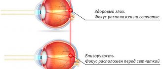
The visual system of a healthy person is binocular or stereoscopic. It forms a three-dimensional image in the brain, which, in turn, is responsible for the qualitative arrangement of objects in the zone of visible space.
The picture obtained with one eye does not give a complete idea of the shape, height, width of the object.
Binocular vision is characteristic not only of humans, but also of fish, birds, and all mammals.
The visual function, in which moving and static objects can be seen only with one eye, is pathological. It is characterized by a narrowing of the boundaries of the field of display of objects, the absence of three-dimensional perception; distant objects can be “lost” in the fog.
There are two types of monocular vision:
- Information constantly enters the brain from only one eye.
- A person sees alternately with his left and right eyes.
In order for the organs of vision to function correctly, the coordinated work of all eye muscles, the correct location of the organs of vision in the frontal and horizontal planes, normal visual acuity, and equal sizes of images on the retina of the two eyes are needed. The transparency of the lens, cornea, vitreous body, and the absence of problems in the parts of the brain responsible for the correct formation of images are also important.
What is monocular and binocular vision?
To navigate in space and see clearly, synchronized, well-coordinated work of both eyes is necessary. The ability to see a picture with both eyes at the same time is called binocular vision. In contrast, there is monocular vision, when the subject can see surrounding objects with each eye separately.
Monocular vision is characteristic of birds, fish and some animals. They are able to perceive visual information with each eye separately. This is one of the tools provided by nature to help living beings survive. With such a vision, the picture turns out to be quite flat: the animal is able to estimate the size of the object and its shape. But due to this feature, the viewing angle increases, which helps to see the potential threat from predators. It is known that the viewing angle of birds reaches 300 degrees: eyes located on the sides allow them to see objects not only from the front and sides, but even from behind. Normally, such vision occurs in all birds, with the exception of some species. For humans, this type of perception is not typical and is considered pathological.
Normal human vision is binocular. That is, we are able to recognize objects and build a visual picture using both eyes at the same time. At the same time, we see a three-dimensional image: human eyes recognize not only shapes and sizes, but also the location of an object in space in relation to other objects. Thanks to this, we distinguish how far an object is from us, whether it is three-dimensional or flat. But there are also vision pathologies in which monocular vision is observed. It comes in two types. In the first case, a person is able to see with only one eye - left or right. Another option is alternate vision, most often found with diplopia. Injuries and congenital diseases are the main causes of this phenomenon.
Binocular vision begins to develop in a child from 3-4 months and completes formation by 12 years. If development occurs late, then there is a high risk of developing strabismus: the organs of vision do not move simultaneously, the child cannot focus with both eyes on an object. If there are other visual abnormalities against this background, such as astigmatism, myopia or farsightedness, the likelihood of strabismus increases.
Modern ophthalmological methods make it possible to restore binocular vision. So, there are surgical and non-surgical methods. The first involves regularly performing special exercises aimed at developing and strengthening the eye muscles, wearing corrective glasses, and following a regimen. The second is surgical intervention when other methods are ineffective.
Types of strabismus
In addition to the ability to see with only one eye, in principle, or with two, but alternately, doctors distinguish the following types of monocular vision:
Monocular strabismus - in this case, the visual function cannot be binocular, since the parallelism of the visual axes is disrupted.
The movements of the eyeballs are not coordinated with each other, so one eye begins to squint to the side.
He hardly takes part in the process of vision, and in the absence of treatment he begins to see worse and worse.
Monocular diplopia (or double vision) – a person sees with different eyes, but the pair of pictures does not combine into a common image. Diplopia develops for various reasons, which are not always associated with ophthalmological diseases or systemic diseases. It can be congenital and acquired. Provoking factors - mechanical injury, problems with the central nervous system, etc.
Transient monocular blindness - an eye disease accompanied by temporary blindness. The condition does not last long - a couple of seconds or up to several minutes. Blindness develops as a result of damage to the optic nerve, retina, and problems with the blood vessels of the brain.
With alternating strabismus, a person can alternately see with the right and left eyes.
The device of a microscope and rules for working with it
A microscope is an optical device that allows you to obtain an inverse image of the object being studied and examine small details of its structure, the dimensions of which lie beyond the resolution of the eye.
The resolution of the microscope gives a separate image of two lines close to each other. The naked human eye has a resolution of about 1/10 mm or 100 microns. The best light microscope improves the capability of the human eye by about 500 times, i.e. its resolving power is about 0.2 µm or 200 nm.
Resolution and magnification are not the same thing. If you use a light microscope to take photographs of two lines located at a distance of less than 0.2 microns, then no matter how you enlarge the image, the lines will merge into one. You can get high magnification, but not improve its resolution.
There are useful and useless increases. By useful we mean such an increase in the observed object that it is possible to reveal new details of its structure. Useless is a magnification in which, magnifying an object hundreds or more times, it is impossible to detect new structural details. For example, if an image obtained using a microscope (useful!) is enlarged many times more by projecting it onto a screen, then new, finer details of the structure will not be revealed, but only the size of existing structures will increase accordingly.
In educational laboratories, light microscopes are usually used, in which microscopic specimens are examined using natural or artificial light. The most common light biological microscopes are: BIOLAM, MIKMED, MBR (biological working microscope), MBI (biological research microscope) and MBS (stereoscopic biological microscope). They provide magnification ranging from 56 to 1350 times. A stereo microscope (MBS) provides a truly three-dimensional perception of a micro-object and magnifies from 3.5 to 88 times.
There are two systems in a microscope: optical and mechanical (Fig. 1). The optical system includes lenses, eyepieces and a lighting device (a condenser with a diaphragm and a light filter, a mirror or an electric light).
Rice. 1. Design of light microscopes:
A - MIKMED-1; B - BIOLAM.
1 - eyepiece, 2 - tube, 3 - tube holder, 4 - coarse aiming screw, 5 - micrometer screw, 6 - stand, 7 - mirror, 8 - condenser, iris diaphragm and light filter, 9 - object table, 10 - revolving device, 11 - lens, 12 - collector lens housing, 13 - socket with lamp, 14 - power supply.
The lens is one of the most important parts of a microscope because it determines the useful magnification of the object. The lens consists of a metal cylinder with lenses built into it, the number of which can vary. The magnification of the lens is indicated by numbers on it. For educational purposes, x8 and x40 lenses are usually used. The quality of a lens is determined by its resolution.
The eyepiece is much simpler than the lens. It consists of 2-3 lenses mounted in a metal cylinder. Between the lenses there is a constant aperture that defines the boundaries of the field of view. The lower lens focuses the image of the object constructed by the lens in the plane of the diaphragm, and the upper one serves directly for observation. The magnification of the eyepieces is indicated on them by numbers: x7, x10, x15. Eyepieces do not reveal new structural details, and in this regard, magnifying them is useless. Thus, the eyepiece, like a magnifying glass, gives a direct, virtual, enlarged image of the observed object, constructed by the lens.
To determine the overall magnification of a microscope, multiply the objective magnification by the eyepiece magnification.
The lighting device consists of a mirror or electric illuminator, a condenser with an iris diaphragm and a light filter, located under the object stage. They are designed to illuminate an object with a beam of light.
The mirror serves to direct light through the condenser and the opening of the stage onto the object. It has two surfaces: flat and concave. In diffuse light laboratories, a concave mirror is used.
The electric light is installed under the condenser in the socket of the stand.
The condenser consists of 2-3 lenses inserted into a metal cylinder. When it is raised or lowered using a special screw, the light falling from the mirror onto the object is condensed or scattered, respectively.
The iris diaphragm is located between the mirror and the condenser. It serves to change the diameter of the light flux directed by the mirror through the condenser to the object, in accordance with the diameter of the front lens of the lens and consists of thin metal plates. Using a lever, you can either connect them, completely covering the lower condenser lens, or separate them, increasing the flow of light.
A ring with frosted glass or a light filter reduces the illumination of the object. It is located under the diaphragm and moves in a horizontal plane.
The mechanical system of the microscope consists of a stand, a box with a micrometer mechanism and a micrometer screw, a tube, a tube holder, a coarse aiming screw, a condenser bracket, a condenser moving screw, a revolver, and a sample stage.
The stand is the base of the microscope.
A box with a micrometer mechanism, built on the principle of interacting gears, is fixedly attached to the stand. The micrometer screw serves to slightly move the tube holder, and, consequently, the lens over distances measured in micrometers. A full turn of the micrometer screw moves the tube holder by 100 microns, and a turn of one division lowers or raises the tube holder by 2 microns. To avoid damage to the micrometer mechanism, it is allowed to turn the micrometer screw in one direction by no more than half a turn.
Tube or tube is a cylinder into which eyepieces are inserted from above. The tube is movably connected to the head of the tube holder; it is fixed with a locking screw in a certain position. By loosening the locking screw, the tube can be removed.
The revolver is designed to quickly change lenses that are screwed into its sockets. The centered position of the lens is ensured by a latch located inside the revolver.
The tube holder carries the tube and the revolver.
The coarse aiming screw is used to significantly move the tube holder, and, consequently, the lens in order to focus the object at low magnification.
The object table is designed to place the drug on it. In the middle of the table there is a round hole into which the front lens of the condenser fits. There are two springy terminals on the table - clamps that secure the drug.
The condenser bracket is movably connected to the micrometer mechanism box. It can be raised or lowered by a screw that rotates a gear that fits into the grooves of a comb-cut rack.
Rules for working with a microscope
When working with a microscope, you must follow the operations in the following order:
1. You should work with a microscope while sitting;
2. Inspect the microscope, wipe the lenses, eyepiece, mirror or electric light from dust with a soft cloth;
3. Place the microscope in front of you, slightly to the left, 2-3 cm from the edge of the table. Do not move it during operation;
4. Open the diaphragm completely, raise the condenser to its highest position;
5. Always start working with a microscope at low magnification;
6. Lower the lens 8- into the working position, i.e. at a distance of 1 cm from the slide;
7. Set up illumination in the field of view of the microscope using an electric light or a mirror. Looking into the eyepiece with one eye and using a mirror with a concave side, direct the light from the window into the lens, and then illuminate the field of view as much as possible and evenly. If the microscope is equipped with an illuminator, then connect the microscope to the power source, turn on the lamp and set the required brightness;
8. Place the microspecimen on the stage so that the object being studied is under the lens. Looking from the side, lower the lens using the macroscrew until the distance between the lower lens of the lens and the microspecimen becomes 4-5 mm;
9. Look into the eyepiece with one eye and rotate the coarse aiming screw towards yourself, smoothly raising the lens to a position at which the image of the object can be clearly seen. You cannot look into the eyepiece and lower the lens. The front lens may crush the cover glass and cause scratches;
10. Moving the specimen by hand, find the desired location and place it in the center of the microscope’s field of view;
11. If the image does not appear, then you must repeat all operations in steps 6, 7, 8, 9;
12. To study an object at high magnification, you first need to place the selected area in the center of the microscope field of view at low magnification. Then change the lens to 40x, turning the revolver so that it takes the working position. Using a micrometer screw, obtain a good image of the object. There are two marks on the micrometer mechanism box, and on the micrometer screw there is a point that must always be between the marks. If it goes beyond their limits, it must be returned to its normal position. If this rule is not followed, the micrometer screw may stop working;
13. After finishing work with high magnification, set the magnification to low, raise the lens, remove the specimen from the work table, wipe all parts of the microscope with a clean napkin, cover it with a plastic bag and put it in a cabinet.
Biological stereoscopic microscope MBS-1 (Fig. 2) provides a direct and three-dimensional image of an object in transmitted or reflected light. It is designed for studying small objects and dissecting them, as it has a large working distance (distance from the cover glass to the front lens).
Rice. 2. Design of the MBS-1 microscope:
1 - eyepiece, 2 - coarse aiming screw, 3 - stand, 4 - mirror, 5 - stage, 6 - stand, 7 - optical head, 8 - lens, 9 - magnification switch handle, 10 - binocular attachment, 11 - lamp .
The main part of the microscope is the optical head. A lens is mounted in the lower part of it, consisting of a system of lenses that can be switched using a handle and thereby change the magnification. Lens magnifications are indicated by numbers on the handle - x0.6, x1, x2, x4, x7. There is a dot on the head body. To set the required lens magnification, you need to align the number on the handle with the dot on the head body.
A binocular attachment is installed on the top of the head. The eyepieces have magnifications of x6, x8, x12.5. To set a distance between the eyepieces that is comfortable for the eyes, you need to move the tubes apart or move them.
A bracket with a rack and pinion movement mechanism is attached to the rear wall of the head housing. The head housing is raised and lowered by rotating the screw. The bracket is placed on a stand attached to the stand.
To work in transmitted light, a light reflector with mirror and matte surfaces is built into the stand body. There is a window on the front side of the case for access to daylight. For artificial lighting, a lamp is used, which is inserted either into a hole on the back of the housing (for transmitted light) or into a bracket mounted on the lens (for reflected light).
The table is installed in a round window on the upper surface of the stand body. It can be either glass (in transmitted light) or metal, with white and black surfaces (in reflected light).
Symptoms
With monocular vision, the patient receives information about the surrounding world, the shape of the visible object, its size, height, width in a distorted form, since only stereoscopic vision allows one to see the world in a three-dimensional manner, correctly. Benefits of binocular vision:
- spatiality of the image, volume, relief;
- expanding the boundaries of vision;
- improvement in the acuity of visual function (binocularity is characterized by binocular summation - the visual function of two eyes is always higher than each one separately);
- determining the distance between visible objects.
These advantages are important at the everyday level and in a number of professions. A person with monocular vision cannot be a professional driver, pilot, or surgeon. Monocular vision is a serious pathology that requires complex diagnosis and proper treatment.
Monocular vision is often a symptom of a serious systemic disease. A thorough, in-depth diagnosis is needed.
Diagnostic methods
To determine monocular vision, various methods are used, some of which can be done independently. Let's look at them.
Sokolov's experience
Another name for the strabismus test is the binocularity test with a hole in the palm. Take a small piece of paper, make it like a spyglass and place it on the sore eye. Turn your left hand with your palm facing your face, placing it from your left eye at a distance of about 15 cm.
If vision is binocular, a hole will be visible in the palm of your hand, through which you can see the same picture that is visible in the paper tube. If there are pathological changes in the visual system, there will be no “hole” in the palm.
Reading experiment
Take a pen/pencil and a book. Place the pen against the background of the open pages of the book so that it covers the lines on the sheet. Start reading closed lines - if you can do this without “peeping”, everything is in order, your vision is normal, binocular. If you have to peep, or you simply can’t read anything, there are disturbances, you can talk about monocular visual function.
Kalfa method
Kalf's method is a miss trial. Take a couple of thin, long objects like a pencil. Hold the first horizontally in your right hand, and the second vertically in your left. Spread your arms in different directions and try to connect the ends of the pencils. With monocular pathology, a person always misses because he cannot give an accurate assessment of the distance between objects and the three-dimensionality of the image. Read about the prevention of myopia in children at this link.
The slip test can also be easily performed independently at home. If necessary, additional diagnostics can then be carried out in an ophthalmology office. This article will tell you about laser correction of myopia.
Four point color test
This technique is used by ophthalmologists, since it involves the need to accurately decipher the meanings. For the test, take a red one, a white one and a couple of green ones. The patient puts on glasses with green and red lenses and begins to look through them at multi-colored objects. If vision is binocular, the subject sees only red and green objects; he perceives white as green-red (or red-green depending on the dominant eye). When monocular, the shade of the ball matches the color of the lens. This technique is highly accurate and can be used to make a diagnosis.
Prism test
The test requires a prism with a power of 8 to 10 diopters. The doctor places the prism in one eye for 20-30 seconds, while observing the other eye. Then the prism is removed, the ophthalmologist looks at the movements of the previously covered eye. In 20-30 seconds, the prism slightly changes the direction of the light rays, as a result of which the image on the retina shifts. When the prism disappears from the field of vision, the picture doubles until the apple takes on its original shape.
And this will happen only if normal binocular vision is developed. When monocular, the gaze will wander. The study must be carried out on both eyes to determine the leading one. Binocular vision is restored by various methods (surgery, therapy, eye gymnastics), the choice of which is made taking into account the specific ailment. The doctor must determine the exact cause of the lack of spatial vision. This material will tell you about the treatment of hidden strabismus in children and adults.
The prism test is the optimal method for diagnosing monocularity in childhood.
Methods for determining monocular visual weakness
Description
The methods for identifying this type of simulation are based on the creation of an illusion in the subject, when the vision of a pseudo-blind eye is determined
, and it seems to the subject that he sees healthy. This is achieved by using the physiological characteristics of the organ of vision. It is known that the visual analyzer, with the same clarity and brightness of the image, is not able to distinguish between right and left images. A person is always convinced that he sees objects on the right with his right eye, and on the left with his left eye. You can create conditions under which the right eye will see objects on the left, and vice versa. A number of devices have been proposed for this purpose.
1. Device proposed by Bertele
, in a box 35 cm long, holes are made on one side for the eyes, and objects for vision research (letters, numbers, signs, drawings) are inserted on the opposite side. In the middle of the box there is a partition with three holes (one in the middle and two on the sides). Using a special device, you can close the middle one (c) or two side ones (a-c) at once. If you close the middle hole and look with both eyes through the side holes at the inserted signs, then indeed, the right eye sees the right half, and the left eye sees the left, as shown in the dotted line in the figure. If you close the side holes and leave one middle one open, the visual axes will change their direction, the eyes will converge and the right will see the left half of the table, and the left will see the right (as shown in the figure with dashes). The malingerer, not knowing these features, will read the signs with a pseudo-blind or amblyopic eye, believing that he is reading with a healthy eye.
Half an hour before the examination (after determining visual acuity and refraction), a drop of eserine is instilled into one eye, and distilled water into the other, and then the vision is examined
.
If the subject shows the same visual acuity, as in binocular examination, simulation has been detected, because now the normal eye, thanks to a spasm of accommodation, has become myopic and, of course, cannot have the visual acuity that it gave before the installation of eserine. Reading the signs of the table in this way is performed by the worst eye, on which adblyopia was simulated. The same experiment can be carried out with atropine, but then vision is examined not with the help of tables, but with the help of small print, 2. Determination of feigned blindness using a stereoscope
. Two complementary pictures are inserted into the latter, for example: for the right eye the picture shows a cage, and for the left eye a bird. Normal eyes looking through a stereoscope will see a bird sitting in a cage; if one eye is blind, either a cage or a bird will be visible (depending on which eye is blind). This experience can be varied by replacing some pictures with others or entire phrases, for example, for the left eye: “I see,” and for the right: “good.” If the subject reads the entire phrase, it means he is looking with both eyes, otherwise he would read either “I see” or the word “good.”
3. Use of glasses of different optical powers
When carrying out this group of methods, it is necessary to carefully monitor the subject
, since sometimes for self-control the patient closes the healthy or diseased eye, which makes assessment difficult.
To create favorable conditions for the study, it is advisable to begin determining vision with a healthy eye.
A weak lens is placed in the trial frame in front of the eye, the vision of which,
(-0.26 D), and in front of the healthy eye there is a convex glass (14 D). Then the subject is asked to read the test signs. If he copes with the task, then he is feigning partial loss of vision.
The doctor performs full vision correction on the subject
y, then he is given the illusion that only the healthy eye is being examined.
To do this, a -1.0 D lens is inserted into a trial frame with the healthy eye and the patient is asked, “Is it true that he has now become worse at seeing the proposed tests?” Then they place 1.0-2.0 D and show signs to check visual acuity and ask the subject: “And now it’s even worse, isn’t it?” The visual acuity of the amblyopic eye is determined. Then they remove the glass and ask: “Can you see better now?” The pretender will answer that his vision has improved.
4. Definition of simulation using colored glasses
The subject is put on glasses with red glass in front of the healthy eye and with green glass in front of the pseudo-blind one.
. Then, on a sheet of white paper, a few words (letters) or his last name are written in red pencil and they are asked to read what was written. If the subject reads, he does so with the eye in front of which a green glass is placed, i.e. one who, according to his statement, is blind or severely amblyopic and previously he could not read with it, since a healthy eye through a red glass cannot see what is written in red pencil on white paper.
The object of study is a table with a black background
, which depicts letters of different sizes, colored red, green and white. The subject is put on glasses with red glass in front of the healthy eye and gray in front of the supposedly blind one. If he is truly blind in that eye, he will only read red and white letters. If he also reads green ones, then this can only be done with an amblyopic eye. This study determines not only the simulation of amblyopia, but also the visual acuity of this eye, since the letters have different sizes, corresponding to the Snellen tables.
There are a special kind of tables on which the letters consist of two parts that complement each other
(both parts together make up one letter, and one part separately makes up another, for example, the letter P is black, and a red hook is attached to it, so that the letter B is formed as a whole, or an additional red stick is attached to the black letter G and the whole is formed letter P, etc.) . If a red glass is left in front of the sighted eye, and gray glass in front of the supposedly blind eye, the subject will only read the letter that is black in color. If he reads the full letter, then he sees it with his “blind” eye and thereby reveals his simulation.
Using colored glasses, you can also determine not only simulation, but also visual acuity
. The study is based on the property of colored glass: colored glass transmits only those rays of the solar spectrum that match or closely match its color. All other colors are delayed and become black or dark gray. Therefore, if you look at red and green letters drawn on a white background through red glass, then the first ones disappear, since they now merge with the red background, and the second ones are visible in the form of black letters, while green letters disappear on a black background, since they are made black as the background of the table, only the red letters are visible. If you look at the same tables through green glass, then green letters disappear on a white background, and red letters disappear on a black background. Successful completion of this exercise depends on matching the color of the glass and the color of the signs.
An important place in determining simulation and aggravation is occupied by differential diagnosis with such pathological conditions of the nervous system as
hysteria, neurosis and others, in which there is an involuntary exaggeration of actual phenomena or a generalization of imaginary violations.
In some cases, injuries to the organ of vision and skull lead to the development of post-traumatic neurosis, so it is very important to promptly identify the presence of a pathological condition
. When simulating post-traumatic changes, there is a clear tendency to preserve and repeat fictitious symptoms; post-traumatic neurosis is characterized by the fact that, along with the phenomena of regression, there is a desire to improve the condition.
It is quite difficult to distinguish hysteria from a fictitious disease, which can lead to false blindness in one or both eyes, double vision, multiple images, changes in the field of vision, etc.
Differentiation of hysteria from fictitious eye diseases is based on the following principles:
1. The simulator is trying
mislead others, but not himself, and the patient subconsciously and without purpose misleads himself and others.
2. When observing a malingerer, it is noted
that he often performs his role with some tension, and when he is supposedly left alone, the tension caused by self-defense and self-control is reduced. Patients with hysteria remain tense even without being supervised.
3. The malingerer tries to avoid various studies
, and the patient willingly and happily undergoes research and even experiences pleasure from demonstrating his suffering.
4. If the malingerer is proven
that the phenomena are imaginary and the damage is intentional, this will cause severe embarrassment or firm defense of imaginary complaints, while the patient with hysteria happily accepts the message about the improvement of the condition.
Situational tasks
Task 1.
A 43-year-old mechanic complains of decreased vision in his right eye, which, in his opinion, prevents him from doing his previous job. He considers the cause of blindness to be a work-related injury to the right Eye received a year ago, and therefore insists on establishing a disability group.
When tested, visual acuity in the right eye and left is 1.0.
Objectively: the refraction of both eyes is emmetric. Examination of the right eye revealed no pathology; the left eye is healthy.
What methods will you use to determine true visual acuity?
Task 2.
A 23-year-old patient, a draftswoman, accidentally pricked her left eye with a compass at work 3 months ago. Currently, he is experiencing decreased vision in his left eye.
An objective examination revealed visual acuity of the right eye to be 1.0, and of the left eye to count fingers near the face.
The right eye is healthy.
Left eye. On the cornea at 6 o'clock a round cloudiness in the form of a “cloud” is detected. The anterior chamber is of moderate depth, the contents are transparent. The iris pattern is not changed. The lens is transparent. On the fundus: the optic nerve head (OND) is pale pink, the boundaries are clear, the caliber of the vessels is not changed.
How do you determine the visual acuity of a pseudo-blind eye?
Problem 3
. A 30-year-old patient notes loss of vision in both eyes after a traumatic brain injury suffered 2 years ago.
When tested, it shows visual acuity of the right eye - 0, left eye - 0.
An objective examination of both eyes: the cornea is transparent, the anterior chamber is of moderate depth, the pupils are wide, d = 0.8 mm, does not react to light. The lens is transparent. No pathology was detected in the fundus.
When observing behavior, tension is noted; the patient walks with outstretched arms and “bumps” into objects. When blindfolded, actions become even more uncertain.
The patient reluctantly agrees to research.
Your tactics. What methods will you use to make a final diagnosis?
—-
Article from the book: Methods for determining visual acuity simulations | Zhaboyedov G. D., Skripnik R. L.
Treatment - how to correct vision
If a person has poor binocular vision, this often causes the development of strabismus. The disease is diagnosed in children and adults. For correction, techniques such as diploptics and orthoptics are used - a system of methods aimed at restoring normal binocular vision and treating strabismus. Find out about high myopia here.
The work of special binocular cells was first studied by the physiologist Hubel (USA), who was given the Nobel Prize for his discoveries.
The main goal of treatment is to combine images obtained from both eyes to normalize binocular visual function. For this, the synoptophore apparatus is used - it gives a separate representation of the parts of the picture and stimulates the eyes to combine them together. Taking into account the angle of strabismus, the specialist changes the localization of the eyepieces. When the patient learns to connect pictures with his eyes, it will be possible to carry out treatment aimed at consolidating the results.
Diploptics is the final stage in the treatment of strabismus.
It is indicated for children over two years of age and adults.
The essence of diploptics is to cause double vision of an object and thereby develop the ability to independently restore binocular vision. The doctor places a prismatic glass in front of the eyes, which causes double vision. When the glass is removed, vision begins to slowly recover. During therapy, the prisms are changed. The final stage of restoring binocular vision is gymnastics and vitamins for vision. Read about electrical eye stimulation in children here.