Central vision
Description
Central vision should be considered the central portion of visible space.
This vision is the highest and is characterized by the concept of “visual acuity”. Visual acuity
- this is the ability of the eye to perceive separately points located at a minimum distance from each other, which depends on the structural features of the optical system and the light-receiving apparatus of the eye. The angle formed by the extreme points of the object under consideration and the nodal point of the eye is called the visual angle.
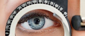
Determination of visual acuity (visometry)
. Normal visual acuity refers to the ability of the eye to separately distinguish two luminous points at a visual angle of 1 minute. It is much more convenient to measure visual acuity not by visual angles, but by reciprocal values, i.e. in relative units. Normal visual acuity equal to one is taken to be the reciprocal of the visual angle of 1 minute. Visual acuity is inversely proportional to the visual angle: the smaller the fenian angle, the higher the visual acuity. Based on this relationship, tables for measuring visual acuity are calculated. There are many versions of tables for determining the severity of fenium, which differ in the test objects, or optotypes, presented.
In physiological optics, there are the concepts of minimally visible, distinguishable and recognizable. The subject must see the optotype
, distinguish its details, recognize the represented sign or letter.
Optotypes can be projected onto a computer screen or display. Letters, numbers, drawings, and stripes are used as optotypes. Optotypes are constructed so that from certain distances the details of the optotype (the thickness of the lines and the spaces between them) are visible at a viewing angle of 1 minute, and the entire optotype is visible at a viewing angle of 5 minutes. The international optotype is the broken Landolt ring
. In Russian ophthalmology, the most common is the Golovin-Sivtsev table, which contains the letters of the Russian alphabet and Landolt rings as optotypes. The table has 12 rows of optotypes. In each row, the sizes of the optotypes are the same, but they gradually decrease from the top row to the bottom. The magnitude of optotypes changes in arithmetic regression. Within the first 10 rows, each row differs from the previous one by 0.1 units of visual acuity, in the last two rows by 0.5 units. Thus, if the subject reads the third row of letters, then visual acuity is 0.3; fifth - 0.5, etc. When using the Golovin-Sivtsev table, visual acuity is determined from 5 m. The lower edge of the table should be at a distance of 120 cm from the floor level.
First, the visual acuity of one eye (right) is determined, then the left eye. The second eye is covered with a shutter. From a distance of 5 m at a viewing angle of 1 minute, details of the optotypes of the tenth row of the table are visible. If the patient sees this row of the table, then his visual acuity is 1.0. At the end of each row of optotypes, the symbol V indicates the visual acuity corresponding to reading this row from a distance of 5 m. To the left of each row, the symbol D indicates the distance from which the optotypes of this row differ at a visual acuity of 1.0. Thus, the first row of the table with visual acuity equal to 1.0 can be seen from 50 m.
To determine visual acuity, you can use the Siellen-Deuders formula
visus = d/D, where d is the distance from which the subject sees a given row of the table (the distance from which the study is carried out), m; D is the distance from which the subject should see this row, m.
Using the above formula, you can determine visual acuity in cases where the study is carried out in an office length, for example, 4.5 m, 4 m, etc. If the patient sees the fifth row of the table from a distance of 4 m, then his visual acuity is equal to: 4/10 = 0.4.
There are people with higher visual acuity
- 1.5; 2.0 or more. They read the eleventh or twelfth row of the table. A case of visual acuity with the naked eye is described: the subject could distinguish the satellites of Jupiter, which are visible from the Earth at an angle of 1 second. If visual acuity is below 0.1, the subject must be brought closer to the table until he sees its first line.
Since the thickness of the fingers approximately corresponds to the width of the strokes of the optotypes in the first row of the table, it is possible to demonstrate spread fingers to the subject
(preferably against a dark background) from different distances and, accordingly, determine visual acuity below 0.1 also using the above formula. If visual acuity is below 0.01, but the subject counts fingers at a distance of 10 cm (or 20, 30 cm), then visual acuity is equal to counting fingers at a distance of 10 cm (or 20, 30 cm). The patient may not be able to count fingers, but detects the movement of the hand near the face, this is considered the next gradation of visual acuity. The minimum visual acuity is light perception (vis = 1/—) with correct or incorrect light projection. Light projection is determined by directing a beam of light from an ophthalmoscope into the eye from different sides. In the absence of light perception, visual acuity is zero (vis = 0) and the eye is considered blind.
To determine visual acuity in children, use the table of E. M. Orlova
. It uses drawings of familiar objects and animals as optotypes. And yet, at the beginning of the study of visual acuity in a child, it is recommended to bring him close to the table and ask him to name the optotypes.
The table for studying visual acuity is placed in a wooden box open at the front, the walls of which are lined with mirrors on the inside. In front of the table there is an electric lamp, covered at the back with a screen for constant and uniform illumination (Roth-Roslavtsev apparatus). The optimal illumination of the table is provided by a regular 40 W incandescent lamp. A light fixture with tables is mounted on the wall opposite the windows. The lower edge of the illuminator is placed at a distance of 120 cm from the floor. The room where patients are waiting for an appointment and the eye room should be well lit. Currently, test mark projectors are increasingly used to study visual acuity. Optotypes of various sizes are projected onto the screen from a distance of 5 m. The screens are made of frosted glass, which reduces the contrast between the optotypes and the surrounding background. It is believed that such a threshold definition is more conducive to real visual acuity.
To determine visual acuity below 0.1, optotypes developed by B. L. Polyak
in the form of line tests and Landolt rings, designed to be presented at a certain close distance, indicating the corresponding visual acuity. These optotypes are specially created for military medical and medical-social examinations carried out when determining fitness for military service or disability group.
There is also an objective (independent of the patient’s indications) method of determining visual acuity, based on optoclistic nystagmus
. Using special devices, the subject is shown moving objects in the form of stripes or a chessboard. The smallest size of the object that caused involuntary nystagmus (seen by the doctor) corresponds to the visual acuity of the eye being examined.
When determining visual acuity, certain rules must be followed
.
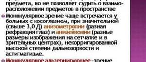
- Examine visual acuity monocularly (separately) in each eye, starting with the right.
- During the test, both eyes should be open, one of them should be covered with a shield made of opaque material. If it is not there, then the eye can be closed with the palm (but not the fingers) of the subject. It is important that he does not press through the eyelids on the closed eye, as this can lead to temporary decrease in vision. The shield or palm is held vertically in front of the eye so that the possibility of intentional or unintentional peeking is excluded, and so that light from the side falls on the open palpebral fissure.
- The study should be carried out with the correct position of the head, eyelids and gaze. There should be no tilting of the head to one or the other shoulder, turning the head to the right or left, tilting it forward or backward. It is unacceptable to squint. In case of myopia, this leads to increased visual acuity.
- When researching, the time factor should be taken into account. In normal clinical work, the exposure time is 2-3 s, in control experimental studies - 4-5 s.
- Optotypes in the table should be indicated with a pointer; its end should be clearly visible; it should be placed exactly with the exposed optotype at a certain distance from the sign.
- The study should begin by showing the breakdown of optotypes in the tenth row of the table, gradually moving to rows with larger characters. In children and people with obviously reduced visual acuity, it is permissible to start testing visual acuity from the top line, showing from top to bottom one character in the line to the row in which the patient is mistaken, after which you should return to the previous row.
Visual acuity must be assessed using a series in which all signs were correctly named
. One error is allowed in the third to sixth rows and two errors in the seventh to tenth rows, but then they are recorded in the visual acuity record. Near visual acuity is determined using a special table, which is calculated at a distance of 33 cm from the eye. If the patient does not see the upper row of the Golovin-Sivtsev table, i.e., visual acuity is less than 0.1, then determine the distance with which he distinguishes the optotypes of the first row. To do this, the subject is brought closer to the table until he sees the first row, and the distance from which he distinguished the optotypes of this row is noted. Sometimes they use cut-out tables with optotypes of the first rad, which bring them closer to the patient.
The presence of vision in a newborn can be judged
by the direct and friendly reaction of the pupils to light, with sudden illumination of the eyes - by the general motor reaction and closing of the eyelids. From the second week, the newborn reacts to the appearance of bright objects in the field of vision by turning his eyes in their direction and can briefly follow their movement. At 1-2 months, the child fixes a moving object with both eyes for quite a long time. From 3-5 months, formal vision can be checked using a bright red ball with a diameter of 4 cm, and from 6-12 months - with a ball of the same color, but with a diameter of 0.7 cm. By placing it at different distances and attracting the child’s attention by swinging the ball, determine visual acuity. A blind child reacts only to sounds and smells.
You can roughly check visual acuity, which is of decisive importance in professional selection, labor and military examination.
Visual acuity may decrease depending on many reasons. They can be divided into three groups.
- The most common cause
is refractive error (nearsightedness, farsightedness, astigmatism). In most cases, visual acuity is improved or completely corrected with the help of glasses. - The second reason for decreased vision
is clouding of the refractive transparent structures of the eye. - The third reason
is diseases of the retina and optic nerve, pathways and visual centers.
It should also be noted that visual acuity changes throughout life, reaching a maximum (normal values) by 5-15 years and then gradually decreasing after 40-50 years.
—-
Article from the book: Eye diseases. Complete Guide | Perederiy V.A.
Article:
Depending on the preservation of visual acuity, several groups can be distinguished among children with visual impairments, characterized by different visual capabilities, different ways of perceiving educational material and orientation in space.
Depending on the degree of visual impairment and methods of perception of educational material, the following groups are distinguished:
a) blind and practically blind (so-called partially seeing) children with visual acuity ranging from O to 0.04, corrected by glasses in the better eye. These children have little or no residual vision. During classes, they mainly use the tactile-auditory method of perceiving educational material, reading and writing in the Braille system. These children learn primarily through touch and hearing. Some of them rely on residual vision when reading and writing. Along with this, if their vision allows them, they master, under certain conditions, the visual method of reading and writing enlarged black and white font. However, residual vision is not sufficiently stable and reliable. It can ensure the correctness and necessary speed of perception of ordinary flat-printed font only when using the sense of touch. Those who are partially sighted are subject to education in special schools for blind children or in special classes for these children at schools for the visually impaired.
During classes, the blind and partially sighted use special teaching aids (published in the Braille system), special technical means, and the necessary didactic material. Partially sighted people also use optics. All this allows you to master the techniques of tactile and visual reading and writing, facilitating the rational use and protection of residual vision;
b) visually impaired children with visual acuity ranging from 0.05 to 0.09, corrected by glasses in the better eye. These children typically have complex visual impairments. Along with a decrease in visual acuity, some of them have a narrowed field of vision and impaired spatial vision. All this complicates the visual perception of educational material. They need to comply with regulated visual load and measures for the protection and rational use of defective vision during classes.
As a rule, they are subject to training in special schools for the visually impaired. When teaching these children, a system of special technical and optical means is used (overhead orthoscopic, line magnifying glasses, etc.) used to correct and compensate for impaired and underdeveloped functions. However, their vision is not stable enough. Under unfavorable conditions it worsens. In this regard, students also need a lighter regime of visual load, skillful alternation of activity, work and rest;
c) visually impaired children with visual acuity from 0.01 to 0.04, corrected with optical glasses in the better eye. Under certain conditions, they can freely read with the help of sight, write, visually perceive objects, phenomena and processes of reality, and visually orient themselves in a large space. Due to the need to use special methods and technical means of teaching, and to comply with certain hygienic requirements, such children are also educated in special schools for the visually impaired. However, some of them, provided the necessary conditions are provided, are able to successfully study in a public school;
d) children with central visual acuity of 0.4-0.5 or higher, corrected with optical glasses. These children do not have pronounced secondary deviations in mental development. Such children are subject to education in regular public school conditions. However, some gentle treatment should be observed in relation to them.
Thus, we see that, depending on the varying degrees of impairment of central visual acuity, students use different ways of perceiving educational material, which presupposes the use of differentiated forms of their education in a special school for the visually impaired and in a public school.
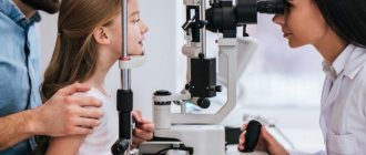
Eye examination methods
Eye examination
includes accurate determination of the patient's visual acuity and refraction, measurement of intraocular pressure, examination of the eye under a microscope (biomicroscopy), pachymetry (measurement of corneal thickness), echobiometry (determining the length of the eye), ultrasound examination of the eye (B-scan), computed keratotopography and a thorough examination retina (fundus of the eye) with a wide pupil, determination of the level of tear production, detailed examination of the patient’s field of vision. If necessary, the scope of the examination can be expanded.
As a rule, the doctor begins the examination by obtaining information from you about your health status and family diseases. Then the doctor will most likely ask you to name the letters or numbers that he points to on a table of letters and numbers arranged in rows of gradually decreasing sizes. It allows you to determine the acuity of central vision.
If you wear contact lenses, your vision will be checked at this time. Additionally, your doctor will use a special instrument called a lensometer to check your glasses to confirm whether your prescription was written correctly.
If the chart test indicates that you need a prescription or contact lens prescription, your doctor will measure your eye's refractive error, or focusing, using instruments that contain a variety of contact lens strengths. To confirm the readings and find out which lenses provide the best vision, you will be given lenses of varying strengths to test.
Peripheral vision examination
External eye examination
Visual coordination test
Biomicroscopy
Dilated pupil examination
Fundus examination
Data from studies of natural and corrected visual acuity
using a slit lamp should be assessed using the symbols of the Snellen or Sivtsev tables. If the patient cannot distinguish large letters, then vision is assessed by determining the number of fingers. Then the patient's perception of finger movements and, finally, the ability to distinguish light from darkness are determined.
Determination of fields of view
performed using a contrast study, which can be used to estimate the approximate degree of visual field loss.
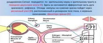
Study of pupil reaction to light
(indirect and involuntary) allows you to assess the state of the visual tract. The absence of a direct light reflex is observed with unilateral damage to the optic nerve and occlusion of the central retinal artery.
A patient with glaucoma has an arcuate scotoma (an isolated area in which vision is weakened or absent along the nerve fibers, along the edges of the optic nerve head). Central scotoma can be observed with optic neuritis. Bitemporal hemianopia/homonymous hemianopia (loss of the right or left halves of the visual fields) and quadrant hemianopia (loss of one quadrant of the visual field of one or both eyes) is observed in patients with neurological pathology.
Intraocular pressure
, as a rule, is measured using a non-contact tonometer. If necessary, intraocular pressure is measured using a Maklakov contact tonometer or Goldman tonometer. To exclude glaucoma, it is possible to conduct computer perimetry, that is, a study of visual fields.
Before any surgical intervention, a refractive examination
, which includes: determination of visual acuity without correction and with optimal correction, biomicroscopy, ophthalmoscopy, tonometry, refractometry (using an autorefractometer), computer topography of the cornea on a computer topograph, ultrasound biometry, ultrasound pachymetry. The data obtained during diagnosis is used by the surgeon when performing excimer laser correction.
Before refractive surgery, patients undergo pachymetry
a device for measuring the thickness of the cornea, which allows you to calculate the maximum permissible depth of laser exposure, which in cases of very high degrees of myopia determines how completely the correction can be carried out.
Any microsurgical or laser interventions are preceded by a complete comprehensive computer diagnostic vision examination
. The examination identifies the range of existing problems and determines treatment tactics.
Any disease of the visual organs requires a special approach to treatment, including at the stage of determining the diagnosis. Many ophthalmological diseases have similar symptoms, so even experienced professionals cannot make a diagnosis, much less determine the extent of the disease, without a thorough examination using specialized equipment. At an early stage, it is advisable to carry out diagnostics as planned. Below is brief information about the main methods for diagnosing eye diseases.
Visometry
– vision testing using special tables and trial lenses. Currently, their place is gradually being taken by more modern projectors and phoropters. This study is subjective, as it largely depends on the state of the visual organs and the body at the time of diagnosis. One of the types of viziometry is the selection of spectacle lenses.
Autorefractometry
- a study of refraction and the strength of curvature of the cornea of the eye, carried out using an automatic refractokeratometer. Using this device allows you to fully automate the process. All that is required of the patient is to look at a special mark, thereby keeping the head in a static position.
Keratometry
– measurement of the curvature of the anterior surface of the cornea. This test is used to calculate the optical power of an artificial lens, and also detects and measures astigmatism. Previously, keratometry was performed manually using a ruler, but in our center to conduct this study, a special device is used - a keratometer.
Tonometry
– measurement of intraocular pressure. Used to diagnose glaucoma and disorders of the optic nerve. The tonometry technique is simple: first, drops are injected into the patient’s eye to reduce the sensitivity of the cornea, and then an ophthalmic tonometer is applied to its surface. The device applies slight pressure to the cornea, and the result of the study is its reverse resistance.
Pachymetry
– measuring the thickness of the cornea at various points. The study is carried out using ultrasound. It is used to diagnose corneal dystrophy and is actively used before laser correction and to assess the condition of the eyes after corneal transplantation.
Biomicroscopy
– visual examination of the cornea, lens, iris and other parts of the eye by creating a contrast between illuminated and unlit areas. Allows you to assess the condition of optical media, blood vessels and optic nerves.
Ophthalmoscopy
– The most common examination using a slit lamp, during which the eyeball and adnexa are examined. It is carried out to diagnose diseases of the cornea, iris, lens and vitreous body.
Gonioscopy
- examination of the anterior chamber angle using a slit lamp and a gonioscope - a special device that is a system of mirrors. It is carried out to detect pathologies of the anterior chamber angle (tumors, foreign bodies, etc.)
Keratotopography
– examined eyes by scanning its surface, the main purpose of which is to determine the sphericity of the cornea. The scanning is carried out using a laser beam, and the results are a topographical “map”, on which areas of different heights are indicated in different colors.
Critical flicker fusion frequency
– a research method used to diagnose pathological processes in the visual system. FCSM allows you to determine the degree of eye fatigue based on the functional state of the cortical part of the visual analyzer and the central nervous system, as well as the degree of inertia of mental processes.
To the list of techniques