What is this
The apple itself consists on the outside of connective tissue, almost protein-free: the white part is almost pure collagen, providing a reliable rounded shape, with a transparent insert in front - the cornea. So transparent that there are not even blood vessels there, the cornea is nourished diffusely due to the suffering of tears and internal aqueous humor. The inside of the eye is lined with the choroid, on which Her Majesty the retina rests.
The cornea is not only a protective cap for the pupil, but also a convex lens: due to its curvature, it does most of the work (two-thirds) in focusing the light beam, which is then corrected by the lens (the remaining third). Thanks to this, laser vision correction operations have become possible, in which the cornea is stratified and its middle layer is given the necessary reduction in curvature by evaporating excess cells with a laser.
Although the cornea does not have blood vessels, it has almost the most sensitive innervation, the sensitivity of which is 600 times greater than the skin and 40 times greater than the dental pulp. [1] That is why neurologists like to check the corneal reflex of comatose patients and those pretending to be them by poking a cotton wool/paper needle directly into the eye.
Slurry
The vitreous body inside the eye itself performs a similar function as the gel in ultrasound research: it prevents light from fading due to the transition of the boundaries of media with different densities/refractive index. A transparent gel-like substance supported by protein fibers (essentially jelly) ensures seamless transit of the light beam from the lens to the retina, and if instead there was air, then interference and diffraction even in such a 3-centimeter area would ruin everything.
In the center of the body, the hyaloid canal runs from the lens to the optic disc, formed by the vitreous membrane. They say it matters for accommodation. [2]
Entoptic phenomena
Video from TED
The vitreous body itself is not of particular interest, but almost everyone is familiar with it in absentia (the older, the more likely it is): if you look at something light and monochromatic, you can see the slow movement following the gaze of some translucent “flies” " in sight. [3] These floaters (“floaters”, “muscae volitantes”) - damage to the gel fibers due to any reasons, usually of no significance, but sometimes (for example, with eye injuries) quite large blood clots can form there, which will obscure the view. In such cases, the body is removed and prosthetics are applied with anything: from saline solution to special artificial gels.
In addition to the described flies, vision often shows another attraction: the blue field phenomenon (Shearer’s phenomenon) - many randomly moving flickering “worms”, especially noticeable when looking at a clear sky. This is the visible movement of leukocytes along the capillaries of the retina - yes, yes, that’s how easy it is to see your own cells without a microscope! Unlike red red blood cells, immune cells are much larger in size and transmit blue light better, and the dark tail visible behind them is an accumulation of red blood cells that give a shadow in the blue spectrum.
Hole
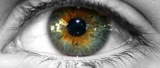
Having peeled off the cornea from the eye, we will see the sphincter of the eye - the colored iris, a membrane with circular (compress) and radial (uncompress) muscles that change the diameter of the pupil depending on lighting conditions and the body’s reaction. Actually, the iris is part of the choroid, but it is logical to talk about it here.
Works like a diaphragm in a camera:
- Miosis: more light - smaller hole, the pupil narrows in a couple of seconds; Suspiciously constricted pupils in the dark may indicate heroin.
- Mydriasis: “fear has big eyes” - fear gives a reaction from the sympathetic nervous system with the release of adrenaline, which causes dilator spasm and dilation of the pupil. It also expands in the dark and under stimulants (cocaine), expansion takes longer, up to 5 minutes.
Don't confuse these two terms on uh
very simple: miosis is small (4 letters), and mydriasis is large.
Lens
Directly under the iris lies a cunning adaptive lens - the crystalline lens (also known as “lens” - lens, objective
), which is not even crystal at all, but quite proteinaceous and not very hard, although elastic.
Transparency is provided by special proteins crystallins
, and it needs elasticity in order to collapse back after being compressed by another intraocular muscle - the ciliary muscle, which changes curvature.
Again, as in a camera: the object is far away - the focal length is at infinity (the lens is relaxed, stretched and thin, the curvature is less), the object is close - the focal length decreases (the lens is tense with the muscle, thick and curved) (spoiler:
Here the analogy with the camera is incomplete, since the technique uses moving the lens away/closer, and not changing the curvature - this is how the eyes of cephalopods, for example, octopuses, work
)
. Actually, this is called “accommodation,” that is, adaptation to changes in the distance of a visible object; Without such a thing, we could clearly see only distant objects, close ones would be blurred.
Like the cornea, the lens does not have feeding vessels, but lives on aqueous humor, although in intrauterine development it is sponsored by the vitreous artery running through that central hyaloid canal. Because of this, the lens tissue is poorly understood by the immune system, which, if its capsule is disrupted, leads to autoimmune problems.
Previously, there was a popular pseudoscientific theory that accommodation is carried out by changing not the curvature of the lens, but the shape of the eyeball itself, which gave rise to the heretical Bates-Shichko system for correcting myopia. As expected, it works at the placebo level, since visual acuity can vary up to 30% during the day depending on the tension of the intraocular muscles.
There are two main problems with the lens: cataracts (clouding) and presbyopia (age-related farsightedness due to decreased flexibility).
Retina
The meatiest part is a ten-story dedicated server with three layers of brain cells carried out from the cranial cavity directly into the eye, which sorts information already on data input: about 130 million light-sensitive cells are ultimately reduced to one nerve of about a million fibers (for comparison: in the auditory nerve only 30 thousand). This work is done by a network of millions of regulatory (bipolar and ganglion) neurons, summing, converting and compressing gigabits of data per second. [4]
The layers of the retina lie turned inside out with the working surface inward: nerve fibers are adjacent to the aqueous humor, then there are 7 layers of support and various neurons, and only then, in the basement of this ten-story building, the light-sensitive cells themselves, sometimes covered by the tenth layer of pigment epithelium, which protects the rods and cones from excessive exposure. Poor light barely makes it through these thickets of cells! Although the nerve fibers here do not have myelin, nature took care of their thinness and transparency. Why exactly this is necessary is still unknown; purely evolutionary theories are proposed (see this); in some mollusks, for example, in the retina of an octopus, photoreceptors come first, and then any support, for the same reason they have no blind spots.
On the side of the nose, there is a blind spot on the retina formed by nerve fibers that form the optic disc; it can be easily seen with peripheral vision if you look with one eye at a fixed point and move some object to the side at the level of the nose of the horizon at a distance of about 20 centimeters (visually).
Sticks and flasks
Rods and cones (“cones and rods”) are special neurons with a special flavoring filler in the form of light-sensitive pigments.
Rods are such a reliable mechanical gearbox: they work only in black and white mode, but in low light conditions, when the cones do not see anything, and also (due to their dense location along the edge of the retina) give us vision at the extreme periphery, “out of the corner of the eye.”
Cones see in daylight and are divided into three groups according to their maximum sensitivity: short-wavelength (blue), medium-wavelength (green) and long-wavelength (red); the loss of any of the types leads to color blindness for that color. [5] Not only the cones themselves are responsible for visual acuity, but also their number and density of arrangement: in general, like in a camera - the more megapixels (cones) and the denser they are, the clearer the picture (sharper vision).
Both in the first and second there are different photopigments that react to photons of light with a chemical reaction and a subsequent nerve impulse sent to our perverted consciousness along the nerves.
Macula
What you look directly
, is projected onto the macula - the most sensitive part of the retina, which contains only cool cones in the amount of about 30 thousand (0.03 megapixels); It is logical that it is located directly opposite the pupil.
There is a small depression in the macula, which is formed by cones exposed from all previous layers for the clearest perception of the HD picture.
Periphery
It is important for all kinds of extreme sports, in order to have time to react to a dangerous object flying from the side and for the development of speed reading: training the receptivity of text outside a clear field of view seriously improves reading speed.
[edit] Functions of the eye
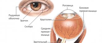
Fig.1.
Functional organs of the eye.[1] Figure 1 shows:
- The optical biosystem of the eye is a system that perceives and encodes received information for the brain, a life support system.
- The cornea is the transparent layer of the eye that covers the front of the eye. It lacks blood vessels and has great refractive power. Part of the optical system of the eye. The cornea borders the opaque outer layer of the eye, the sclera.
- The anterior chamber of the eye is the space between the cornea and the iris. It is filled with intraocular fluid.
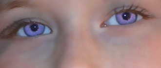
- The iris is shaped like a circle with a hole inside (the pupil). The iris consists of muscles that, when contracted and relaxed, change the size of the pupil. It enters the choroid of the eye. The iris is responsible for the color of the eyes: if it is blue, it means there are few pigment cells in it, if it is brown, it means there are many. It diaphragms the passage of light rays, adjusting the light flow similar to the diaphragm in a camera.
- The pupil is an opening in the iris. Its dimensions depend on the level of illumination. The more light, the smaller the pupil.
How it works
It’s extremely simple: reflected from something we look at, light passes through the cornea, aqueous humor, lens (here it turns over), the vitreous body and all layers of the retina, reaching the penultimate one, where special cells, through simple chemical reactions, generate a nerve impulse in depending on the possibility of your perception, which is divided into b/w rods and three colors of cones.
The human eye is often cited as an example of one of the wonders of nature by all sorts of creationists: supposedly it is so complex that it could not have appeared without an original plan and “intelligent” design. They would crap themselves when they found out how shitty and unprocessed the RAW picture gets from the organ of vision to the brain (see right). Clear and color vision transmits to us only 15 degrees (cone) of an inverted world with a hefty blind spot and surrounded by dull, faded indistinguishable spots in the periphery of vision. It is from this stuffing that the brain compiles a living and voluminous world with the images living in it. The eye is just a lens that has no special meaning without the brain’s ability to recreate conscious images from pictures: highlight a chair and a window, estimate sizes and distances.
The beginning of the calculation of information from the eye begins in the retina itself, from where it then enters data processing centers - visual analysis centers, the main of which is located in the part of the head that is not closest to the eye - in the back of the head. There, the information is first split into sections of the visual fields (central/peripheral, right/left, top/bottom), for which certain zones of the cortex are responsible, and then the impulses are analyzed for content. In the same subconscious, beloved by Freud, numerous image adjustments take place, eliminating aberrations, distortions, overlapping blind spots and other defects in RAW vision.
What’s interesting is that this entire army of signals is processed by the brain simultaneously, and not sequentially as in a computer: although the cortex has zones that are responsible for lower or higher images (lines/shapes/movement and recognition of faces/objects), information between them is transmitted in parallel , therefore, sometimes a substitution occurs when we see optical illusions (the brain mistakenly supplements the lower elements with higher elements, as when you imagine some other object in a cloud).
Movements
The direction of gaze is controlled by twelve muscles, six for each eye, working more harmoniously than Russian synchronized swimmers. For each of the three perpendicular axes of movement, there are two antagonist muscles, which simultaneously relax and tense: to look up, the upper muscle tenses and the lower muscle relaxes. In addition, the approach of the observed object leads to an automatic convergence of the axes of both eyes - convergence - and constriction of the pupils.
The secret is that Mother Nature (thank God, lol) did not trust you to manage this delicate process, but hard-coded the setting into the BIOS. Thanks to this, the processes of tracking, merging a stereoscopic image, approaching/removing the gaze and many unconditional reflex reactions (such as turning the gaze to a sudden movement) occur without your inhibited and inept intervention. While the impulses travel their long way from the eye to the visual center and your consciousness, the subcortex will already have time to react to signs that are critical for it, processing simple signals before thinking: this is how we can train ourselves to react to something without even having time to think about it, for example, in sports, where the reaction to a ball flying straight into the face is brought to automaticity.
The remaining few muscles inside the eye are controlled not even by the subcortex, but by the autonomic nervous system: commands to dilate and constrict the pupil are given by the sympathetic and parasympathetic, respectively, which is programmed to change the amount of light entering the eye and the need for an urgent reaction. In addition, the accommodation of the lens works without your stupid instructions, although we can influence it with an effort of will, consciously focusing our gaze on the breasts of selected objects or even without them.
Camber/toe
When looking into the distance, the axes of the eyes converge somewhere in infinity, which is why the approach of the object being looked at causes double vision; in order to cross these almost parallel lines to merge into one picture, the eyes use vergence - a friendly movement in opposite directions (convergence - ascension eyes) around the vertical axis, because around the horizontal the eyes move in one direction. In addition to the convenience of looking at one instead of two pictures, the brain can calculate the distance to objects based on this correction. Try pressing your finger on one eye through the eyelid so that double vision occurs: in this state it will be much easier to bump into objects.
It is believed that incorrect vergence may indicate the possibility of developing schizophrenia. [6]
Fusia
The brain compares two slightly different (by distance - 6 cm between the axes of the right and left eyes, and by viewing angles) images into a single one, which gives us a three-dimensional picture of the world, including the ability to estimate depth/distance due to doubling (parallax effect) those objects that are out of focus (i.e. closer or further than what we are looking at), the so-called. eye gauge
We essentially see two pictures, each hemisphere of the brain has its own, which the brain constantly corrects by adjusting the eye muscles unnoticed by us (after all, the eyes move not only up/down simultaneously, but also in opposite directions), orienting the projections of the observed objects exactly in the central fovea. This can be noticed if you put a weak lens on one eye - the vision will be double at first, but then it will adjust by turning the eye. You see the result of merging images from the right and left macula (“binocular summation”) as a clear text that you are reading right now; this occurs through a complex comparison of right and left pictures in the brain itself.
This and other technical details provide us with binocular vision, which makes it possible to perceive the volume and relief of objects. If only one eye is working, then estimating the distance is also possible, but it will be based on size, perspective, experience and other secondary signs, which is much less accurate, so one-eyed people cannot drive a car.
Paths
After the retina has processed the light that has fallen on it into nerve impulses, the latter rush along the optic nerve to the intersection with its counterpart, where these two fraternally exchange 50% of the fibers: now on the left
fibers come from both
left
halves of the retina, and on the right, respectively, the right.
Thus, due to the reversal of light by the lens and the chiasm, the left
side of the brain looks
with the left
halves of the retinas at
the right
field of vision, and the right side vice versa.
After the chiasm, the optic nerve becomes the optic tract and enters the midbrain, where preprocessing of the video signal by the lateral geniculate body continues (still unconscious to us). After LCT there is a wide bundle of fibers under the beautiful name “visual radiance”, which finally enters the cortex of the occipital lobes, where thinking begins to recognize it, highlighting the things it needs.
The symbol of the eye and its meaning
The eye is not only an organ of vision, through whose function we have approximately 80% of information about the world around us. The eye feeds our perception through the light received by this instrument and interpreted by our brain. Rhythmology calls the eye “the brain brought out.”
In addition to the fact that the eye connects us with fire (light) , it is also akin to the water element (tears , tear fluid). This property shows the analogy of identifying the eyes with the luminaries - the fiery Sun (right) and the water Moon (left). At the same time, the right solar eye is emitting, influencing, and the left lunar eye is perceiving; it is into it that someone else’s will, someone else’s influence can penetrate.
The eye symbolizes the active influence of one creature on another , at the physical, psychological, and magical levels. It is a tool for projecting the power of a being, a personality, into the outside world.
Of the positive images, here one can recall the eye of Ra and the eye of Horus - as attributes of the power, strength and power of the all-seeing deity of the Egyptians, their supervision of their human civilizations.
Negative images include the gaze of the Gorgon or Basilisk jellyfish, Gogol’s Viy, the all-seeing eye of Sauron (“The Lord of the Rings” by Tolkien). The ability to influence and destroy with the help of internal evil transmitted through the eyes is the concept of the “evil eye,” the evil eye.
This organ of vision connects a person with the reality of the external matrix, with the usual picture of the world. The eye limits our perception, and when we want to tune in to more subtle information, we usually close our eyelids. For example, in the books of Carlos Castaneda, some magicians, at the transition between worlds, saw an eye hanging in space and simply dived into it with all their perception.
And the symbol of the eye in the palm means clairvoyance, supersensible perception, the development of spiritual vision, when a person sees the essence and has access to direct knowledge.
We also know that in addition to the usual two that we can see in the mirror, we have a “Third Eye” - internal vision associated with the perception of internal or higher information, space and time of unmanifested worlds.
The third eye (epiphysis + pituitary gland) is spiritual consciousness, wisdom and intuitive perception that allows you to look behind the curtain of illusion and see the essence.
Mythical creatures have three eyes - the ability to see in three worlds : dense (ordinary), subtle and fiery. Often in such creatures the third eye on the forehead was a symbol of direct influence, for example in Shiva it is an instrument of destruction and punishment.
Conversely, creatures who looked with only one single eye, for example, the Cyclops, had only a limited vision of the ordinary world, they were characterized by spiritual underdevelopment.
But often, one-eyedness or even blindness could indicate special magical and spiritual power (Odin, chosen by Neo in “The Matrix-3”, soothsayer Vanga).
One - who exchanged his right eye for wisdom
In various mythologies, there are stories about the theft of the eyes of the gods, blinding and their healing, which becomes a symbol of rebirth. For example, among the ancient Hittites, there lived a Thunder God, from whom the Serpent took not only his eyes, but also his heart. In connection with this, the God of Thunder had to give birth to a man, marry him to the daughter of the Serpent, and only after that, with the help of the man and his persuasion, dad’s sight and heart were returned.
A very similar story is also told about the Jupiterian character – the Egyptian Horus. Hawkeye of Horus (left) - also removed by the demonized Set and later revived, which allows us to associate this story with the lunar cycle, symbolism of the changing phases of the moon .
One of the semantic symbols of the eye is perception, attention and control . For example, the constantly awake Kronos in ancient Greek mythology had 4 eyes: two of them were closed, possibly sleeping, and two were always open - apparently, they were on duty.
Often the image and symbol of the eyes is associated with a divine figure and his presence, for example:
- Sumerian Enki - “lord of the sacred eye” is rightfully considered a symbol of wisdom and omniscience;
- the divine Eye of the Egyptians, the winged eye are symbols of the power, strength and wisdom of the all-seeing God. Another version is a symbol of the pineal gland, the pineal gland, through which you can communicate with higher dimensions;
- the peacock with its plumage of many eyes is a zoomorphic image of the meditating Buddha;
- in Christianity, the eye was depicted on the wings of cherubs and seraphim, as a sign of insight and wisdom;
- The eye of the Lord is an eye in a triangle, originally a sign of Jehovah, or God the Father, who is everywhere, sees everything, knows everything. And later this symbol already belongs to the entire divine Trinity (God the Father, God the Son and God the Holy Spirit) in the radiance of holiness;
- the eye of the heart is in Islam a symbol of clear and true vision, which can only be achieved through the heart's spiritual center, the eternal here and now, through the point of the present.
And finally, the eye is a symbol of the Observer .
This term has several interrelated meanings.
The first meaning is that those who are on the spiritual path know that isolating the Observer and strengthening the Observer effect is a certain stage in the development of awareness.
The second meaning is from quantum physics:
The observer effect (observer consciousness) is a group of hypotheses about the possibility of an observer influencing elementary particles. According to Bohr, without an observer, the surrounding reality is only a probabilistic form. Concrete reality appears only with the arrival of the observer. Some scientists equate the observer, man, and human consciousness.
Thus, the meanings of the eye symbol are:
- vigilance and protection, control and surveillance;
- perception of the visible and visible, limiting a person to this or gaining the Observer effect;
- providence, foresight, intuition, insight, supersensible knowledge;
- light and luminaries, Moon and Sun, as well as stars (especially Polaris);
- divine presence and penetration into human life;
- transition point, portal.
More
- Myopia (coming soon);
- Amaurosis - blindness;
- Home eye test.
Home reading
- Eye diseases, ed. V. G. Kopaeva (caution: fuflomycins are found in treatment);
- A good blog from an ophthalmologist;
- ABC of eyes;
- D. Hubel. Eye, Brain, Vision - a cosmic book on neuro-ophthalmology from Nobel laureates D. Hubel and T. Wiesel;
- Practical neuroophthalmology. Gustov A.V.;
- Read more about binocular summation.
| [ + ] The eye is an integral part of a person. | |
| Physiology | Physiological: Blood (Complement) • Urine • Feces • • Hormones • Death • Immunity • Carcinogenesis • Atherosclerosis • Pathogenesis Higher: Automatism • Sleep • Stress • Will (Laziness) Sexual: Sex • Orgasm • Sex hormones • Menstruation • Sperm • Pregnancy |
| Pathanatomy | Fibrosis • Cancer |
| Pathphysiology | Syndrome • Pain • Heredity • Mental automatism • Jaundice • Diarrhea • Myopia |
| Chemistry | Urea • Uric acid • Dihydrogen monoxide |