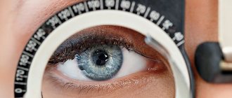
Determining the quality of vision in modern ophthalmology is carried out in several ways. This parameter is determined by a person’s ability to clearly distinguish objects located at a given distance. Currently, there are two methods: subjective - using printed tables, and objective - using precision instruments.
For such measurements, eye refraction is important, although visual acuity does not always depend on it. For example, a person with farsightedness may have difficulty seeing close up, but have good visual acuity and correctly name all the letters or symbols on a test chart. Let us explain what refraction is. Our organs of vision are a complex optical system that has its own focus. Its location relative to the retina is called the refraction of the eyes. If the visual organs do not have pathologies, then the focus is clearly on the surface of the retina, this is called emmetropia, or normal vision. With hyperopia (farsightedness), the focus is located behind the retina, and with myopia (nearsightedness) it is located in front of it. Moreover, all children at birth have a weak degree of farsightedness, which is due to the anatomically shortened diameter of the eyeball. Only after some time, as the whole organism grows, the eyeball also enlarges, and vision indicators return to normal, if there are no other pathologies.
Indicators below 1.0 what does this mean?
In order to understand the reason for the sudden change, comprehensive examinations are carried out. An acuity score below 1.0 usually indicates advanced disease. The most common ailments include:
- Myopia. Focusing occurs in front of the retina. Changes in visual acuity reduce the ability to distinguish objects in the distance, a feeling of rapid fatigue, pain, and headaches appear.
- Farsightedness. The focus of the image is behind the retina. Decreased visual acuity makes a person unable to see well at close range. Accommodation is impaired, blurred vision is observed, and strabismus occurs.
- Astigmatism. The reasons for the sharp decrease are irregular shapes of the lens or cornea. Images are distorted, objects appear in two, and headaches occur.
- Glaucoma. The disease appears due to abnormalities in intraocular pressure. When eye pressure decreases, deformation of the internal structure of the visual organ and retinal degeneration occurs. If the optic nerve is severely damaged, then with this diagnosis the disease leads to complete blindness.
- Cataract. There is clouding of the lens. As the disease worsens, a person begins to react painfully to light and has difficulty distinguishing colors. There are difficulties with reading and orientation at dusk.
The specifics of work can often lead to vision deterioration: improper working conditions, hazardous production, constant strain of attention. However, eye diseases are also observed in children. In this case, the loss of good vision is often associated with inherited ailments.
Basic Rules for Testing Eye Clarity
Visual acuity impairments are determined using special tables that display letters or numbers. The procedure is performed in an ophthalmologist's office or in specialized stores for the selection of glasses and lenses. Any first sign of an ocular disorder should prompt further diagnosis and treatment.
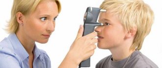
For an adult, tables with letters are used, for a child who cannot yet read, with symbols. The norm is when the 10th line out of 12 is clearly visible and readable. Based on such a diagnosis and further examination, it will be possible to understand what affects the deterioration of vision and what treatment will be optimal.
Basic verification rules:
- A man sits at a distance of five meters from the table.
- The images are located from the window on the opposite side.
- The 10th line of the diagram is located strictly opposite the eyes.
- The table must be illuminated with special lamps.
- Each eye is diagnosed separately, one is open, the other is closed, but not closed (this affects the severity of symptoms and the veracity of the results).
- You need to recognize a letter or sign within 2-3 seconds; more time will indicate a deviation.
How to do a home inspection
If you are interested in a decrease in human visual acuity, you can conduct a preliminary diagnosis at home. Various online tests will help with this, where the result is given at the end.
You can also use tables for checking, as in an ophthalmology office. Examples of tables can be found on the Internet and printed on a standard A4 sheet. The finished sheet with images is placed on the wall. A fluorescent lamp or two lamps (40 Watt) on the sides are installed above the table. The presence of the first changes and violations is indicated by the inability to examine the letters or symbols of the 10th line. In this case, you should immediately make an appointment with an ophthalmologist.
How to check visual acuity
One form of VA testing was used 1000 years ago when desert Bedouins used the ability to identify double stars to evaluate vision. The development of the Sivtsev table was an important milestone in the standardization of VA measurement.
Diagnosis is carried out by opticians, ophthalmologists, scientists, and nurses.
The Sivtsev test is a table of letters or symbols commonly used in schools and eye doctors' offices. It includes 11 lines of capital letters. Each line has an increasing number of letters that become progressively smaller in size. A person views the diagram while sitting or standing, from a distance of 6 meters.
In the eye doctor's office, the chart is projected or shown as a mirror image. One eye is covered with a simple occluder, card, or cloth. The letters are read aloud with an open organ of vision, from the top of the diagram down until the person can accurately distinguish them. The test is then repeated with the other eye.
Checking is carried out using Landolt rings, Snellen or Orlova tables.
Dangerous symptoms
There are the first signs of vision deterioration, which may indicate the emergence and development of diseases:
- A black curtain covering the view. This symptom indicates retinal detachment. If you do not immediately pay due attention to the deterioration and decline of eye health, the situation will soon worsen. Immediate hospitalization is required.
- Redness of the mucous membrane, sharp pain, fog obstructing vision. In particularly difficult cases, nausea and vomiting are observed. All this may indicate glaucoma. What to do if vision deteriorates, urgent therapy will be required immediately; if complications arise, surgery will be required.
- Reduced visibility. The severity of the symptoms differs in damage to the optic nerve. This disease is dangerous because it can lead to glaucoma and even removal of the visual organ.
- Distorted, blurry image. A straight line appears curved. To understand what is causing the visual impairment, you will need to consult a doctor. Distortions can be caused by both age-related changes and retinal damage.
- A decrease in visual acuity is often expressed in a lack of brightness and contrast, and the appearance of fog. The reason is clouding of the lens, which is a sign of cataracts. The disease can lead to blindness.
- Dark spots, cloudiness. Such symptoms often include retinal lesions and retinal hemorrhages. The resulting losses lead to complete blindness.
- Dryness, burning, lacrimation. In this case, the reasons for the decrease in visual acuity are constant fatigue and overstrain. This is especially true for people who spend a lot of time at the computer and working with documents.
Loss of visual acuity makes life joyless and causes the development of numerous pathologies. In order to detect deviations in a timely manner and successfully solve the problem, you need to regularly visit an ophthalmologist (at least twice a year). At risk are people with a genetic predisposition to diseases that have been in their family.
Visual acuity often decreases after injuries, for example, when the upper cervical vertebrae are damaged. Many ailments develop due to diabetes mellitus, cervical osteochondrosis, and previous genetic diseases. The organs of older people are subject to changes. Timely consultation with a doctor and diagnosis is the most important step towards a speedy recovery.
How to maintain visual acuity?
Our visual acuity is influenced by many factors. And if we are unable to control heredity and environmental influences, then the conditions of our daily life can be made comfortable and safe for the eyes.
Basic recommendations
:
- Balanced diet, consumption of sufficient vitamins (see article on vitamins for vision)
- Organization of comfortable and convenient lighting in the workplace and at home
- Compliance with general rules (distance to the object, illumination, duration of eye work) when reading, watching TV, working at the computer
- Quitting bad habits that negatively affect visual acuity - primarily smoking and drinking alcohol
- Regular examination by a doctor, at least once a year.
The last point is especially important, since only your attending physician can give effective and individual recommendations. Drug or hardware treatment and eye exercises may also be prescribed. Following your doctor's orders will help not only maintain, but also improve your visual acuity.
What is myopia?
Myopia is a refractive error in which a person loses the ability to clearly see objects distant from him.
This happens because the image is focused not on the retina, but in front of it. The shape and size of the eyeball change. It becomes more elongated. This leads to the fact that the outlines of objects become blurry and foggy. The reasons for the development of myopia are varied. Its occurrence is often associated with a genetic factor. If both parents are myopic, then it is likely that the child will inherit this pathology. It happens that the eyeball changes in size even before the baby is born. In this case, the cause of the development of myopia is intrauterine development disorders. Myopia is often a consequence of birth injuries. Increased visual load has a particular influence on the development of myopia. Reading in low light and playing computer games are all factors that contribute to the onset and progression of myopia. Most often, refractive error develops at school age, when visual load increases significantly. Pathologies such as:
- keratoconus;
- spasm of the ciliary muscle;
- subluxation of the lens;
- decreased immunity;
- hypovitaminosis.
Many people suffer from visual impairment. Myopia is one of the main reasons for the decrease in its severity. World statistics show that myopia affects approximately 30% of the population. The peak development of pathology occurs at school age. Correcting it is very important, but before that it is necessary to determine the degree of myopia. This will help you choose the right ophthalmic products.
Visual acuity diseases
The main visual impairments are nearsightedness, farsightedness, astigmatism and a condition known as keratoconus.
Nearsightedness, also known as myopia, is a condition in which a person can see near objects more clearly than distant objects. Light rays are focused in front of the retina. This results in blurred vision. Myopia can be inherited from parents.
Its development is affected by frequent or prolonged work at a computer at close range. Typically found in school-age children, worsens during adolescence, and usually stabilizes between 20 and 40 years of age.
Farsightedness is a condition in which a person sees distant objects better than those located at a close distance. Hypermetropia involves focusing light rays at the back of the eye. As the eye grows and lengthens, farsightedness decreases. Symptoms:
- blurred vision;
- inability to distinguish objects located nearby.
Astigmatism develops in combination with myopia and farsightedness. The cornea and lens of the eye, as a rule, have a spherical shape. When one or both are more steeply curved in one meridian, astigmatism is diagnosed. Objects near and far appear blurry due to uneven curvature, which prevents light rays entering the eye from focusing on a single point on the retina.
Symptoms include blurred vision at near and far distances. Eye strain and fatigue.
Age-related changes in the eye include:
- age-related farsightedness;
- cataract;
- glaucoma.
How is visual acuity determined?
To undergo an eye examination and find out if you are nearsighted, you need to visit an ophthalmologist. The doctor conducts an examination using special computer equipment. Almost all modern clinics are equipped with this. Used for vision diagnostics and tables. The most popular among them is the one developed by ophthalmologist Sivtsev. Other techniques are also used. The examination, which is carried out using instruments, is very important. So the ophthalmologist can examine the fundus of the eye, the condition of the retina, and diagnose the degree of myopia or other refractive error. Computer equipment allows you to accurately calculate the optical power. This will be required in order to write out a prescription for glasses or contact lenses.
How to determine myopia using the Sivtsev table?
The most famous tool for testing vision and diagnosing myopia is the Sivtsev table. Every person who has been to an ophthalmologist’s office at least once in their life and undergone an examination is familiar with it. The table was developed by the Soviet ophthalmologist Dmitry Aleksandrovich Sivtsev. Doctors began using his work in medical practice in 1925 and continue to use it to this day. In the ophthalmologist’s office, Sivtsev’s table is placed on the wall. It is illuminated by lamps with a lighting power of 700 lux. This is the generally accepted meaning. If it is less or more, the results of the vision test will be incorrect. During the diagnosis, the patient is asked to sit on a chair. Your back must be kept straight. The visual organs are checked one by one. The eye that is not involved in the test is covered. Sivtsev's table contains 12 lines, which consist of block letters. Their sizes decrease downwards. At the beginning of each line the distance in meters is indicated. It is from here that the patient should consider which letters are indicated on the poster. For the top row the distance is set at 50 meters, for the bottom at 2.5 meters. Being at such a distance from the table, a person with normal vision should consider its elements.
The value is indicated on the right side of each line. It is marked with the letter V. This indicates vigilance in case of myopia, if the patient reads letters from a distance of 5 meters. If errors are made, they speak of a refractive error. If the value is less than 0.1, the patient is diagnosed with myopia. When indicators exceed the previous value of farsightedness.
Sivtsev table
It was proposed by the Russian physician D. A. Sivtsev in 1925, and we are familiar with it from childhood, since it hangs in every ophthalmology office. In the Sivtsev table, unlike the Snellen table, there are 12 rows, consisting of 7 letters of the Russian alphabet: Ш, И, М, Н, Б, И, К. To carry out a test using this method, a distance of five meters is required, not six. In addition, the lighting in the office is important - this can also affect the results. So, the brightness of the light should be approximately 700 lux. Too bright or, on the contrary, insufficient light can affect the diagnostic results.
The top line of the tables consists of two large letters Ш and Б. Normally, a person without eye pathologies should be able to see them from a distance of 50 m. Under office diagnostic conditions with normal vision, the patient should correctly name the letters on the 10th line from five meters. In this case, his visual acuity will be equal to 100%, or one (1.0). If he is not able to name the optotypes on the 10th line, the table is brought closer by 0.5 meters until the letters become distinguishable. At the same time, there are certain requirements for errors during verification: from lines 4 to 6 - only one mistake is allowed, from lines seven to ten - a couple of errors, and from lines 1 to 3 all letters must be named correctly.
The Sivtsev table is a convenient way to determine visual acuity quickly and accurately. However, in some cases, the Golovin table is additionally used. The Soviet ophthalmologist S.S. Golovin used Landolt optotypes - rings, one of the sides of which has a gap. His table also consists of 12 rows, and the diameter of the rings decreases proportionally towards the bottom row. The person being tested must correctly name the side of the rupture in the ring indicated by the doctor. Why is such additional diagnostics needed? The fact is that when passing a driver's or military commission, some people being tested are cunning, remembering the location of the letters in Sivtsev's table. This way they name them correctly even if they can't see them. But in Golovin’s table it is almost impossible to remember the location of optotypes. The size of the gap is very small, and with poor vision it will not be possible to see it: it will seem to the patient that the circle is solid. In the ophthalmologist’s office with a Sivtsev table there is almost always a Golovin table hanging - for a cross-sectional examination method, if necessary.
How to check the degree of myopia using the Golovin table?
Sergei Selivanovich Golovin is a famous Soviet ophthalmologist who developed his own vision testing system, which was called the Golovin table. In modern medicine, it is used as often as that proposed by Sivtsev. Both tables are similar to each other. The difference between them is that instead of letters, Golovin used Landolt rings, black circles with a gap on one side. Golovin's table has 12 rows. They depict Landolt rings, each of them torn on one side. The largest rings are located on the top row. The next one is smaller. And so on until the end of the table, the last line of which shows the smallest rings. On the left line is the distance in meters, indicated by the letter D. From this distance, a person with normal vision should recognize the optotypes. The values are indicated on the right side. They are designated by the letter V and indicate visual acuity in diopters. If the value exceeds 1.0, a diagnosis of farsightedness is made. If less than 1.0, then myopia. During the check, the backlight should be directed at the table. Often incandescent lamps or 2 fluorescent lamps are used for this purpose. Lighting should not be less than 700 lux.
Doctors consider the method of testing visual acuity for myopia, which was developed by Golovin, to be more reliable. This test is difficult to pass from memory, since it is almost impossible to remember the rings and their location. This scale is more often used to test vision in adult patients. Children have a harder time remembering complex shapes. According to the Golovin table, visual clarity is often checked when passing a military commission.
Determination of myopia using the Orlova table
Sivtsev and Golovin tables are usually used in tandem to test the vision of adult patients and adolescents. To diagnose myopia in preschool children, the Orlova table is often used. Ophthalmologists use this method to examine children from 3 to 7 years old. Its main purpose is to test vision in children who cannot read. Determining the degree of myopia of a child using Orlova’s table is very simple. In many ways, this method is similar to those proposed by Sivtsev and Golovin.
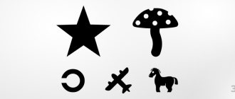
The difference is that to clarify the degree of myopia, the child is asked to recognize and correctly name the images on the poster. Especially for kids, the table contains pictures of animals and toys, as well as signs familiar to them: circles and plus. When checking myopia using the Orlovy table, the doctor takes the child to the poster in advance and asks him to name the objects depicted on it. This is done so that the baby can understand what is required of him. Young children do not always speak clearly and may mispronounce words. Preliminary familiarization of the child with the table helps the ophthalmologist understand how the patient names objects.
Visually, the methods of Sivtsev, Golovin and Orlova are similar. At the beginning of each line on the left side of the table is the letter D, which represents the distance in meters. From this distance, a child with good eyesight should be able to see the images on the poster. On the right side of the table is the letter V. It indicates the value of visual acuity in conventional units and allows you to determine the degree of myopia.
Visual acuity in children
The main difficulty in assessing VA in infants is that they cannot be tested using standard diagnostic methods used for adults. The second difficulty is that the norm of acuity in an infant and an adult is different. Third, their vision is not static; as a rule, it quickly improves in the first year of life.
Because of the immaturity of the infant's visual system and the dynamic nature of visual development in the first months, any program for assessing visual status in infants must recognize two important points:
- The results of the visual assessment should be compared with normative data from infants of the same age tested by the same diagnostic methods. Comparison of results with norms based on data from adults or older children, or infants examined using another procedure, leads to misdiagnosis of visual impairment.
- Results from visual assessments performed in infancy do not necessarily predict visual status later in life. A child whose vision appears normal in the first months after birth may later develop problems if the optical system does not undergo significant development, which usually occurs between infancy and adulthood.
Between infancy, which is usually considered over at age 1, and the child's entry into the school system at age 5–6, there is a period during which the child demonstrates significant development in both vision and cognitive skills. As a result, the instruments that can be used to assess visual perception in preschool-age children vary depending on age and cognitive ability.
To measure health in preschool age, tables with depicted animals or figures are suitable.
In general, children of normal intelligence who reach 5 or 6 years of age can be tested using the same procedures used to assess visual function in adults. However, their results tend to be lower than those of adults, and it is therefore important to compare the results of school-age children with those of children of the same age.
Imaging screening tests for infants and preschool children:
- reaction to light;
- pupil reaction;
- the ability of the eyes to follow a finger or toy.
Developmental problems in children with poor visual acuity
A child's vision affects his performance in school. They may have serious developmental problems. Poor health affects a student’s ability to learn and adapt in the classroom. School-age children spend a lot of time in health centers to improve visual perception, this affects relationships with peers in the class.
Blurry vision not only affects learning, but also causes other problems. For example, headaches, increased fatigue.
According to the American Academy of Child and Adolescent Psychiatry, mental health disorders affect the child's psyche. The mental state is disturbed in 1 out of 10 schoolchildren.
Children develop attention deficit hyperactivity disorder (ADHD).
If a child turns letters over when reading or writing, has poor handwriting, does not like to read or has difficulty reading fairy tales, or behaves inappropriately in society, he may have a vision problem.
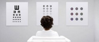
Vision diagnostics using the Snellen scale
One of the most accurate tables that allows you to determine the degree of myopia was developed by the Dutch doctor Hermann Snellen. It consists of 11 lines and letters of the Latin alphabet. The letters gradually decrease in size from line to line from top to bottom. The largest letters are located on the top line. A person with normal vision will be able to see them from a distance of 60 meters. In some clinics the top line is taken as the first line, in others the bottom line.
In the absence of myopia, the patient is able to clearly see the lines that are located below the 11th or 1st. It depends on the rules adopted in the clinic. Each line has a specific distance. The distances are 36, 24, 18, 12, 9, 6 and 5 meters. The distance decreases as the size of the optotypes decreases. The right side of the table shows the distance from which a patient with good vision should distinguish letters. It is measured in pounds. 20 pounds equals 6 meters. The patient being tested must sit at this distance. During diagnosis, one eye must be covered with a special pad. Similarly, the degree of myopia is checked using the tables of Sivtsev, Golovin and Orlova.
Vision testing using Polyak optotypes
Boris Lvovich Polyak is a Soviet ophthalmologist who contributed to the diagnosis of refractive errors. He developed his own methodology specifically for the military commission and medical and social examination. The optotypes that were proposed by Polyak are used to identify disabilities and suitability for military service. This diagnostic method is rarely used in clinics. Optotypes are designed to determine central visual acuity less than 0.1. The Pole did not set out to create a special table.
He developed a set of posters that depict different geometric shapes: rings, straight lines. Landolt rings were also used. Polyak's technique differs in many ways from the tables presented by other ophthalmologists. To test vision using Polyak's optotypes, the patient does not need to be at a distance. The cards created by the doctor are small in size. They are offered to be viewed at close range. Determining the degree of myopia is based on the width of the gaps between the strokes and the thickness of the lines. Doctors determine visual acuity in the range from 0.04 to 0.09.
Is it possible to determine the degree of myopia at home?
You can test your vision at home. To do this, you need to print out the tables of Sivtsev and Golovin; they are available on ophthalmological websites. The most convenient way is to place the table on the wall and move 5 meters away from it. It is from this distance that it is recommended to carry out the test at home. You can start from the bottom row. If you can correctly name all the images, then there are no refractive errors.
There are many online services that offer visitors vision tests. This option will be convenient for those people who do not have a printer or do not want to use Sivtsev and Golovin tables. The online test is carried out using special programs. Each of them has its own algorithm of action. Some offer to determine the direction of the figures, others contain a list of questions.
All possible advantages of different retinometer models
The very first retinometers had one significant drawback. They did not have light intensity controls. The halogen lamp used in the examination blinded the patient and caused quite a lot of discomfort. Modern models of devices for determining ROS are equipped with a device with which you can adjust the brightness level of the lamp.
The advantages of the method include:
- reliable results that do not depend on the research conditions;
- speed of the procedure;
- the light weight of some devices, their portability, which allows them to be used on the go.
Features of children's vision and congenital farsightedness
People of different ages have their own vision standards. So, for older people it will be lower, but all children are born with a slight degree of farsightedness. The reason for this is the shortened size of the eyeball. In an infant it is 17-18 mm, while in an adult it is already 23-25 mm. Light rays are not focused on the retina due to the insufficient size of the eyeball, but fall into the area behind it. The amount of farsightedness in young children is approximately 2.5-3 diopters. However, by about 7 years, vision is normalized in the absence of other pathologies.
To study children's visual acuity, either the Sivtsev-Golovin table or the Orlova method is used, depending on age. Orlova’s table depicts simple figures: a mushroom, a car, a Christmas tree and other objects that children who do not yet know their letters can name.
Vision 0.4 and 0.7: which is close to normal, which is weaker?
Flashka
If, after visiting an ophthalmologist, you see in your card a record of a vision level of 0.7, this means that you see the seventh line from the top, and this is much better than 0.4 (you only see the fourth line from the top). But it is definitely better to have vision at the level of one. But with regular work on a computer, this is practically impossible.
Olegk
Normal or one hundred percent vision is considered to be visual acuity when a person can read the tenth line without making more than two mistakes. This vision is designated 10/10, or 1.0.
This means that if the norm is 1.0, it follows that 0.7 is “closer” to the norm than 0.4, and therefore weaker than 0.4.
Clip
It depends on what it is measured in. If expressed as a percentage of absolute vision (one), then 0.7 is closer to 1.0 than 0.4.
But if these are diopters, then 0.4 is less myopia or farsightedness. In this case, an indicator of 0.4 is better than 0.7.
bolshoyvopros.ru
Features of vision in old age
Upon reaching the age of 40-45, aging mechanisms are launched in our body, including those affecting the visual organs. Even with previously normal vision, its quality gradually begins to decline. In 95% of people, presbyopia appears, an age-related farsightedness associated with the loss of elasticity of the lens and a decrease in its accommodation. When trying to see nearby objects, the eyes quickly become tired and headaches occur. You have to resort to glasses or contact lenses for correction, even if your vision was 1.0 or 100%.
According to statistics, by the age of 60, normal visual acuity remains in approximately 70% of people, and after 80 years only in 45-48%. In older people, central peripheral vision and color perception deteriorate. Eye diseases develop: glaucoma, cataracts, age-related macular degeneration. Often, older people undergo a lensectomy to replace the lens clouded by cataracts with an intraocular lens. This operation will allow you to maintain visual acuity for the rest of your life.
Impaired visual acuity in pathologies
Visual acuity depends on the refraction of the eyes - the refractive power of their optical system, expressed in diopters. Depending on the refractive power of the eye, the refraction may be normal or have deviations. Widespread and well-known eye refractive errors are:
- Farsightedness (hypermetropia). With a shortened ocular axis, the image is formed behind the retina. The refractive power of the optical system of the eye is insufficient
- Myopia (myopia). The image is focused in front of the retina. Causes: elongated ocular axis, refractive power too high
- Astigmatism is a violation of the sphericity of the cornea.
Other pathologies that affect visual acuity develop mainly with age. These primarily include:
- Presbyopia (age-related farsightedness) is a partial loss of elasticity of the lens, when it is no longer able to change its curvature (accommodate). This occurs due to loss of flexibility of the ligaments and compaction of the lens itself.
- Cataract - disturbances in the nutrition of the lens, a decrease in its transparency
- Glaucoma is an increase in intraocular pressure with possible damage to the optic nerve (if diagnosed and treated early, minimal damage can be avoided).
These eye pathologies that pose a threat to our health can be eliminated with the help of conservative, laser and surgical treatment. If for glaucoma it is possible to use all three methods of treatment (depending on the degree), for presbyopia (age-related farsightedness) hardware treatment, laser vision correction and replacement of the lens with a multifocal lens for refractive purposes are possible, then for cataracts only surgical treatment is possible.
Verification using visometry
Subjective methods for assessing the quality of visibility include visometry using Sivtsev-Golovin tables. Visual acuity is considered to be the ability to see two points located at a distance of 5 meters from the eyes. In this case, the distance between them should be 1.45 mm. If a person clearly distinguishes the desired points at a given distance, then his visual acuity is normal and equal to 1.0.
Sivtsev’s table has been familiar to everyone since childhood. It is a set of seven letters: M, K, N, Sh, Y, I, B, arranged in 12 rows and decreasing towards the bottom. The test is carried out from a distance of 5 meters in turn for both eyes. If a person is able to distinguish letters starting from the tenth line, his visual acuity is normal and equal to 1.0. Based on the results of visometry, the following can be revealed:
- if you can see the letters only from the ninth line, then your vision is 90% normal;
- if lines 8 to 5 are visible, this indicates the ability to see normally by 60%;
- from lines 5 to 3, visual acuity is approximately 40%.
If the patient sees only the letters on the top two lines, then the situation is catastrophic: his vision only works at 10%. Recording the results of visometry may look like this: V (visus, visual acuity) OD/OS = 1.0/0.4, which means the norm is 1.0 in the right eye (OD) and its acuity is only 40% in the left eye (OS ).
Visometry is a method of subjective assessment of the quality of vision, based on human sensations. Usually, objective methods for determining visual acuity are used together with it.
Interpretation of visometry indicators
At the end of the test, the specialist calculates the result. To the right of each row is a value characterizing visual acuity - V, or visus. On the left side of each row there is a D value - the distance in meters from which the patient must recognize the optotypes in it. The doctor, based on the data obtained during the study, calculates the final result: V = d/D, where d = the distance from which the study is carried out. A value of 1.0 indicates that the person recognized the letters on all 10 rows of this table. The entry V OD = 0.3 indicates that the patient sees symbols with the right eye only in the top three lines and has 30% vision. The final record might look like this: V OD/OS = 1.0/0.6, which means normal vision in the right eye (OD) and only 60% good vision in the left eye (OS) or V OD/OS = 0.7/0.4, which means 70% of normal in the right eye and 40% in the left.
The last two lines in the Sivtsev-Golovin tables can only be seen by people with vision exceeding the norm and equal to 1.5 units (150%) or 2 units (200%). Such unique people are able to see inscriptions on road signs at very distant distances, and some of them even see the rings of the planet Saturn. On the contrary, there are individuals with microscopic vision who can see very small objects in detail without the use of magnifying optics.
After diagnosis using tables, visometry is sometimes continued with correction if vision in any of the eyes is less than one. To do this, glasses with different diopters are inserted into a special frame.
After isometry with correction, the record may look like this: V OD = 0.3, sph –0.6 = 1.0, that is, with visual acuity in the right eye of 0.3 units (30%) when a lens with optical power is placed on it –0.6 diopters a person sees all the lines. This is how lenses for glasses are selected.
In modern ophthalmological practice, computer technologies are used to determine visual acuity. All that is required of the patient is to look carefully, without moving his head or blinking, at the dot in the center of the screen. This takes a little time, and in the end the device produces a result with data on the lenses with what optical power you need to purchase for glasses. However, this diagnostic method has some drawback: it is impossible to determine with absolute certainty whether a person will be comfortable wearing these lenses.
Autorefractometry method
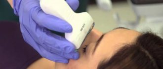
This study is carried out through the pupil, so for better visibility, drugs are used to dilate it: Irifrin, Cyclomed, Atropine or others. A wide pupil allows you to reliably identify visual pathologies such as hidden farsightedness, spasm of accommodation or strabismus in children.
The procedure itself is quite simple and takes minimal time. The patient sits in front of the autorefractometer. He must rest his chin on a special support to avoid involuntary head movements, which may affect the results. Then the gaze needs to be fixed at a given point. In modern devices, a bright picture is used instead; it is easier for young children to focus on it for a while. During the procedure, you should sit quietly, trying to blink as little as possible.
The doctor directs the infrared beam exactly to the center of the pupil and turns on the device. The beam passes through the lens and cornea, is refracted and, reflected from the retina, returns back. In this case, the computer program, starting from the given normal values and comparing them, calculates the clinical refraction of the eyes. Upon completion of the calculations, the device produces a printout with the received data. Here's what you can learn from the results of autorefractometry:
- optical power of the left and right eyes (in diopters);
- distance between pupils;
- the distance from the top of the eyeball to the back surface of the glasses lens (vertex distance) should be approximately 12-14 mm;
- optical power of a cylindrical lens (for astigmatism);
- axis of a cylindrical lens.
In modern ophthalmology, light devices are gradually being replaced by wave devices: they have even greater accuracy of readings, recording the slightest nuances in determining refraction. Based on the results obtained using wave instruments, it is possible to select vision correction products more correctly.
Means of increasing visual acuity for low vision
Description
Low vision
is considered a visual condition in which no means of correcting ametropia can achieve visual acuity higher than 0.3. This boundary was chosen on a social basis - with such vision, most information sources are inaccessible to a person. However, difficulties arise even when reading an ordinary book or newspaper becomes impossible.
According to the international classification, the degree of visual impairment is divided into four categories
:
- Category I - visual acuity ranging from 0.33 to 0.1;
- Category II - from 0.1 to 0.05;
- III category - from 0.05 to 0.02;
- Category IV - from 0.02 to light perception.
Since a common outcome of lesions in the macular region of the retina is a sharp decrease in the resolution of the eyes, the only way to improve vision in case of low vision is only by increasing the image on the retina.
Thus, all optical means of enhancing vision for low vision are magnifying systems. Based on the method of application, correction means are divided into devices for improving distance vision
and devices
for working at close range
. The need to improve near vision is a more frequent and, one might say, more important task, since near vision is an integral part of the labor rehabilitation of persons suffering from low vision.
Optical magnification can be achieved using relatively simple optical means, such as various magnifying glasses
. Magnifiers mounted on a spectacle frame or other headband for ease of use are often called hyperoculars.
A more complex way to obtain the required magnification is to use telescopic systems. Systems used to help with low vision can be built like the Galilean telescopic system or the Kepler telescopic system. The importance of the existence of telescopic systems lies in the fact that their use is the only way to improve the vision of a visually impaired person when looking into the distance.
Galileo system
consists of a positive objective lens and a negative eyepiece lens. The Kepler system (Fig. 2) is built from two positive lenses, one of which is the objective, and the other is the eyepiece* In both the first and second cases, the lenses are located relative to each other at a distance at which the rear focal plane of the first component is aligned with the front focal plane of the second.
In both the first and second cases, the apparent magnification of the telescopic system is determined by how many times the tangent of the angle at which the observer sees the image of the object when observed through the system increases, compared with the tangent of the angle at which the object is visible to the naked eye: Galileo's system is advantageous differs in that it gives a direct image, which is why it is more often used in practice. In addition, since a necessary condition for creating a Galilean system is the conjugation of the real focus of the positive lens-objective with the imaginary focus of the negative lens-ocular, such a system turns out to be shorter. To obtain a direct image when using the Kepler system,
it is necessary to additionally use a wraparound lens or a combination of prisms, which, of course, increases the size and weight of such glasses.
However, using the Kepler system
at equal magnifications with the Galileo system, more can be achieved
The exit pupil in telescopic glasses is the pupil of the observer's eye, therefore the diameter of the eyepiece lens should be slightly larger than the width of the pupil under conditions of near visual work. In turn, to fully utilize the magnifying capabilities of the telescopic system, the ratio of the diameters of the lens and eyepiece must be equal to the required magnification of the entire system. Further magnification of the objective lens does not lead to greater magnification (with the same eyepiece lens), but can significantly expand the field of view of the device. To achieve minimal weight for telescopic glasses, the lens and eyepiece are most often made from single lenses. Therefore, the angular value of the field of view rarely exceeds 30-40°. From the point of view of ease of use, they try to limit the system length to 25-30 mm, the lens diameter to 30-35 mm and the eyepiece diameter to 10-15 mm. However, in some cases it is necessary to go beyond these typical values.
Of course, a visually impaired eye can simultaneously have ametropia. Correction of ametropia in telescopic glasses can be done by adding a correction lens immediately behind the eyepiece or by changing the distance between the lens and the eyepiece. In the first case, the refraction of the additional lens must correspond to the refraction of the spectacle lens of ordinary distance glasses. In the second method, for myopia, the distance between the lens and the eyepiece is reduced, and for hypermetropia, it is increased.
Monitoring remote objects
For distance vision, telescopic systems can be used both as a monocular device and as a binocular one. Like monoculars, telescopic systems are usually used in cases where increased distance visual acuity is only occasionally required. When using binocularly, you also have to remember about anisoeikonia
— inequality of retinal images, which can be observed both due to the anisometrogy inherent in the patient’s eyes, and when equality of magnification is not observed in both branches of the magnifying telescopic system. With sufficiently high vision, binocular vision is negatively affected by the presence of anisoeikonia of 2%. It is difficult to predict what amount of anisoeikonia will be tolerated for a particular degree of vision loss. But for practical use, an approximate judgment is sufficient - each table row “lost” for reading increases the permissible degree of anisoeikony by another 2%.
To determine the possibility of using telescopic glasses for their selection, there are “low vision correction kits”
.
Almost every company that produces aids for the visually impaired produces similar sets. Within the CIS, one could most often come across the HKC-t set produced by the Leningrad Optical-Mechanical Plant, and the .
Both sets include two Galileo telescopic systems
with a magnification of 1.7-2x and a field of view angle of 12°, which can be mounted on a metal frame with elastic temples (NKS-1) or inserted into a standard trial frame (set
).
Correction of ametropia of the observer's eyes when viewing distant objects is carried out by installing eyepiece attachments of the required refraction on each of the systems. These attachments are either included in the set (HKC-t), or they are used as lenses from a standard set of spectacle lenses (set ).
The same ones included in the set
, telescopic systems can be used to construct glasses for near vision, for which the set is equipped with special lens attachments for near vision and transparent threaded fasteners for telescopic systems. These transparent clamps perform a dual role in near vision - they provide the necessary convergence of the right and left branches of the glasses, and also enable vision for orientation in space and viewing without magnification.
The result of selecting a telescopic means of increasing real distance visual acuity may be ordering one of the following devices
:
- Telescopic glasses.
Such glasses, as a rule, are assembled on the basis of a standard reinforced spectacle frame and also represent the Galileo system - the negative eyepiece lens is strengthened behind the main positive lens (closer to the eye). The result is a design that is quite acceptable from a cosmetic point of view. However, it cannot maintain cosmetic acceptability at greater than 4x magnification. - Combined glasses.
In this case, the Galileo telescopic system is created from positive lenses mounted in a conventional frame and a contact lens, which, together with the ocular media system, creates a diverging lens. Along with high cosmetic properties and ease of wearing, one cannot deny a significant drawback - the need to wear a contact lens, which visually impaired people cannot easily apply and remove. - Telescopic glasses
based on the factory telescopic system. Most often, such glasses have to be made simultaneously in two versions - glasses for distance and glasses for near. Glasses for near are distinguished by a relatively large magnification of the basic telescopic system (5-6 times versus a 2-fold system for distance) and the introduction into the design of glasses of elements that provide artificial convergence to the nearest point of fixation.
In some cases, a minor lesion of the foveoli of the bright retina creates an absolute scotoma in the center of vision.
The preserved parafoveal retina could provide the patient with higher vision than the peripheral areas of the retina, but object fixation is hampered by the stereotype of the mouse's oculomotor action. In these cases, vision can be increased by attaching a prism of 6-8 prism diopters to the eye or by decentering a hyperocular lens in the frame. Binocular systems
for distance viewing they can also be used for viewing objects up close. However, this requires sacrificing binocular vision. This is due to the fact that binocular systems for distance do not have convergence, and one branch of the system when viewed close up has to be closed to avoid double vision. Most often, the lens of the worse-seeing eye is covered with a special frosted glass plate - an occludor. If the visual acuity of both eyes is approximately equal, it is recommended to alternately work with the right and left eyes. This technique can also be used in the treatment of amblyopia.
Telescopic systems
, modified for near vision, are often called
"telemicroscopes"
. Special cases of such a telemicroscope are ophthalmic slit lamp and operating microscope.
Observing nearby objects
Optical devices for people with reduced visual acuity, designed for observing objects located at close range with magnification, are, in their optical essence, magnifiers or low-magnification microscopes. They are used either as magnifying glasses for monocular observation, or, supplemented with prisms that provide a certain angle of convergence, for binocular observation.
The range of manufactured magnifiers is quite large. Both “familiar” hand-held magnifiers with low magnification and complex desktop devices with clamps for objects (books, parts) are available. Therefore, you can always choose the model that is most suitable for this type of work.
Of particular interest are plastic Fresnel lenses in sizes ranging from “pocket” format to sheets equal in area to half a newspaper sheet. Almost weightless and easily rolled into a tube, such lenses are very convenient to transport and can always be carried by the patient when traveling. The difference in image quality between “traditional” loupes and Fresnel lenses (which give lower image quality in polychromatic light) is practically not noticeable for the visually impaired. In addition, from the point of view of the dimensions and weight of the device, and often from economic considerations, in most cases it is more advisable to have two or three magnifiers, specialized for certain distances to the object, than to use one universal device. The magnification of various magnifying glasses can range from 2-25 times. Up to 7-8 times magnification, hand-held magnifying glasses are usually used for briefly viewing an object.
At high magnification levels, holding a magnifying glass on an object for a long time becomes tiring, and in this case, tripod magnifying glasses are most often used. In the third and fourth categories of low vision, the use of a magnifying glass or telemicroscope is practically the only way to help patients using optical means, since the magnifications created by such devices can be very high.
As a magnifying system, for tripod magnifiers it is more advisable to use devices based on
the Kepler system
, since a “telemicroscope” with the Kepler system allows you to increase the distance from the lens to the object under consideration, which, at equal magnifications, is often more important than reducing the linear field of view. At the same time, in the case of using a stationary device, its weight and dimensions are of secondary importance.
Some visually impaired people, suffering not only from retinal lesions, but also other diseases, are forced to lead a bed or semi-bed lifestyle. In this case, the magnifying device must be combined with prism systems that change the direction of gaze, which may be required not only for reading, but also for viewing distant objects.
Trial use of the telescopic system
Many doctors consider telescopic glasses to be an incredibly technical way to improve vision for the visually impaired. Often this idea leads to the fact that patients who need magnifying systems for vision adopt this superstitious horror and do not try to seek help in this way of improving the quality of their life. A paradoxical situation arises - precisely in those cases where the degree of visual impairment is still low, but it is already clear that the therapeutic effect does not lead to success or is for some reason impossible, the patient is actually denied a long-known, proven method of help.
Meanwhile, you can demonstrate the capabilities of a magnifying system to a patient in a fairly simple way. Each ophthalmologist’s workstation includes a set of trial glasses for selecting glasses with a trial frame. This is the required minimum (Fig. 11-12) to create telescopic glasses for distance. Insert a -12 l and g lens into the glass socket closest to the patient’s eye (into the socket where the diaphragm is usually placed, simulating the desired pupil width for mydriasis). Insert the +12 liter lens into the middle slot of the clip. Telescopic glasses with magnification t .3-1.6 times (depending on the patient’s refraction) are ready. By moving the positive lens forward, it is possible to compensate for the patient's slight hyperopia. By moving the positive lens back, closer to the eye, myopic refraction can be compensated.
Of course, ametropia of any kind can be compensated by inserting additional glasses between the positive and negative lenses. After compensating for ametropia, +3.0-3.5 diopters glass can be inserted in front of the positive lens. Now the system turns into telescopic reading glasses, monocular of course, since the trial frame does not allow the creation of convergence angles. However, by adding prisms in a base-to-nose position, binocular reading can also be achieved.
In addition to demonstrating to the patient the capabilities of the telescopic system, such glasses can be useful in other cases, for example, in identifying malingering. The simulator cannot predict in advance what kind of magnification will be created by the telescopic system. And if he, for example, read the second line of the table without glasses, then reading any other lines with glasses, except for the third or fourth, will indicate a simulation.
Television aids for the visually impaired
The rapid development of radio electronics has led to the creation of virtually a new special branch of technical aids for the visually impaired.
Almost every household TV can be used as a display device for a closed-circuit television system for the visually impaired. To do this, just connect a video camera to it. In this case, the possible achievable magnification of the object is primarily influenced by the size of the TV screen. The video camera can additionally be equipped with pancratic optical attachments or zoom lenses, which allows you to select the magnification necessary for this type of close-up work.
Of course, computer-based television devices are much more promising. In this case, in addition to the possibility of scaling the image by distance and the properties of the camera's optics, an additional possibility of “electronic zoom” appears. It should be noted that some computer software manufacturers (for example,
) specifically include in their products tools for people with hearing and vision disabilities (so-called “accessibility”).
No matter how perfect the device for improving vision in case of low vision is, it still deprives a person of many habitual stereotypes. Therefore, in some cases, one should consider “switching” the vision function when reading, for example, to perception from the voice. That is, the previous centralized method of “reading text”
and creating a public music library for the blind and visually impaired can be replaced with individual text databases. This is especially important if the visually impaired person has any specialty, the literature on which is not included in record libraries due to its rarity. This transition is relatively easy to implement using a computer. Special programs can convert text stored in “electronic form” into voice, and this problem has already been solved for the English language. We must assume that in the near future, as computers and their programs improve, language restrictions will disappear.
—
Article from the book: Macula. Treatment methods, main lesions, laser treatment, low vision (clinical essay) | Skitsyuk S.V., Pristash I.V.
Skiascopy procedure
Another objective method of assessing vision, which does not require the use of complex instruments, yet shows fairly accurate results. Before skiascopy, mydriatics are also used to dilate the pupil. The procedure is carried out in a darkened room. The ophthalmologist directs a beam of light from a special lamp into the patient's eye, then takes a skiascope. This instrument is a mirror with one side concave and the other flat. The doctor rotates the mirror around a vertical or horizontal axis, and the light reflex shifts from the pupil and a shadow appears.
The specialist monitors its movement relative to the rotation of the mirror through the hole in the center of the skiascope. By the direction of the shadow, you can determine the refraction of the organs of vision, and accordingly, its acuity. If you use converging and diverging lenses inserted into a skiascopic ruler, you can measure the optical power of each eye with an accuracy of 0.25D. Taking this into account, you can choose glasses or lenses as correctly as possible.
Preventive measures
Vision is a precious gift, and you need to take care of it, avoiding overload on the eyes. Parents should monitor their child, paying attention to external signs that may indicate the presence of visual pathologies. It is especially important to focus on this at the beginning of schooling: it is during this period that myopia most often begins to progress due to increased visual loads. You also need to limit your child’s spending too much time with gadgets or at the computer.
Adults themselves need to monitor their eye health and promptly contact a specialist if discomfort occurs. Many ophthalmological pathologies are detected already at a critical stage. For example, it is not for nothing that glaucoma is called the silent thief of vision; its symptoms become noticeable when intraocular pressure reaches a critical level of 40-50 mmHg. That is why for every person after 40 years of age, visiting an ophthalmologist to check visual acuity should become the norm.
Table for determining visual acuity
For normal visual acuity (1.0 or unity), the distinguished elements must have an angular size of 1, while the entire optotype has an angular size of 5; this international agreement on the size of parts formed the basis for the creation of optotypes.
The method of checking vision according to the Golovin Sivtsev table, placed in a Roth apparatus, is the most common in Russian clinics (in commercial ophthalmological centers they often use sign projectors from foreign manufacturers with a different set of signs).