The rapid, short-term closing of the eyelids, which occurs in response to a delicate touch on the cornea, is called the corneal reflex. The touch is carried out using a small piece of clean cotton wool. If a person is not diagnosed with one or another pathology, then as a result of testing the cornea, both organs of vision are closed.
The examiner should try to touch only the cornea above the iris of the organ of vision. The area above the pupil is taboo.
What is a reflex
Reflex is the response of the central nervous system to irritation of the recipes, which ensures the body's response to changes in the internal or external environment. The implementation of reflexes occurs due to irritation of nerve fibers, which are collected in reflex arcs. Manifestations of the reflex are the occurrence or cessation of activity on the part of the body: contraction and relaxation of muscles, secretion of glands or its stop, constriction and dilation of blood vessels, changes in the pupil, etc.
Reflex activity allows a person to quickly react and properly adapt to changes around him and within. It should not be underestimated: vertebrate animals are so dependent on the reflex function that even its partial disruption leads to disability.
The value of reflex inhibition
Since a nerve fiber can maintain excitation for a long time, inhibition is an important adaptive mechanism of the body. Thanks to it, nerve cells do not experience constant overexcitation and fatigue. Reverse afferentation, thanks to which inhibition is realized, is involved in the formation of conditioned reflexes and relieves the central nervous system of the need to analyze secondary tasks. This ensures the coordination of reflexes, such as movements.
Reverse afferentation also prevents the spread of nerve impulses to other structures of the nervous system, maintaining their functionality.
Types of reflexes
All reflex acts are usually divided into unconditional and conditional. Unconditional ones are transmitted hereditarily; they are characteristic of every biological species. Reflex arcs for unconditioned reflexes are formed before the birth of the organism and remain in this form until the end of its life (if there is no influence of negative factors and diseases).
Conditioned reflexes arise in the process of development and accumulation of certain skills. New temporary connections are developed depending on conditions. They are formed from unconditioned ones, with the participation of higher brain regions.
All reflexes are classified according to different criteria. According to their biological significance, they are divided into nutritional, sexual, defensive, orientation, locomotor (movement), postural-tonic (position). Thanks to these reflexes, a living organism is able to provide the main conditions for life.
In each reflex act, all parts of the central nervous system are involved to one degree or another, so any classification will be conditional.
Depending on the location of irritation receptors, reflexes are:
- exteroceptive (external surface of the body),
- viscero- or interoreceptive (internal organs and vessels),
- proprioceptive (skeletal muscles, joints, tendons).
Depending on the location of neurons, reflexes are:
- spinal (spinal cord),
- bulbar (medulla oblongata),
- mesencephalic (midbrain),
- diencephalic (diencephalon),
- cortical (cerebral cortex).
The reflex acts carried out by neurons of the higher parts of the central nervous system also involve fibers of the lower parts (intermediate, middle, medulla oblongata and spinal cord). In this case, the reflexes that are produced by the lower parts of the central nervous system necessarily reach the higher ones. For this reason, the presented classification should be considered conditional.
Depending on the response and the organs involved, reflexes are:
- motor, motor (muscles),
- secretory (glands),
- vasomotor (blood vessels).
However, this classification applies only to simple reflexes that combine certain functions within the body. When complex reflexes occur that irritate the neurons of the higher parts of the central nervous system, different organs are involved in the process. This changes the behavior of the organism and its relationship with the external environment.
The simplest spinal reflexes include flexion, which allows you to eliminate the stimulus. This also includes the scratching or rubbing reflex, knee and plantar reflexes. The simplest bulbar reflexes: sucking and corneal (closing of the eyelids when the cornea is irritated). Mesencephalic simple ones include the pupillary reflex (constriction of the pupil in bright light).
The importance of the corneal reflex for the diagnosis of neurological disorders
If a doctor's examination of the corneal reflex shows that there is no response to an external stimulus, this may mean a deep coma. In addition to coma, the absence or weakness of the corneal reflex may indicate a number of pathological conditions.
- Deformation of one of the areas of the brain, which is called the Varoliev bridge.
This part of the brain is responsible for transmitting information from the spinal cord to the brain, and also controls the facial and largest cranial nerves. This pathology may be indicated by an absent corneal reflex in combination with disturbances in the functioning of the extraocular muscles.
- Neuroma (benign tumor) of the auditory nerve.
This pathology is characterized by hearing loss on one side, impaired swallowing function, and pain. With a neuroma, the corneal reflex weakens.
They can cause both a weakening and a revival of the reflex reaction of the cornea. The option of reflex inhibition is considered dangerous for the patient, which may indicate falling into a coma. If the reflex weakens gradually, the doctor may suspect the presence of an internal hematoma, which is gradually increasing in size.
It is worth noting that the corneal reflex or its absence is not a clear reason for making a diagnosis. A diagnostic study of the corneal reaction is auxiliary in a comprehensive examination of the patient.
Features of the structure of reflex arcs
A reflex arc is the path that nerve impulses travel, carrying out unconditioned and conditioned reflexes. Accordingly, the autonomic reflex arc is the path from irritation of nerve fibers to the transmission of information to the brain, where it is converted into a guide to the action of a specific organ. The unique structure of the reflex arc includes a chain of receptor, intercalary and effector neurons. Thanks to this composition, all reflex processes in the body are carried out.
Reflex arcs as parts of the peripheral nervous system (the part of the nervous system outside the brain and spinal cord):
- arcs of the somatic nervous system, which provide nerve cells to skeletal muscles,
- arcs of the autonomic system that regulate the functionality of organs, glands and blood vessels.
Structure of the autonomic reflex arc:
- Receptors. They serve to receive irritating factors and respond with excitation. Some receptors are presented in the form of processes, others are microscopic, but they always include nerve endings and epithelial cells. Receptors are part of not only the skin, but also all other organs (eyes, ears, heart, etc.).
- Sensory nerve fiber. This part of the arc ensures the transmission of excitation to the nerve center. Since the nerve fiber bodies are located directly near the spinal cord and brain, they are not included in the central nervous system.
- Nerve center. Here, switching between sensory and motor neurons is ensured (due to instantaneous excitation).
- Motor nerve fibers. This part of the arc transmits a signal from the central nervous system to the organs. The processes of nerve fibers are located near internal and external organs.
- Effector. In this part of the arc, signals are processed and a response to receptor stimulation is formed. The effectors are mostly muscles that contract when the center receives stimulation.
The signals of receptor and effector neurons are identical, since they interact following the same arc. The simplest reflex arc in the human body is formed by two neurons (sensory, motor). Others include three or more neurons (sensory, intercalary, motor).
Simple reflex arcs help a person involuntarily adapt to changes in the environment. Thanks to them, we withdraw our hands if we feel pain, and our pupils react to changes in lighting. Reflexes help regulate internal processes and help maintain a constant internal environment. Without reflexes, homeostasis would be impossible.
Physiological properties of nerve centers
The physiology of the nervous system is variable at its different levels. The later the department is formed, the more complex its work and hormonal regulation. There are six properties that are inherent in all nerve centers, regardless of their topography:
- Conducting excitation only from the receptor to the effector neuron. Physiologically, this is due to the fact that synapses (the junctions of neurons) act only in one direction and cannot change it.
- The delay in the conduction of nervous excitation is also associated with the presence of a large number of neurons in the arc and, as a consequence, synapses. In order to synthesize a transmitter (chemical stimulus), release it into the synaptic cleft and thus initiate excitation, it takes more time than if the impulse simply propagated along the nerve fiber.
- Summation of excitations. This happens if the stimulus is weak, but constantly and rhythmically repeated. In this case, the transmitter accumulates in the synaptic membrane until there is a significant amount of it, and only then transmits the impulse. The simplest example of this phenomenon is the act of sneezing.
- Transformation of the rhythm of excitations. The structure of the reflex arc, as well as the features of the nervous system, are such that even to a slow rhythm of the stimulus it responds with frequent impulses - from fifty to two hundred times per second. Therefore, muscles in the human body contract tetanically, that is, intermittently.
- Reflex aftereffect. The neurons of the reflex arc remain in an excited state for some time after the cessation of the stimulus. There are two theories on this matter. The first states that nerve cells transmit excitation for a fraction of a second longer than the stimulus acts, and thereby prolong the reflex. The second is based on a reflex ring, which closes between two intermediate neurons. They transmit excitation until one of them is able to generate an impulse, or until an inhibitory signal arrives from outside.
- Drowning of nerve centers occurs with prolonged irritation of receptors. This manifests itself first as a decrease, and then as a complete lack of sensitivity.
How the reflex works
A nervous process can provoke or increase the activity of an organ. When nervous tissue receives irritation, it goes into a special state. Excitation depends on differentiated concentrations of anions and cations (negatively and positively charged particles). They are located on both sides of the membrane of the nerve cell process. When excited, the electrical potential on the cell membrane changes.
When a reflex arc has two motor neurons in the spinal ganglion (nerve ganglion), the cell's dendrite will be longer (a branched process that receives information through synapses). It is directed towards the periphery, but remains part of the nervous tissue and processes.
The excitation speed of each fiber is 0.5-100 m/s. The activity of individual fibers is carried out in isolation, that is, the speed does not transfer from one to another.
Inhibition of excitation stops the functioning of the site of stimulation, slowing down and limiting movements and responses. Moreover, excitation and inhibition occur in parallel: while some centers fade away, others are excited. Thus, individual reflexes are delayed.
Inhibition and excitation are interconnected. Thanks to this mechanism, the coordinated operation of systems and organs is ensured. For example, the movements of the eyeball are carried out by alternating the work of muscles, because when looking in different directions, different muscle groups contract. When the center responsible for muscle tension on one side is excited, the center on the other slows down and relaxes.
In most cases, sensory neurons transmit information directly to the brain using a reflex arc and several interneurons. The brain not only processes sensory information, but also stores it for future use. In parallel, the brain sends impulses along the descending pathway, initiating a response from effectors (the target organ that performs the tasks of the central nervous system).
Visual path
The anatomical structure of the visual pathway is represented by a number of neural links. In the retina, these are rods and cones, then bipolar and ganglion cells, and then axons (neurites that serve as a path for impulses emanating from the cell body to the organs).
This circuit represents the peripheral portion of the visual pathway, which includes the optic nerve, chiasm, and optic tract. The latter ends in the primary visual center, where the central neuron of the visual pathway begins, which reaches the occipital lobe of the brain. The cortical center of the visual analyzer is also located here.
Components of the visual pathway:
- The optic nerve begins at the retina and ends at the chiasm. Its length is 35-55 mm, and its thickness is 4-4.5 mm. The nerve has three sheaths and is clearly divided into halves. The nerve fibers of the optic nerve are divided into three bundles: axons of nerve cells (from the center of the retina), two fibers of ganglion cells (from the nasal half of the retina, as well as from the temporal half of the retina).
- The chiasm begins above the area of the sella turcica. It is covered with a soft shell, length is 4-10 mm, width is 9-11 mm, thickness is 5 mm. This is where fibers from both eyes connect to form the optic tracts.
- The visual tracts originate from the posterior surface of the chiasm, go around the cerebral peduncles and enter the external geniculate body (the unconditional visual center), the visual thalamus and the quadrigeminals. The length of the optic tracts is 30-40 mm. The fibers of the central neuron begin from the geniculate body and end in the sulcus of the bird's spur - in the sensory visual analyzer.
Pupillary reflex
Let's consider the reflex arc using the example of the pupillary reflex. The path of the pupillary reflex passes along a complex reflex arc. It starts from the fibers of the rods and cones, which are part of the optic nerve. The fibers cross in the chiasm, passing into the optic tracts, stop in front of the geniculate bodies, partially twist and reach the pretectal region. From here, new neurons go to the oculomotor nerve. This is the third pair of cranial nerves, which is responsible for the movement of the eyeball, the light reaction of the pupils, and the raising of the eyelid.
The return path begins from the oculomotor nerve to the orbit and the ciliary ganglion. The second neuron of the link emerges from the ciliary ganglion, through the sclera into the perichoroidal space. A nerve plexus is formed here, the branches of which penetrate into the iris. The sphincter of the pupil has 70-80 radial neuron bundles entering it sectorally.
The signal for the muscle that dilates the pupil comes from the ciliospinal center of Budge, which is located in the spinal cord between the seventh cervical and second thoracic vertebrae. The first neuron goes through the sympathetic nerve and sympathetic cervical ganglia, the second starts from the superior ganglion, which enters the plexus of the internal carotid artery. The fiber that supplies the pupillary dilator nerves leaves the plexus in the cranial cavity and enters the optic nerve through the trigeminal ganglion. Through it, the fibers penetrate the eyeball.
The closedness of the circular work of the nerve centers makes it perfect. Thanks to the reflex function, the correction and regulation of human activity can occur voluntarily and involuntarily, protecting the body from changes and danger.
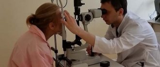
Checking for preservation of the corneal reflex
Only an ophthalmologist during an examination can confirm its presence or absence. This verification procedure is quite simple, absolutely painless for the patient and does not take much time. When performing it, the doctor must be careful and gently touch the eyelid and the eye itself. The examination is carried out in several stages:
- the patient is placed on the couch;
- then the doctor lifts the patient's eyelid;
- Next, the specialist uses a damp piece of cotton wool to touch the cornea and check for a reaction.
If the corneal reflex is normal, then the eyeball should immediately roll back, as in a healthy sleep. If nothing happens, then the patient may have problems with the trigeminal and facial nerves.