Congenital ptosis is the inability of the eyelid to fully open the eye from birth, i.e. it is always in a lowered state.
[toc]
In general, pathology is observed in people of different sexes and ages and can be caused by a variety of reasons. With ptosis, the eyelid muscle cannot raise it to the proper height and the eye always remains closed halfway or more. The pathology has varying degrees of severity; at the last stage, the eye is almost completely closed.
It would seem that this problem is minor and is of a cosmetic nature, but in severe forms of the disease the eyelid closes the eye so much that the person is always forced to strain the eyebrow muscles to keep the eye half open, and sometimes throws back the head to see through the affected eye. This pose even received a name in medicine - “stargazer pose.”
The essence of pathology
Most often, ptosis is bilateral, that is, both upper eyelids are drooping, while acquired ptosis is usually unilateral, as it develops due to injury or some disease.
Congenital ptosis of the upper eyelid is inherited from one of the parents, although it is not a fact that a father or mother with ptosis will necessarily have a child with underdeveloped muscles of the upper eyelid.
The cause of pathology of the upper eyelid in children may be a disease of the oculomotor nerve that developed in utero and, accordingly, is considered congenital. This nerve not only moves the eye, but also controls the raising and lowering of the eyelid.
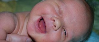
A fairly rare cause of congenital ptosis is the so-called palpebromandibular syndrome. It is expressed as follows: the nerve impulse into the muscle that lifts the eyelid enters from the ternary nerve during chewing. That is, ptosis in a child appears only in a calm state, but when he chews, the eyelid rises to a normal level. With this syndrome, strabismus and amblyopia often develop.
The rarest congenital pathology is blepharophimosis. The disease is characterized by a short palpebral fissure. This type is usually bilateral, often resulting in the effect of inverted lower eyelids. The presence of blepharoptosis prevents the child from closing his eyes even when he is sleeping.
Upper eyelid ptosis
Classification
1. Ptosis can be congenital or acquired.
Congenital ptosis occurs due to underdevelopment of the muscle that lifts the upper eyelid (levator) or disruption of its innervation associated with hereditary genetic abnormalities or pathology of pregnancy and childbirth. Congenital ptosis in a large percentage of cases is combined with other visual anomalies: strabismus, amblyopia, anisometropia.
Acquired ptosis, depending on the cause that caused it, is divided into:
- aponeurotic – associated with stretching or weakening of the aponeurosis of the muscle that lifts the upper eyelid. This includes senile (involutional) ptosis as a manifestation of the natural aging process of the body; ptosis that occurs after ophthalmological operations and injuries of the levator aponeurosis;
- neurogenic - when the nervous system is damaged as a result of any diseases or injuries (multiple sclerosis, consequences of a stroke, etc.) Ptosis can develop, for example, with paralysis of the cervical sympathetic nerve, since the muscle innervated by the sympathetic nerve is involved in raising the eyelid. In this case, simultaneously with the drooping of the upper eyelid, retraction of the eyeball (enophthalmos) and narrowing of the pupil (miosis) are observed. This symptom complex is called Horner's syndrome.
- mechanical ptosis – occurs when the eyelid is deformed by scars, tears, foreign bodies
- apparent (false) ptosis – with excessive skin folds of the upper eyelid (blepharochalasis), with severe hypotony of the eyeball, ptosis with strabismus.
2. Ptosis can be unilateral or bilateral.
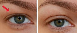
3. According to the degree of severity, they distinguish:
- partial ptosis – the edge of the upper eyelid is located in the upper third of the pupil;
- incomplete ptosis – the edge of the upper eyelid reaches the middle of the pupil;
- complete ptosis - the upper eyelid completely covers the pupil.
With ptosis, the mobility of the upper eyelid may be reduced or completely absent. A drooping upper eyelid mechanically impedes vision, so the usual position of raised eyebrows arises; in severe cases, especially in children, a forced compensatory position develops: the head is raised, the eyebrows are raised, the forehead is wrinkled. Ptosis in children interferes with the normal development of the visual analyzer, contributing to the development of amblyopia (“lazy” eye) and strabismus, and narrowing of visual fields. Depending on the severity of the drooping eyelid, one or another degree of visual impairment is noted.
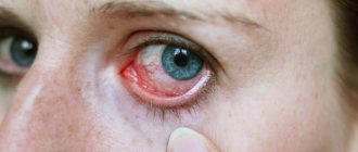
Other manifestations of ptosis include: eye irritation, fatigue due to constant muscle tension, and possible double vision. If ptosis is accompanied by the inability to completely close the eyes, then symptoms of dry eye, chronic conjunctivitis and keratitis are observed.
In rare forms of ptosis, there may be other symptoms. For example, with Marcus-Gunn syndrome, ptosis disappears when the mouth is opened and the jaw is clenched tightly.
Acquired ptosis
Speaking about ptosis, we must not forget about situations where drooping eyelid occurs as a result of injury or disease, that is, acquired ptosis. This type of disease is observed much more often than congenital:
- When the oculomotor nerve is paralyzed, neurogenic ptosis occurs. It can be caused by diabetic neuropathy or a tumor compressing the oculomotor nerve. If the cornea is injured, or if it is covered with ulcers, neurogenic ptosis can be artificially induced to protect it.
- Myogenic ptosis is characterized by increased manifestations of the disease over time. For diagnosis, endorphin is used, which can relieve signs of pathology for a short time.
- Aponeuric ptosis is common in older and older people and develops when the tendon of the eyelid muscle stretches or becomes detached from the bone to which it was originally attached. This phenomenon leads to weak muscle tension and the eyelid does not rise completely.
- In mechanical ptosis, the eyelid is shortened as a result of swelling or scarring.
Acquired ptosis
Ptosis of the eyelid: symptoms
Often with ptosis, eyelid mobility is reduced or completely absent. Sometimes, due to a drooping eyelid, a person involuntarily raises his eyebrows and wrinkles his forehead. Some Patients - often children - in order to see better, tilt their heads back and take the so-called “stargazer pose”.
Drooping of the eyelid interferes with blinking movements, and this leads to overwork, dryness and soreness of the eyes. Sometimes amblyopia (“lazy eye”) develops, since the eye under the constantly closed eyelid does not work. Chronic conjunctivitis and keratitis also occur with ptosis.
Symptoms of pathology
The main manifestation of ptosis is drooping of the eyelid, and this is typical for any age of the patient. However, the disease has a number of other signs, which determine the final diagnosis:
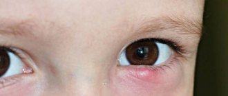
- The eyes are irritated and there is redness.
- When closing an eye, a person makes significant efforts.
- The eyes quickly get tired, since the patient is forced to always keep the muscles of the eyelid in a tense state.
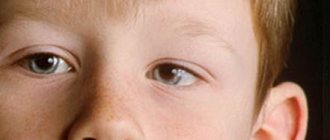
- In children, the “stargazer pose” is observed.
- Children with congenital ptosis are often diagnosed with strabismus.
Development of ptosis
Diagnostics
It is almost impossible to confuse ptosis with anything or not notice it, especially for an ophthalmologist. It is much more important (and in some cases more difficult) to establish the immediate cause of drooping eyelid, since further treatment measures depend on this. In particular, the congenital/acquired nature of ptosis, the presence of concomitant chronic diseases or abnormalities of intrauterine development, the presence of a hereditary factor and other anamnestic data are important. Thus, congenital ptosis is often accompanied by the so-called. semilunar fold of skin (epicanthus) in the inner corner of the eye, sometimes muscle paralysis and paresis, etc. The degree of drooping, residual motor activity of the eyelid, coordination of movements of the eyeballs, facial symmetry, and eyebrow mobility must be examined and taken into account.
The increased risk of the previously mentioned complications, obvious and latent, necessitates visimetry (accurate measurement of visual acuity in each eye), ophthalmotonometry (measurement of intraocular pressure), ophthalmoscopy (examination of deep ocular structures, in particular the fundus. In some cases, where there are reasons suggest a pathology of the central nervous system (strokes, tumors, multiple sclerosis, and many others), tomographic diagnostic methods are prescribed; if the presence of a foreign body or post-traumatic complications is suspected, orbital radiography or CT may be indicated. If necessary, related specialists are involved, most often neurologists (and also oncologists, infectious disease specialists, geneticists, etc.).
Diagnostic measures
To treat ptosis, it is necessary to understand what exactly caused it; only after this a treatment regimen and strategy is developed. To find out the reasons, the patient is carefully examined:
- An anamnesis is being collected. During a conversation with the patient, the doctor finds out whether there were similar cases in the patient’s family. What illnesses did the person suffer from childhood and did he have any injuries to the eyelid or head. A well-collected anamnesis can reveal the cause of the disease without the use of laboratory tests.
- An examination by an ophthalmologist reveals myopathy, strabismus, or increased pressure inside the eyeball.
- If, during an eye examination, weakness of the superior rectus muscle of the eyelid is revealed, then a conclusion is drawn about the congenital nature of the pathology.
- The oculomotor nerve and the pathology that caused its paralysis can be detected by magnetic resonance imaging of the head.
Degrees of ptosis
Features of drooping upper eyelid in newborns
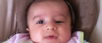
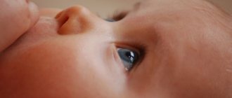
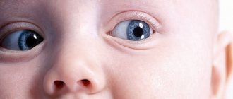
Ptosis is a drooping of the upper eyelid that occurs due to weakening of the muscles. It can develop in one eye or in both eyes at once.
However, if the eye is half-covered by the eyelid, it is not at all necessary that the child has ptosis. There are other reasons for this, which your doctor will tell you about. The specialist will evaluate visual acuity, the condition of the pupils, the ability of the eyes to move, the elevation of the eye muscle and the height of the eyelid.
If the doctor nevertheless diagnoses ptosis of the upper eyelid in an infant, do not panic. This is not a visual defect, but just a cosmetic defect that can be eliminated. Surgery is not performed on newborn babies: all that can be done is to regularly consult an ophthalmologist who will monitor the condition of the eyes. Correction of ptosis will be possible later, when the child is a little older. Surgery is possible only at three years of age, but in severe cases the operation is performed immediately.
In some cases, a drooping upper eyelid can block vision. In such cases, medical intervention is necessary, as the quality of vision may deteriorate significantly. Ptosis also causes the child to have to tilt his head back to see objects. This has an adverse effect on motor skills.
Treatment of ptosis
Ptosis in a child can be cured in various ways. Everything depends on the form and severity of the disease, in addition, the cause that caused the pathology matters. Thus, an individual treatment strategy is developed in each individual case.
Treatment may be conservative. During its course, the muscle of the eyelid is affected by medication. It helps only in mild forms of the disease and is rarely used due to its poor effectiveness.
Therapeutic treatment is usually used for neurogenic ptosis. During treatment, the function of the oculomotor nerve is restored. For this purpose, UHF therapy, galvanotherapy and other methods of influence are used. In some cases, it is necessary to fix the patient’s eyelid with a special plaster, which reduces the patient’s social activity, since in this state it is difficult to maintain life and communication at the same level. Surgical treatment is used in cases where conservative and therapeutic treatment has failed.
Surgical treatment of ptosis in children
Therapy should not be delayed, especially in children. As soon as a child shows signs of ptosis, he is referred for examination and immediate treatment. After all, even the slightest deviation of the eyelid can cause such manifestations in a child - curvature of the spine (since he will constantly throw his head back and to the side), strabismus and myopia.
And these side effects are sometimes more difficult to cure than the pathology itself. Adult patients cannot have such severe complications, since their body has long been formed, and it cannot change significantly during illness.
During surgery, the eyelid muscle is sutured to the frontalis muscle in order to increase its mobility. This kind of intervention slightly increases the mobility of the eyelid and the cosmetic effect is rather weak. However, it is simple, which cannot but affect the postoperative period; the patient recovers in a matter of days.
The other method is more complex, but much more effective. This is a resection of the muscle that holds the eyelid up. During this operation, through an incision in the skin, the surgeon gains access to the desired muscle and sutures it, making it shorter. After the wound heals, the sutured eyelid muscle successfully lifts and holds it. In addition, the postoperative scar is sutured with a cosmetic suture, so that after complete healing it is practically invisible.
Sutures from the skin of the eyelid are removed after 4-5 days, and with proper treatment of postoperative injuries, a person can return to normal life within 2 weeks. The only disadvantage of such an operation is its complexity, so only an experienced surgeon can perform it.
There is a surgical operation to install a duplicator of the muscle aponeurosis. This procedure also shortens the muscle that controls the eyelid and restores the person's ability to see equally in both eyes. The operation is complex and is performed only in specialized clinics by specialized specialists.
Acquired unilateral ptosis
Prices for surgery
The cost of surgical removal of upper eyelid drooping in our ophthalmology center, depending on the category of complexity, ranges from 30,000 (1st category) to 40,000 (2nd category) rubles per eye.
The complexity of the operation and the choice of method for correcting ptosis is determined by the surgeon at the initial consultation.
In conclusion, it is necessary to once again emphasize the importance of timely contacting an ophthalmologist at the first signs of ptosis, no matter what caused it. This is especially true for childhood ptosis, given the speed of all processes (including the pathological developments described above) at this age. A fairly simple and safe operation to eliminate blepharoptosis is a much more reasonable solution than long-term therapy for amblyopia, strabismus, anisometropia, or other complications directly related to “just” a drooping eyelid, which sometimes takes years and does not guarantee one hundred percent results.
Diagnosis of ptosis in a child
Despite the bright external manifestation, ptosis of the upper eyelid requires professional diagnosis. This is necessary to establish the cause of its occurrence, which will help determine how to treat ptosis of the upper eyelid.
Diagnosis of ptosis is performed fairly quickly. To do this, the specialist measures the height of the eyelids, checks their symmetry, and determines the completeness of movement. The ophthalmologist also pays attention to visual acuity and the condition of the fundus.
As a rule, the following diagnostic procedures are prescribed:
- Ultrasound of the eyeball;
- computer perimetry, electroencephalography;
- OCT.
In rare cases, an MRI is necessary to make an accurate diagnosis. Also, the ophthalmologist must prescribe a consultation with a neurologist.
In addition, the specialist will find out information about the baby’s family, taking into account heredity. If ptosis is acquired, the doctor may ask about the presence of other diseases that can cause ptosis.
Treatment of the impending century
Differentiation of congenital and acquired forms, determining the causes of the disease help the doctor select a treatment regimen. Conservative treatment methods include the following physiotherapeutic procedures:
An important part of the treatment of such pathology is special gymnastics.
- physiotherapy;
- exposure to direct electric current (galvanization);
- ultra-high frequency therapy;
- massage;
- application of paraffin applications.
If after 6-9 months conservative methods of therapy do not produce an effect, then, in the absence of contraindications, surgical intervention is prescribed. If a disease has been identified that provoked ptosis, then the underlying disease is treated first. Surgical intervention is performed for partial drooping of the eyelid at the age of 13-16 years, and for complete drooping in preschool children due to the threat of amblyopia.
Before surgery, the affected skin fold is fixed with a plaster to prevent the appearance of strabismus. In children, surgery is performed under general anesthesia. Excess skin is removed, the corresponding muscles are tightened, a cosmetic suture is applied, and then a bandage is applied, which is removed after a few hours. Postoperative hematomas and swelling on the face disappear in about a week.
Congenital ptosis of the upper eyelid
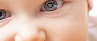
Congenital ptosis occurs in newborns if the baby is predisposed to hereditary diseases. Sometimes ptosis in a baby can occur even in cases where the parents had mild ptosis. In rare cases, a newborn develops the following pathologies, accompanied by drooping of the upper eyelid:
- Palpebromandibular syndrome: during it, when the masticatory muscles move, the upper fold of the eyelid moves.
- Horner's syndrome is congenital: it is accompanied by drooping of the upper eyelid, different eye colors, sunken eyeball and an almost complete absence of pupillary reaction to light.
- Blepharophimosis: a genetic pathology in which the palpebral fissure is too small and the muscles responsible for the work of the upper eyelids are underdeveloped.
All these diseases are called pseudoptosis. In each of them, excess skin folds hang over the eye, but the eyelids remain raised. They do not require treatment in themselves, but they resort to it in cases where they cause discomfort in an older child.