A person’s ability to simultaneously see with both eyes and obtain a three-dimensional view of surrounding objects is called binocular vision. If this ability is impaired, it affects the sharpness, clarity and other features of visual perception. Special exercises will help restore binocular vision in adults.
In this article
- How does binocular vision work?
- Is it possible to restore binocular vision?
- How to restore binocular vision at home?
- Effective exercises to restore binocular vision
- Simple exercises at home
- Effective Focusing Exercises
- Looking at stereograms is useful for binocular vision
How does binocular vision work?
With normal binocular vision, each human eye receives a separate image, after which the brain converts them into a single three-dimensional image. For a number of reasons, the eyes of adults can work separately, and then the merging of two images into one does not occur. This negatively affects the acuity of visual function and makes it difficult to adequately perceive the distance between objects and evaluate them in a three-dimensional perspective.
Binocular vision disturbances can occur if there is a large difference in the optical power of the right and left eyes, the eyeballs are located asymmetrically, the functioning of the nervous system or eye muscles is disrupted, and pathological changes are observed in the retina, cornea or lens. The most common ophthalmopathologies that accompany binocular vision disorders are strabismus and amblyopia.
Lack and failure of binocular vision
Binocular vision impairment develops in various ophthalmological pathologies. Almost all of them can cause the condition if the transmission of nerve impulses to the visual centers of the brain is disrupted. There are main pathologies in which monocular vision most often appears:
- diseases in which the refraction of light passing through the eyeballs differs;
- pathologies leading to damage or dysfunction of the muscles located around or inside the eyeball (inconsistency of actions is formed);
- penetration of a bacterial agent into the surface and internal structures of the eyes;
- the development of benign and malignant tumors that are located inside or near the eyes, compressing internal tissues;
- mechanical damage and burns leading to disruption of the passage of the light beam through the eye, displacement of the eyeball in the orbit;
- diseases leading to bone displacement, which disrupts the normal structure of the orbit:
- cataract in one eye - clouding of the lens due to an increase in the fractions of insoluble proteins;
- glaucoma in one eye - increased secretion of intraocular fluid, which leads to an increase in chambers pressing on the retina and optic nerve;
- retinal disease (blood malnutrition, thermal and chemical burns, mechanical damage, detachment).
Monocular vision can be caused not only by pathologies of the eyeballs, but also by the brain and nervous tissue:
- diseases of the optic nerve (inflammation, damage);
- intoxication of the body, which causes brain damage;
- intracranial hematomas compressing the center of vision:
- mechanical impact (bruises, fractures, concussions, knife and gunshot wounds) damaging the center of vision;
- neuralgia;
- infections and viruses penetrating the brain, spreading to the center of vision.
To restore binocular vision, the doctor must identify the root cause of the condition. If this point is not met, the disease will return again.
We recommend reading: Differences between binocular vision and monocular vision
Is it possible to restore binocular vision?
Today in ophthalmology there are two main methods of restoring healthy binocular vision: orthoptics and diploptics.
- Orthoptic treatment is aimed at improving the way the eyes work together as a single organ of vision. To achieve this goal, special exercises are used to stimulate the eyes to connect two pictures together.
- Diploptics is a technique for restoring vision, the essence of which is to cause the patient to experience double vision of the object of perception. This method stimulates a person’s ability to independently restore binocular stereoscopic vision. To perform diploptic exercises, the level of strabismus should not exceed 7 degrees.
The final stage of treatment for binocular vision disorders is special therapeutic exercises for the eyes, the task of which is to increase the mobility of the eyeballs.
Mechanism and conditions for binocularity
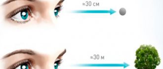
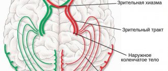
If a person can combine two images from both retinas into one picture, he has developed binocular vision. This unification occurs in the cerebral cortex and is ensured by the fusion reflex. To do this, it is necessary that the brain receives two absolutely identical pictures from both eyes, that is, they must match each other in size and shape. For spatial vision to function, light rays must fall on identical points on the retina. These points are called corresponding points. Each point on one retina has a corresponding point on the other retina. If light falls on them, the pictures come together, as if superimposed on each other. If focusing occurs at different points, then the pictures are different. This leads to diplopia - double image.
Vision will be binocular if:
- there is the ability for fusion (merging images in the brain);
- the eyeballs move in concert and are located symmetrically and parallel in the same frontal and horizontal plane;
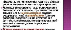
- visual acuity is sufficient for normal visual perception of the environment (not lower than 0.3-0.4 diopters);
- there is no aniseikonia, when the eyes see images of different sizes;
- there are no opacities of the cornea, lens or vitreous body, which are accompanied by a decrease in visual acuity in one eye;
- there are no pathologies of the central nervous system.
There are many conditions for the formation of stereoscopic vision. Moreover, the above factors apply not only to the organs of vision, but also to other systems of the body. Lack of binocularity can be a symptom of both eye diseases and non-ophthalmological pathologies.
How to restore binocular vision at home?
Restoring binocular vision at home can only be done if such therapy has been prescribed by an ophthalmologist. After diagnosis, the specialist decides whether standard therapy will help you or whether serious surgical treatment will be required.
Often, binocular vision disorders can be treated either on an outpatient basis in the clinic or at home, following the doctor’s instructions. As a rule, they include wearing special glasses, performing a set of exercises and procedures aimed at restoring balance between the eyes.
Strabismus is a type of binocular vision disorder.
According to experts, the most common disorder of binocular vision today is strabismus. It consists in the deviation of the visual axes of one or simultaneously both visual organs from the point of direction of the object in question. Due to strabismus, there is a mismatch in the functioning of the eyes, and binocular vision needs correction. The most common type of pathology today is the so-called monocular strabismus, which doctors can also call dysbinocular amblyopia.
With this visual impairment, usually only one eye squints, the optical power of which is often reduced, and therefore it is not used by the brain, since it is practically impossible to read information from it. The second type of this disorder is called alternating strabismus, or, more simply, alternating. In this case, both organs of vision squint with the same deviation and can look in turn. Simply put, both eyes are used in the visual process, but alternately.
Effective exercises to restore binocular vision
Regular training of binocular vision is an important condition for its restoration. There are several training systems that were developed by specialists in the field of ophthalmology - Kashchenko, Bates, etc. Only an ophthalmologist during an in-person examination can select the optimal set of exercises for a particular patient.
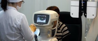
Basic eye training is carried out in a clinical setting using special equipment. But, in addition to hardware treatment, experts recommend performing special gymnastics at home as an additional measure. Below we present several exercise options that are often prescribed to restore binocular vision in an adult and are suitable for home use.
The influence of binocular vision on career choice
Impaired binocular vision not only entails certain limitations in the normal perception of the outside world, since in connection with it it is impossible to correctly, and most importantly, quickly assess the spatial relationships of surrounding objects. In most cases, people suffering from this pathology face a serious question in choosing a profession. Unfortunately, with such vision it is impossible to work as an astronaut, pilot, driver, surgeon, etc. In extreme cases, a medical occupational examination can be carried out to identify the severity of the pathology.
Simple exercises at home
Draw a vertical line 10 cm long and 1 cm wide with a marker on a sheet of paper. Attach the sheet to a well-lit wall at eye level and move one meter away from it. Now tilt your head so that you see the drawn line in a single copy, that is, it does not double before your eyes. After the double image merges into one, begin to slowly move your head down, without taking your eyes off the strip. Continue until the image begins to double in size. Repeat the same steps from the very beginning, only moving your head up and then alternately to the sides. You need to do this exercise at least three times a day for five minutes. In the same way, you can train binocular vision by using any small object instead of a drawn line.
Diagnostics
The ability for binocular vision begins to develop in infancy (approximately the 3rd month of life) and begins with the consolidation of a special reflex - binocular fixation. It is finally formed by the age of 12, then gradually improves and continues to change throughout human life. For a reliable diagnosis, the functional ability of both eyes must be tested simultaneously.
There are several methods for testing binocular vision. Of these, non-hardware methods can be distinguished, which have both advantages (speed, ease of diagnosis and lack of cost) and disadvantages (low accuracy):
- 1. Test with the appearance of double vision when the eye is displaced. The easiest way to diagnose: is done by lightly pressing your finger on the eye through the eyelid.
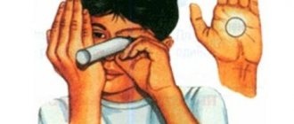
- 2. Sokolov's test. It is carried out using a hollow tube with a diameter of about 3 cm and a length of 20-30 cm. The patient places the tube to one eye, and places his palm on the side of the second eye near the far end of the tube. If the images are combined and the patient sees as if through a 'hole' in the palm, then he has normal binocular vision.
- 3. Kalfa method (miss test). The patient holds two sharpened pencils (one vertically, the other horizontally) in his arms outstretched in front of him and tries to connect their tips. With binocular vision, subjects easily cope with the task. If there are misses, the patient's vision is monocular.
- 4. Reading with interference. The patient is asked to read a short text in a book and a reading obstacle is placed 3-5 cm from his nose on the side of either eye. A pencil is usually used as a hindrance. With binocular vision, despite the interference, the images are combined and the patient can easily read the entire text. With monocular vision, part of the text is blocked and inaccessible for viewing.
- 5. Test for installation movement. A pencil or other small object is placed in front of the patient's eyes at a distance of 20-30 cm. When a person covers one eye with his hand, that eye makes a movement, deviating towards the outer corner. After the patient removes his hand, with binocular vision the eye makes a return (setting) movement in the opposite direction. In the absence or slight movement, monocular vision is diagnosed. In children, the presence of such movements is checked using a special prism of 10-12 diopters. The baby's attention is attracted to some interesting object. During examination, the prism is quickly placed and removed in front of one eye, then in front of the other. With binocular vision, both eyes will make reciprocal movements.
More accurate diagnostic methods are hardware research methods, which are carried out using special instruments:
- 1. Belostotsky-Friedman test. It is carried out using a four-point color test (TsT-1 device). It is based on dividing the fields of vision of both eyes using filters of different colors. Easy to use, it makes it possible to determine the nature of vision, the angle of strabismus and the dominant eye. This method of determining and assessing binocular vision involves 'soft' field separation ('soft' haploscopy) using color (red and green) filters in conditions closer to natural ones.
- 2. Synoptophore. It works on a similar principle. Allows you to identify the correspondence ability of the retina and determine the angle of strabismus.
Synoptophore
To test binocular vision in 3-4 year old children, color tests are performed in the form of objects that are well known and familiar to children’s perception: tree, fruit, car, etc.
Effective Focusing Exercises
- Option 1.
Place a visual object on the wall and move away from it at a distance of two meters. Extend your hand in front of you and raise your index finger up. It should be located in the same visual axis with the object on the wall. First, focus your attention on a visual object and observe it through your fingertip. It will seem to you that your finger is splitting into two. Then change your focus and focus it on the tip of your finger. Now you should see the hand as a solid image, and the object on the wall will split into two. Keep in mind that the clearest image will be on the side of the eye with the best vision. Sometimes you can train with one eye closed so that the other one works at maximum efficiency at this time. This exercise needs to be repeated several times. Alternately switching focus from near to far objects trains binocular vision, and over time you will notice that image clarity increases.
- Option 2.
Take a bright drawing and first examine it in detail in its entirety. Then select a small element of the image and try to concentrate on it, outline its outline with your eyes, examine the inside, not paying attention to the main background. Then do the same thing, but choose an even smaller element to focus on.
Checking binocular vision using the Kalf method
Another simple but effective way to test binocular vision is the Kalf method. To carry it out you will need two pencils, two knitting needles or other objects of a similar shape with sharp edges. Take them in different hands. Place one pencil vertically and the other horizontally, then try to connect them so that you get a right angle. With monocular vision, patients usually miss the mark and fail to achieve the correct connection even after several attempts. If stereoscopic vision is normal, such a test will not cause difficulties.
What is strabismus?
Strabismus is an abnormal position of the organ of vision, in which deviation of one or both eyes is detected in turn when looking straight. With a symmetrical arrangement, the image falls on the central part of the retina of each eye. Then two separate patterns in the cortical part of the organ of vision are combined into one.
With the development of strabismus, fusion does not occur and as a result, the central nervous system, trying to protect itself from diplopia, “crosses out” the picture received from the squinting eye. If a person remains in this state for a long time, amblyopia begins to develop (exclusion of the damaged eye from the visual process).
Depending on the type of strabismus, the disease is divided into converging, divergent, superior or inferior. Strabismus is not only a cosmetic defect; it interferes with the ability to fully perceive the environment. If the pathology develops in children or the elderly, it is often accompanied by diplopia.
| When strabismus is detected in children, it is necessary to take urgent measures. Otherwise, vision problems may occur. This is due to the fact that the central nervous system gets used to ignoring the data received from the squinting eye. As a result, the child does not learn to see with this eye and amblyopia appears. |