Amblyopia - what is it?
Amblyopia
is a decrease in visual acuity in one or both eyes due to various pathologies of the visual apparatus in children and adults, which cannot be optically corrected with glasses or contact lenses. The simpler term for amblyopia, “lazy eye,” speaks for itself. The eye with the pathology sees worse; the entire visual load is transferred to the healthy organ of vision, unless, of course, the amblyopia is bilateral. Gradually, an eye with amblyopia, if untreated, can completely “switch off” from the vision process: the brain blocks blurry images coming from the amblyopic eye, which, in turn, leads to a sharp decrease in visual acuity of the healthy eye. Thus, in the most advanced cases, amblyopia can lead to complete blindness.
The diagnosis of “amblyopia” does not imply a universal medical treatment program for Patients, due to the multiplicity of causes and types of manifestations of the disease.
Amblyopia symptoms
Most patients with a slight degree of amblyopia are not aware of the presence of this disease (with the exception of patients with strabismus).
The main symptoms of amblyopia are the following:
- different optical powers of the eyes;
- disorientation in unusual places and unfamiliar conditions;
- impaired coordination of movements;
- deviation of the eyes when reading;
- closing an idle eye;
- tilting the head when looking at an object of interest;
- color vision impairment;
- decrease in the level of light sensation;
- inability to fix the eyes steadily;
- rapid fatigue;
- headache;
- tinsurroundings.
If you notice similar symptoms, consult a doctor immediately. It is easier to prevent a disease than to deal with the consequences.
Main types of amblyopia:
- Anisometropic amblyopia - occurs when there is a difference in the refraction of the two eyes: it can develop even with a difference between the eyes of 1 spherical diopter. At the same time, the worse-seeing eye perceives the image unclearly, which over time leads to addiction and loss of high visual acuity.
- Refractive amblyopia occurs with uncorrected myopia, hypermetropia or astigmatism. When treating this type of amblyopia, it is necessary to prescribe glasses or contact correction, and conduct courses of hardware treatment for amblyopia to stimulate the visual analyzer. When refraction is stabilized and after the age of 8-9 years, it is better to perform excimer laser correction of refractive errors in the affected eye.
- Obscurational amblyopia occurs when vision loss occurs due to an eye disease, such as cataracts or grade II ptosis.
- Strabic amblyopia is characteristic of strabismus. In this case, amblyopia and strabismus are treated simultaneously.
In any case, programs for the treatment of amblyopia are prescribed by the doctor after a thorough diagnosis and taking into account concomitant eye pathologies. Before treating amblyopia, it is necessary to clearly determine its cause, the complex effect on which will determine the success of amblyopia correction. If the Patient consults a doctor on time, in the vast majority of cases, amblyopia can be treated with hardware and does not require surgery.
Courses of hardware-computer treatment are the only effective method of treating amblyopia, as they force the amblyopic eye to work, which leads to an increase in visual acuity and a decrease in the degree of amblyopia in children and adults.
Amblyopia - symptoms and treatment
To understand the pathogenesis of amblyopia, it is necessary to know the pathway of the visual analyzer, which allows you to perceive and analyze all visual stimuli.
After a visual stimulus enters the retina, the impulse from the photoreceptors travels along the ganglion nerve cells through the optic nerve to the chiasm, where the visual pathways partially intersect. After the chiasm, the optic tracts begin and end in the lateral geniculate body of the thalamus. Most of the fibers from the thalamus go as part of the visual radiation to the primary visual cortex - area V1, which is located in the occipital lobe of the brain. All these connections have a strict topographical organization, that is, each previous structure is sequentially projected onto the next.
The cortical layers of the primary visual cortex consist of groups of cells called “ocular dominance columns.” The cells within these columns respond to certain visual stimuli. In addition, each such column responds more to a stimulus coming from the right or left eye. Next, the information is transmitted to other parts of the visual cortex for subsequent processing.
The formation of ocular dominance columns depends both on the correct anatomy of the visual analyzer, formed in utero, and on adequate visual experience. Synaptic connections between the eye and brain are either strengthened by interconnected activity or weakened by uncoupled activity.
At birth, the foveal cones of the retina, which are responsible for converting light stimulation into nervous stimulation, are just beginning to differentiate. Their development actively occurs during the first 24 months of a child’s life and continues throughout the entire critical period of development.
Monocular deprivation (when one eye is completely “turned off” from vision) during a critical period of development leads to a pronounced decrease in the representation of the affected eye in the primary visual cortex and an increase in the representation of the healthy eye. If the visual system is affected by factors such as blurred visual stimuli or lack of binocular vision during this critical period of development, visual acuity will progressively decline. This process will continue until the end of the critical period, after which visual acuity will stabilize.
In isotropic amblyopia (when the refractive error is the same in each eye), a blurred image is present on both retinas. The resulting blurred image disrupts the normal neurophysiological development of the visual system. This leads to the fact that even when the refractive error is eliminated, the image blur remains.
With anisometropic amblyopia (when a blurred visual image comes from only one eye), the development of the visual analyzer of only the affected eye is disrupted. Over time, the visual system begins to actively suppress the blurred image, causing spatial changes in the ocular dominance columns, which leads to loss of visual acuity and disturbances in binocular vision.
In the case of dysbinocular amblyopia (caused by strabismus), the central areas of the retinas receive different visual information. As a result, the image from the deviated eye is suppressed. A long-term deviation of the eye leads to anatomical and functional changes in the visual analyzer, which is clinically manifested by low maximum correctable visual acuity of the affected eye.
Extensive experiments conducted on animals have shown that with amblyopia, neuroanatomical disorders occur in various parts of the visual analyzer pathway. The initial change is atrophy or loss of neurons in the lateral geniculate body and the visual cortex corresponding to the affected eye. Therefore, the goal of treating amblyopia is to create equal representation of both eyes in the central nervous system. If amblyopia is successfully treated, these changes may undergo partial or complete reversal.
Amblyopia in children: causes, features, treatment
Amblyopia in children is not so rarely diagnosed. It manifests itself as a consequence of congenital or acquired strabismus, other eye pathologies, and also as an independent disease, which can be caused by a whole range of unfavorable factors:
- Prematurity or low fetal weight (less than 2 kilograms)
- cerebral palsy
- Unfavorable pregnancy, including infectious diseases suffered by the mother during pregnancy, alcohol consumption and smoking
- Genetic prerequisites for the development of eye diseases.
Parents should remember that it is necessary to diagnose and begin treatment for amblyopia in children as early as possible, since by the age of 7, that is, by the time the child goes to school, the formation of his visual apparatus is actually completed. This means that all subsequent treatment and correction of amblyopia will be difficult and, possibly, will produce less results than at an earlier age. That is why you should not neglect regularly checking your child’s vision with an ophthalmologist, especially if one of the child’s immediate relatives has vision problems.
Diagnostics
To diagnose amblyopia, the following manipulations must be performed:
- conduct comprehensive ophthalmological examinations (the ophthalmologist examines the palpebral fissure, the position of the eyeball, evaluates the reaction of the pupil to light);
- perform ophthalmological tests (visual acuity testing, color testing, perimetry, refraction test);
- ophthalmoscopy;
- biomicroscopy;
- examination of the fundus with a Goldmann lens;
- determination of the transparency of refractive media;
- Ultrasound of the eye;
- measurement of the strabismus angle on the synaptophore;
- carrying out refractometry and skiascopy;
- tonometry;
- electroretinography;
- consultation with a neurologist.
Hardware treatment of amblyopia in children and adults
Hardware treatment of amblyopia in children is generally similar to hardware treatment of amblyopia in adults - it is necessary to undergo vision diagnostics and repeat courses of treatment according to an individually prescribed scheme by a doctor: once every six months or more often. The success of amblyopia treatment also largely depends on careful adherence to recommendations and the patience of parents.
At the Eye Clinic of Dr. Belikova, special computer simulators await little patients, which will turn eye training into an exciting activity, and the treatment of amblyopia and strabismus into a game.
Despite the fully formed visual system, hardware vision treatment in adults using modern ophthalmological equipment also gives good results. Doctors at leading eye clinics have at their disposal every opportunity to improve a Patient’s vision with amblyopia and strabismus without surgical interventions using gentle and effective hardware treatment.
In one visit to the Eye Clinic of Dr. Belikova, you can undergo a complete diagnosis of visual acuity, receive recommendations from leading ophthalmologists and begin treatment.
Causes
The main causes of amblyopia are the following:
- strabismus (in this case, dysbinocular amblyopia is observed);
- high degree of farsightedness or myopia;
- severe astigmatism (refractive amblyopia);
- the presence of obstacles in the eye for the normal passage of sunlight (eyesore, cataract, scar after injury);
- damage to the oculomotor muscles;
- constriction of the pupil;
- drooping upper eyelid;
- emotional shock.
Software and computer techniques
Ophthalmologists, together with IT specialists, have developed many training and educational programs for the treatment of strabismus and amblyopia. They acquire particular significance for the treatment of children, since correction occurs in a playful way. Fascinating exercises help the child (who finds it difficult to explain in words the need for eye exercises and instructions for its implementation) to carry out the treatment course regularly and with interest. Proper systematic training helps restore binocular vision and activate the “lazy eye”.
What is "lazy eye"?
The problem of amblyopia (the “lazy eye” phenomenon) appears in the first years of a child’s life, when visual areas are actively forming in the brain. If this process is disrupted, vision deterioration occurs, which cannot be corrected by wearing contact lenses or glasses.
In this case, disturbances occur not at eye level, but in the cerebral cortex: the “lazy eye” does not focus on the object and a split image occurs. To avoid this, the brain learns to block signals from the “non-working” eye, and eventually it ceases to participate in the visual process.
Amblyopia usually begins to appear in infancy or early childhood. But it is difficult to diagnose: children rarely complain of poor vision, quickly adapting to it, and there are no pronounced symptoms. Therefore, parents need to be observant and suspect vision problems when indirect signs appear:
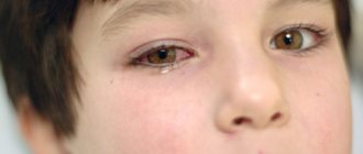
- Strabismus
- Anxiety in a child when closing one of the eyes
- When looking at an object, the child tilts or turns his head towards it