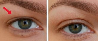
Epimacular membrane, or epiretinal fibrosis, is a consequence of collagen accumulation. As a result, a kind of membrane appears on the inner surface of the retina in the central region, leading to visual impairment.
The largest volume of the eyeball is accounted for by the vitreous substance, which consists of a jelly-like compound. Due to this, the shape of the eyeball is preserved. It contains a large number of fibers, which at one end are tightly attached to the retina of the eye. Over time, the volume of the vitreous decreases and it shrinks and then moves away from the surface of the retina. Most often this does not lead to any consequences. However, sometimes the surface of the retina is damaged, which triggers regeneration processes. As a result, scar tissue forms and becomes tightly attached to the retina. If the scar tissue contracts, the shape of the retina changes. If it is located in the central macular region, then the clarity of central vision is reduced.
Causes
The formation of cellophane retinopathy occurs due to damage to the surface of the retina. Active cell division begins, resulting in the formation of cells capable of absorbing and digesting foreign bacteria, collagen cells and fibrocytes.
The development of fibrosis occurs more often in old age. Typically, the pathological condition is found in patients over 65 years of age.
A web-like whitish formation is associated with:
- posterior vitreous detachment;
- vascular pathologies of the retina;
- diabetic microangiopathy;
- postoperative changes;
- intraocular inflammation;
- retinal tears.
Often this disease develops for no reason. This epiretinal membrane (EMM) is called idiopathic. That is, it is of unknown origin and is associated with age-related changes in the body.
Etiology
The appearance of a fibrous membrane is provoked by damage to the superficial layers of the retina. Neuroglial cells migrate to the surface of the fundus and form a fibrous film, vaguely similar to cellophane. The epiretinal membrane interferes with the passage of light rays, which is the main etiological factor in vision loss.
Epiretinal fibrosis is associated with the following diseases:
- retinal tears and detachment,
- diabetes mellitus types I and II,
- occlusion of the central vein by blood clots,
- post-traumatic and postoperative changes,
- intraocular inflammatory processes.
If a specific causative factor has not been identified, epiretinal fibrosis is called idiopathic. The risk of developing this disease increases with age, and men and women get sick with approximately the same frequency. The risk of developing pathology significantly increases due to tobacco smoking, alcohol abuse, and the predominance of excessively fatty foods in the diet. In addition, the initial signs of the disease are often detected in people who spend long periods of time looking at computer or television screens.
Symptoms
The clinical picture of the disease at the first stage of development is hidden. The formation of MEM resembles a thin transparent film. Gradually it begins to deform the retina, tightening it. The retina becomes wrinkled with folds.
In the absence of therapy, cellophane retinopathy thickens and becomes harder. Changes in tissues lead to increasing sclerosis of the vessels of the fundus, then to the formation of edema and cause ruptures, or rather a small flat detachment. The condition is accompanied by a significant deterioration in visual perception. Formal vision suffers to a greater extent. This occurs due to the location of the hymen above the macula.
Retrolental fibroplasia leads to blurred visual perception, distortion of the contours of objects and the image as a whole, and double vision.
Fibrosis of the CT and retina can be characterized by retinal and preretinal hemorrhages of varying sizes, which cause opacification of the posterior vitreous.
Intraocular sclerosis affects the set of cellular nutrition processes that ensure the vital activity of tissue cells and organs of the visual system. This process leads to retinal degeneration, the emergence of sequential posterior subcapsular (cup-shaped) cataracts, post-traumatic aseptic uveitis and a secondary increase in intraocular pressure.
The disease is serious and requires timely diagnosis and therapy. Fibrosis leads to a number of complications if help is not provided on time.
What is epiretinal fibrosis of the eye?
Epiretinal fibrosis is an ophthalmological disease accompanied by the appearance of a cobweb-like film in the middle of the retina, which subsequently leads to deformation of the retina, as well as the formation of folds and wrinkles on it.
Most often, the pathology develops unnoticed by a person, is progressive in nature and occurs in people over 65 years of age. The transparent membrane, tightening the retina, begins to thicken over time and becomes rigid, which can lead not only to swelling, but also to rupture of the retina.
Foci of fibrosis negatively affect the vessels of the retina, cause swelling of the macula, and disrupt the functioning of the eye, which can lead to detachment of the inner membrane.
Diagnostics
To diagnose a pathological condition, an ultrasound examination, determination of optical power and intraocular pressure, and OCT are performed (a diagnostic examination that uses a beam of light, which is both a scanner and a microscope).
There are 4 stages of development of the pathological condition, which are determined by examining the visual organs using an ophthalmoscope:
- the first shows a yellowish formation;
- on the second - the structure of the retina is disrupted, it is up to 0.04 cm in diameter;
- on the third - the defect covers an area of more than 0.04 cm;
- on the fourth, Weiss rings are formed.
An ultrasound is performed if ophthalmoscopy turns out to be uninformative (for example, when the doctor discovered corneal opacities, cataracts, or vitreous opacities).
Additionally, FA (modern X-ray contrast examination of the optical system) is performed. It is necessary to assess the severity of edema and abnormalities of the choroid of the affected organ of vision. The structure of the film and macula can be studied in detail by OCT.
Clinical picture
As a rule, the initial stage of the disease is latent, but over time the fibrous membrane grows, displacing the retina. Swelling occurs, which is one of the key symptoms.
Over time, a macular hole is formed - a hole in the macula in its central part, which leads to a decrease in visual function.
Common symptoms of the disease are:
- distortion of the “picture” perceived by the eye,
- diplopia – splitting of visible objects,
- difficulties in reading small print, studying small details, etc.,
- complaints of fog, blurred vision,
- blurred vision, especially in poorly lit conditions.
In difficult cases (with diabetic retinopathy, arterial hypertension, severe injuries, etc.), a complication such as hemophthalmos may develop, in which there is profuse hemorrhage from the newly formed retinal vessels into the vitreal cavity, as a result of which the vitreous body becomes saturated with blood. Hemophthalmos is clinically manifested by a sharp decrease in visual acuity and the appearance of floating spots before the eyes.
Most often, with retinal fibrosis, changes in vision occur rather slowly. But depending on the presence of aggravating factors, rapid loss of visual function is possible. If you seek medical help in a timely manner and register with an ophthalmologist, there is a possibility of stopping the negative dynamics.
Treatment
The pathological condition is treated in two ways - traditional and surgical. Sometimes a cobweb-like whitish formation does not require therapy. Treatment is usually not required when visual function is only slightly impaired.
Traditional methods of treatment
Traditional treatment will not help get rid of vitreous fibrosis, but it helps improve symptoms.
Healers recommend using pharmaceutical chamomile, calendula flowers and lingonberry leaves. Medicinal herbs can be used individually or in combination. A decoction is prepared from them, infused and taken 2 times a day. The course of treatment is 45 days.
This decoction is useful because it relieves inflammation, pain, and prevents the development of dystrophy or its progression.
You can use tinctures prepared with vodka. 1 head of peeled garlic is poured into 1 liter of vodka. The medicine is infused for 2 weeks. Use for oral use within 2 months.
Diuretic folk remedies are useful if the disease is accompanied by dystrophy. Knotweed, violet and bearberry are mixed together. Take 1 tbsp. l. medicinal collection and pour a glass of boiling water (not boiling water). Use the infusion for 10 days.
Surgical method of therapy
This method is based on the removal of cellophane retinopathy. Surgery is prescribed when there is a strong decrease in the optical power of the diseased eye.
To remove the membrane, the patient sees a dentist, endocrinologist, and otolaryngologist. Conducts laboratory tests of urine and blood.
The operation is performed under local or general anesthesia (at the request of the patient or the doctor’s indications). It is performed in stationary conditions on the operating table.
After it, there are no large scars left on the face or eyes, since small punctures are made in the mucous membrane and white membrane. Having gained access to the vitreous body, it is first removed by performing a vitrectomy.
Then access to the membrane itself is opened, using a microscope and a diffusing lens, a device for supplying fluid to the eye and other special instruments, the epiretinal membrane is removed.
Immediately after surgery, the patient's visual perception will not be restored. Improvement occurs gradually, over several months or more. Vision is restored in 80–90% of cases.
The operation prevents blindness. After its completion, it is recommended to use antibacterial and anti-inflammatory drugs. The former will prevent infection from developing, the latter will relieve inflammation, which often develops after surgery.
If necessary, medications are prescribed for rapid tissue regeneration.
↑ 9. Tuberculous uveitis
Tuberculosis accounts for about 0.2% of all cases of uveitis. The risk group includes persons in contact with tuberculosis patients. The causative agent of this disease is Mycobacteria tuberculosis. There are three known ways of developing ocular tuberculosis: hematogenous, exogenous (through the conjunctiva), and contact. Most often, the pathogen enters the choroid via a hematogenous route and then spreads to adjacent tissues. The inflammatory focus is formed by a granuloma containing epithelioid cells, lymphocytes, and giant cells.
Complaints of pain, redness of the eye, floaters, decreased vision.
Characteristic symptoms : phlyctenulous conjunctivitis and sclerokeratitis develop in the anterior segment of the eye. interstitial keratitis, granulomatous iritis with infiltration of the iris and the formation of Coerre and Busacca nodules. When the posterior segment of the eye is affected, patients experience symptoms of necrotizing retinitis with periphlebitis, tuberculous granuloma, exudative retinal detachment, retinal vasculitis, miliary choroiditis, juxtapapillary chorioretinitis (Jensen), chronic subactive panuveitis (endophthalmitis), optic neuritis. The choroid is most often affected. If the patient is not prescribed proper therapy, the process takes on a chronic progressive course.
The diagnosis is made on the basis of the clinical picture, x-ray examination of the lungs, tuberculin test data, immunological research (detection of antibodies to the pathogen), bacteriological examination (determination of the pathogen). The tuberculous nature of the disease is confirmed by the positive effect of specific therapy.
Differential diagnosis is carried out with idiopathic uveitis, capcoidosis, toxocariasis, toxoplasmosis, syphilis, fungal choroiditis, choroidal metastasis of malignant tumors.
Complications
After surgery, the disease can lead to serious complications. Consequences that await the patient after surgery:
- increased IOP;
- secondary glaucoma;
- cataract;
- repeated retinal detachment;
- clouding of the cornea;
- infection.
The recovery time and the development of complications depend not on the doctor who performed the operation, but on the patient. It is important to follow all doctors’ instructions and regularly visit an ophthalmologist, not to use medications not prescribed by a doctor, and especially not to use traditional methods unless the doctor has approved them.
Forms
Experts to this day have not come to definite classification schemes for the forms of fibrotic liver damage, but many distinguish periportal, -venular, -cellular, -ductal, septal, and mixed types.
The form of fibrosis is not visible on ultrasound; only a biopsy and examination of tissue under a microscope is considered an informative diagnostic technique. At the same time, the degree of liver fibrosis is determined and the location of the pathological tissue is determined, after which the specialist makes a conclusion.
Pericellular, -venular, -portal type
Fibrous tissue in the pericellular type is located around hepatocytes, which is observed in alcoholic illness, as well as infectious hepatitis.
Venular is characterized by the replacement of the central vein with fibrous tissue. If the latter is located between the vein and the liver cells, it is worth talking about the perivenular form. This type of pathology occurs with alcohol damage and cardiac fibrosis of the liver.
In the area of the portal tracts, fibrosis is located in the portal form of pathology. This is observed in infectious, alcoholic, autoimmune, and toxic hepatitis. Often the hepatocytes that are closest to the affected tracts die. As the number of dead cells increases, periportal fibrosis develops.
Periductal, septal, mixed type
If the outflow of bile is impaired, against the background of stagnation, fibrous tissue is localized along the biliary tract (periductal type).
This is observed with biliary cirrhosis, sclerosing cholangitis, as well as various anomalies of the bile ducts.
When fibrous septa are visualized in the liver lobules, massive death of hepatocytes should be suspected. These partitions may vary in size and thickness. Each of them changes the lobular structure of the gland.
The consequence of this is the formation of false lobules, disruption of normal blood flow and death of hepatocytes. Similar pathological processes are observed in chronic hepatitis.
In the mixed type, combinations of the listed forms are found in the liver tissues. This form is most often found with fibrous lesions of the gland. It can occur in various liver pathologies.
Prevention
There is no proven evidence of the benefits of preventive measures. Detachment of the SD in the posterior part occurs in 75% of people over 65 years of age.
However, since the web-like whitish formation is a protective reaction to OCT, further inflammation, scar tissue and exudate formation are possible. These complications can be prevented by going to the hospital.
Fibrosis can be avoided by following a number of the following recommendations:
- avoid injury;
- go to the hospital in a timely manner;
- treat eye diseases correctly;
- carry out the prevention of ophthalmological problems in old age regularly.
↑ 2. Pseudogistoplasmosis syndrome
Pseudogistoplasmosis syndrome is named because of the similarity of its ophthalmoscopic appearance to histoplasmosis, which is common in some endemic areas, particularly the Midwestern United States.
Complaints may be absent until the central zone of the fundus is involved in the process.
Characteristic symptoms: the presence of a triad of signs (peripapillary atrophy with pigment deposition, CNV in the macular zone and multiple atrophic foci in the posterior pole and on the periphery of the fundus). In the central zone, four to eight small foci (200-500 µm) with clear boundaries are usually found. Pigment is usually absent in the lesions, but in some cases it is deposited in the center of the lesion. On the periphery of the retina, the lesions may be larger or smaller in size, or sometimes they are absent.
The subretinal neovascular membrane is located in close proximity to the atrophic focus. This syndrome is characterized by the absence of inflammation in the CT and anterior segment of the eye. The subretinal neovascular membrane is determined on FA. Atrophic lesions begin to fluoresce in the early phase and stain in the late phase, similar to pigment epithelial defects. The eye damage is bilateral. The disease occurs in people over 40 years of age. In the presence of CNV, the prognosis is poor.
The etiology of the disease has not been established, the pathogenesis has not been fully studied.
The diagnosis is made based on the clinical picture and angiographic findings.
Differential diagnosis is carried out with multifocal choroiditis and panuveitis, acute posterior multifocal placoid pigment epitheliopathy, shotgun retinopathy, cryptococcal chorioretinitis, tuberculous chorioretinitis. sarcoidosis, myopic retinal degeneration.
Central serous retinopathy
This is a disease of a toxic-infectious nature, characterized by vasospasm of the choriocapillary plate of the choroid and manifested by a sudden deterioration of vision (decreased acuity and narrowing of visual fields) with the development of a central scotoma, opacification and swelling of the retina in the macula area. May lead to retinal detachment.
Treatment of central serous retinopathy
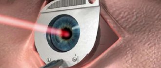
Laser coagulation of the retina

