Main symptoms:
- Eye fatigue
- Headache
- Double vision
- Visual impairment
- Blurred picture
- Nausea
- Motion sickness in transport
Ametropia is a refractive error of the eyeball, which is characterized by failures in focusing light rays on the retina. Normally, light rays are focused on the retina, but when the focus is lost, this process occurs behind the retina or in front of it. As a result, a person sees the world around him with distortions, blurry and unclear.
- Etiology
- Classification
- Symptoms
- Diagnostics
- Treatment
- Possible complications
- Prevention
This ophthalmological pathology occurs very often in humans, regardless of gender and age. There are several varieties of this disease, which is diagnosed during examination by an ophthalmologist. Depending on the degree and type of the disease, therapeutic measures will be carried out, lenses and drops will be prescribed. Methods are selected individually; the use of surgical correction is possible.
The treatment prognosis is favorable - it is possible to normalize existing visual disturbances.
What is ametropia: general concepts
Light rays, reflected from objects and passing through the optical system of the eye, with normal vision are focused precisely on the central zone of the retina - the macula. At the same time, a person clearly and contrastly distinguishes distant objects. If the rays are focused in front of or behind the retina, then the quality of vision is impaired, objects look blurry near or far, depending on the type of ametropia. This deficiency is corrected with contact lenses or glasses. In difficult cases, surgical or laser vision correction is prescribed.
The following eye refractive errors are classified as ametropia:
- nearsightedness (myopia);
- farsightedness (hypermetropia);
Astigmatism and presbyopia are also common refractive errors, although in these cases poor vision is not associated with the focusing of the light beam on the retina and the size of the eyeball.
The most common is myopic ametropia of the eyes. This is usually a hereditary or congenital disease, although recently the percentage of people who acquire myopia during their lifetime has increased significantly. Let's talk about the forms of ametropia in more detail: causes, symptoms, degrees, methods of treatment.
Myopia
Myopia is the most common form of pathology, which usually begins to develop in childhood. There are two main reasons for the development of this type of ametropia.
- Axial myopia. It develops when the eyeball is enlarged along the anteroposterior optical axis, as a result of which light rays do not reach the retina when focusing.
- Refractive myopia. Caused by irregular curvature of the cornea or lens. When refracted, the rays are scattered in the wrong way, also not reaching the central part of the retina. As a result, the image turns out fuzzy and blurry.
Due to the listed physiological reasons, a person has difficulty distinguishing between objects located at a distant distance, but clearly sees close objects. As a rule, acquired myopia develops due to non-compliance with the rules of visual hygiene. Schoolchildren spend the vast majority of their time with gadgets, and they also have high visual stress at school. In addition, a prerequisite for the development of this type of eye ametropia, especially with a hereditary factor, can be poor nutrition, ophthalmological and traumatic brain injuries and other factors.
In medicine, there are three degrees of myopia:
- weak from 0.5 to 3 diopters;
- average from 3 to 6 diopters;
- strong from 6 diopters or more.
A high degree of myopia can reach very significant values - up to 30 diopters. In this case, surgery is usually recommended, and if there are contraindications, soft or hard lenses (including orthokeratological ones) can be prescribed. It is difficult to wear glasses with a strong negative optical power of the eyes: thick glasses magnify them and distort the outlines of objects, severely limit peripheral vision, and besides, the eyes in such glasses quickly get tired, and the head begins to hurt. With significant myopic ametropia, degenerative changes in the retina, its detachment, and other disorders in the eye structures can also be observed.
Correction with laser
Source: bolezniglaznet.ru Laser vision correction is the correction of eye refraction by changing the thickness of the cornea, carried out using an excimer laser.
By changing the thickness of the cornea, its optical power is changed, due to which the light is focused on the retina - a person sees surrounding objects clearly and clearly. Laser vision correction is an advanced trend in modern ophthalmology.
There are 2 types of laser vision correction:
- photoreactive keratectomy (PRK)
The laser removes the surface layers of the cornea, changing its thickness. With the help of PRK, you can correct myopia (up to -6 diopters), hypermetropia (up to +3 diopters), astigmatism (up to -3 diopters).
After PRK, the recovery period is quite long - up to several months it is necessary to instill special drops into the eyes. The advantages of PRK are that the procedure is completely painless, short exposure time, and stable results.
- LASIK (laser keratomileusis, lasik)
It is a combination of microsurgical excimer-laser stages. During the operation, under the guidance of an ophthalmologist, a corneal flap is folded back using an automated special medical instrument - a microkeratome.
Then the thickness of the cornea, pre-calculated using computer technology, is removed with a laser. The bent flap is returned to its place. The operation takes 1 - 1.5 minutes. Within a few hours you can return to your normal lifestyle. With LASIK it is possible to correct higher degrees of ametropia.
Indications for laser vision correction:
- different visual acuity in the left and right eyes;
- professions that require quick response (for example, pilots or firefighters);
- the desire of the patient himself.
Contraindications to laser vision correction:
- age less than 18 years;
- progressive myopia;
- pregnancy and lactation (breastfeeding);
- general severe diseases of the body (for example, diabetes mellitus - a disorder of glucose (sugar) metabolism);
- acute infectious diseases;
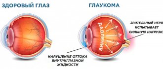
- a number of eye diseases: inflammatory in nature, cataracts (clouding of the lens), glaucoma (increased intraocular pressure), history of retinal detachment.
In the presence of severe presbyopia and lens opacities:
- replacing a cloudy lens with an artificial one through surgery - currently preference is given to soft intraocular (intraocular) lenses, which require a minimal incision in the eye and are placed inside in a folded state;
- correction of amblyopia (decrease in visual acuity, not amenable to optical correction, occurring in the absence of organic disorders, usually in one eye; amblyopia is common among children and adolescents);
- occlusion of a healthy eye - gluing or establishing a special occlusion (shutter) of a healthier eye for 2 - 6 hours a day in order to train the weaker eye.
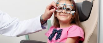
Symptoms of myopic ametropia in children
A child, especially a preschooler, is not always able to understand what is happening to his vision. Parents should be attentive to his behavior. Here are some signs that may indicate the development of childhood myopia.
- Frequent complaints of headache. This symptom is attributed to fatigue, and at this time myopia progresses.
- Irritation and eye fatigue. With constant tension in an attempt to see distant objects, a spasm of accommodation occurs (false myopia). Pain when blinking, dryness and irritation of the mucous membrane may occur.
- Increased tear production. With myopia, the eyes are more sensitive due to the dilated pupil. More light enters it, and lacrimation is the body’s protective reaction to bright light.
The listed signs should be an alarming signal for parents. If ametropia is not corrected in a timely manner, negative consequences may occur, because other eye structures inevitably suffer.
Symptoms
Symptoms of visual impairment include the following:
- eyes get tired quickly;
- double vision occurs;
- the child looks at objects from a close distance, but does not pay attention to distant ones, or vice versa;
- blurring the contours of an object;
- headaches due to eyestrain;
- nausea, motion sickness in transport.
All of the above symptoms require immediate response and appropriate measures to improve and normalize vision.
Hypermetropia
Farsightedness is a visual impairment when a person sees objects close up blurry and cannot read small text, but at long distances he distinguishes objects clearly and clearly. This happens because light rays are focused not on the retina, but behind it. There can be several reasons for farsightedness. In general, all children are born with a degree of farsightedness of approximately +2.5-3 diopters, and this is a normal physiological phenomenon. This ametropia is due to the anatomical size of the eyeball. It has a diameter of 17-18 mm, whereas in an adult it is 23-25 mm. As the baby grows, the size of the eyeball increases. Normally, by 12 months of life, the power of farsightedness should be no more than 2 diopters, and by approximately 14-18 years the eye reaches an adult size of 23-25 mm, the optical focus completely moves to the retina and ametropia disappears.
However, if farsightedness is pathological, then it poses a danger to the health of the visual organs and requires timely treatment. Let's tell you more about the varieties of this disease.
Treatment methods
The most common way to improve vision is to wear glasses with specially selected lenses or use contact lenses. However, this means wearing them frequently or constantly, which is not always convenient. Therefore, other methods have been developed. These are ophthalmic surgeries that often use a laser. Subsequently, wearing glasses is no longer required.
Today the following operations are carried out:
- keratotomy;
- conductive keratoplasty;
- replacing the lens with a donor one;
- implantation of special intraocular lenses.
All such types of surgical interventions are paid and require high professionalism of doctors. Therefore, having decided on this method of treatment, you should carefully select a clinic working in this area.
Congenital farsightedness in children
The following reasons can provoke this type of ametropia:
- the presence of strong degrees of the disease in both parents;
- a woman’s use of alcohol, nicotine, inappropriate medications during pregnancy, as well as a history of severe infectious disease;
- genetic abnormalities.
If in the first two cases hypermetropia can be predicted, then medicine is not yet able to influence genetic mutations. A child with a deviation can be born to completely healthy parents if they are carriers of the defective gene.
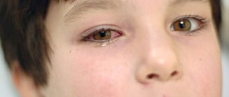
Signs of farsightedness in children
Parents of babies who are at risk due to hereditary factors should pay attention to some signs that may indicate vision problems in the child. The following manifestations should be warning signs:
- the child often squints or opens his eyes wide, rubs them;
- moves books and toys further away from the face;
- after doing creative work with small objects (for example, drawing, assembling construction sets), his eyes turn red and watery;
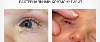
- The baby has frequent inflammatory diseases of the organs of vision - conjunctivitis, blepharitis, etc.
If at least two of the listed symptoms of ametropia are present, then parents must take the child to an ophthalmologist for an examination. The doctor will detect abnormalities, if any, and prescribe suitable and effective therapy.
Causes of acquired farsightedness
This form of pathology occurs due to the influence of certain factors already in adulthood. Here are the main ones.
- Incorrectly performed surgical or laser surgery on the eyes. An inexperienced doctor may incorrectly adjust the surface of the cornea or lens. Light rays passing through these optical media end up being refracted incorrectly into the area behind the retina.
- Internal tumors. Often, a neoplasm in the eye is imperceptible in the initial stages. Only after a noticeable deterioration in vision does a person turn to an ophthalmologist. The tumor grows and puts pressure on the eyeball, changing its shape, resulting in farsightedness.
- Eye injuries. When exposed to the outside, the shape of the lens or cornea may change, and, accordingly, their refractive ability may be impaired.
Acquired hyperopia is practically not correctable with glasses or contact lenses and is usually treated surgically.
Astigmatism
Astigmatism is a refractive error in which a person sees objects blurry, indistinct, as if in a fog. This refractive error is caused by the curvature of the smooth, spherical shape of the cornea or pathology of the lens. Light rays passing through these optical media are projected not in the form of points, but of lines, circles, ovals, so the image turns out fuzzy and blurry, and also elongated horizontally or vertically. At the same time, astigmatism is characterized by poor vision at both long and near distances.
Concomitant strabismus
With concomitant strabismus, eye mobility is not limited, and the angle of deviation of the primary and secondary parameters coincides along all nine meridians (the differences do not exceed five diopters).
This type of strabismus can be divided into:
- Accommodative;
- Non-accommodative;
- Partially accommodative.
The novelty of the classification is aimed at dividing the accommodative type of strabismus into refractive, combined, non-refractive and decompensated.
Degrees of astigmatism
There is a classification of this eye pathology depending on its severity:
- weak to 3D. Subject to contact or spectacle correction, and also corrected using microsurgery;
- average from 3 to 6D. With such values, it is best to correct vision with toric lenses or undergo surgery;
- strong - over 6 diopters. This high degree of astigmatism occurs as a result of significant changes in the surface of the cornea. It is recommended to correct this form of pathology with rigid gas-permeable lenses or laser correction. In this case, it is very difficult to use glasses, the eyes get tired, and peripheral vision is narrowed.
Many people do not even suspect that they are developing astigmatism, since its symptoms resemble eye fatigue from visual strain. Meanwhile, here are some manifestations that may indicate it:
- impairment of near vision clarity;
- asthenopia - rapid eye fatigue;
- difficulties with orientation in space;
- frequent headache;
- dry mucous membrane.
If the listed symptoms recur with a certain frequency, then this is a reason to visit an ophthalmologist to find out the cause of the discomfort. Such phenomena may well indicate developing astigmatism, in which case it will be necessary to take measures to correct it. In most cases, types of ametropia can be corrected. There are different ways to treat this form of pathology.
Principles of ametropia correction
Moderate and weak degrees of farsightedness, myopia, and astigmatism can be successfully corrected with contact lenses or glasses, but high degrees of ametropia are usually treated through surgery or laser intervention. In the presence of anisometropia - a significant difference between the optical power of both eyes, exceeding 3 diopters - only contact lenses or surgery are recommended. Hardware treatment and special visual gymnastics also provide good results for mild pathologies. The treatment method is determined by a specialist.
It is worth remembering that early diagnosis is important for the successful correction of existing ametropia. In order not to miss the onset of the disease, especially in a child, you should undergo preventive examinations with an ophthalmologist at least once a year, and for those who have already been diagnosed with refractive error, every six months. Only careful attention to your health will allow you to maintain normal vision for a long time.
MagazinLinz.ru team
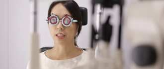
Diagnostics
The disease is diagnosed at the first examination by an ophthalmologist during an external examination of the eye.
Additionally, the following activities may be prescribed:
- automatic refractometry – helps determine the point of the best image, taking into account the retina;
- visometry - it is used to determine visual acuity;
- cycloplegia – helps to identify true or false myopia;
- ophthalmometry - it is used to examine the cornea, its curvature and refractive power;
- pachymetry – the thickness of the cornea is determined using ultrasound;
- ophthalmoscopy - it is used to check the condition of the fundus, optic nerve and vascular functionality;
- A-scan of the eye - check the length of the axis.
Pachymetry
After a comprehensive study, the patient is selected the basic principles of ametropia correction.