Causes of dilated veins in the fundus
Eye diseases and decreased visual acuity associated with dysfunction of the vascular network are increasingly being diagnosed. According to obaglaza.ru, this consequence is caused, first of all, by late seeking medical help and ignoring the symptoms of systemic diseases.
Nature of the vessels
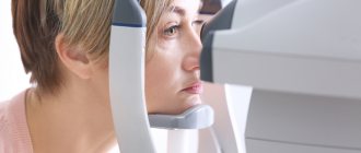
During a medical examination, it is very important to undergo an ophthalmoscopy. Depending on the degree and nature of the disturbances in the venous network of the fundus, the doctor can determine the disease that caused the pathology:
- dilation of venous vessels and narrowing of arterial vessels - evidence of the initial stage of hypertension;
- microaneurysms and hypertrophied branching of venous vessels, which can sometimes be accompanied by retinal edema - develops in diabetes mellitus;
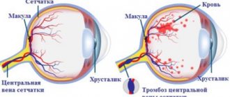
- vascular spasms, thrombosis of the central vein and artery, with a background of the fundus of the eye with a whitish-milky tint (insufficient blood supply) - speak of vascular diseases;
- a bright dot in the area of the macula and swelling of the retina - according to obaglaza ru, indicates a bloodless central artery of the eye;
- retinopathy (insufficient blood supply to the retina) is a complication that is a consequence of hypertension, vascular disease and diabetes;
- swelling of the retina near the projection of the beginning of the optic nerve, strong branching of the veins - indicates a disruption in the functioning of the central vein of the eye or its blockage (with hypertension, renal and vascular pathologies).
ABOUT VISION
Full-blooded and tortuous retinal veins are what parents often read in reports and what is presented to them as some kind of retinal vascular angiopathy.
What is angiopathy? Literally translated, the term “angiopathy” itself means that there is a pathology “pathy” of the vessels “angio”, but what kind of pathology this term does not indicate. This term is used in modern ophthalmology, but with clarifications. Example: diabetic retinopathy (Diabetic Angiopathy). However, in the cases that we discuss, the word “angiopathy” is used in isolation. In addition, this diagnosis is made very often and on the basis of it the neurologist draws some conclusions - usually nootropics are prescribed (which in fact never makes sense).
The term: “Retinal angiopathy” simply does not carry any meaning without specification and is often used by pediatric ophthalmologists in relation to children who, in their opinion, have blood vessels that are somehow different from the norm. How they differ is also an uncertain question. No criteria other than personal impression are used. And most importantly, these “changes” do not reflect any pathological process. In fact, such a diagnosis is made only in our country and the countries of the former USSR; it does not carry any specifics and usually children who receive such a diagnosis are absolutely healthy.
Unfortunately, this “diagnosis” still occurs quite often. Unfortunately, neurologists also take this “diagnosis” into account. I even know one doctor who was reprimanded for not diagnosing “retinal angiopathy” during consultations with a large number of children. Needless to say, nothing serious was missed and this was an argument against which it is difficult to find objections. Although, in my opinion, research should be the argument for a doctor.
How it all started:
Diagnosis of increased intracranial pressure is in particular based on the condition of the fundus. In the fundus of the eye, the ophthalmologist sees part of the optic nerve - the nerve that carries information about what a person sees in the brain. Essentially this is the part of the brain that is accessible to inspection. The ophthalmologist sees, as it were, a section of this nerve and it appears to him in the form of a circle or oval.
From the center of this circle, artery and vein blood vessels emerge onto the surface of the retina, which take part in the blood supply to the periphery of the retina. With intracranial hypertension, swelling of the optic nerve is possible, which leads to changes in the shape of the visible disc and its edges. The disc becomes uneven, part of it protrudes anteriorly. Arteries and veins are compressed; Accordingly, arterial blood enters in less quantity and the arteries become narrow, and venous blood cannot flow out in full and the veins become plethoric (thick) and tortuous.
Those. there is some kind of sequence: changes in the optic nerve - narrowing of the arteries - dilation of the veins.
This condition is called: “Congested optic disc”. With a stagnant disc, the venous congestion is pronounced and there is a narrowing of the arteries. Such changes are secondary to papilledema and this is a very serious symptom, and children with such a symptom should be urgently hospitalized.
Fortunately, increased intracranial pressure is not common, but many parents are faced with the fact that the ophthalmologist refers the child to a neurologist because of plethora of veins. This is explained by the fact that over time, the plethora of the veins itself began to introduce oculists into sacred awe. Those. plethora without changes in the optic nerve head and narrowing of the arteries.
Congestion of the retinal veins is a very subjective sign. This sign may be assessed differently by different doctors. The plethora of the veins can change depending on the child’s position (standing, lying), physical activity, and other things. A lot depends on the subjective assessment by the doctor.
It is impossible to talk about the caliber of the retinal veins as something constant and diagnostically significant.
Unfortunately, in Russia, medical knowledge is transmitted by so-called regional schools and it is passed on by word of mouth. For a long time, ideas about a particular diagnostic sign may not change and even become overgrown with fiction. IN
In Moscow, you can still find ophthalmologists who suggest checking with gastroenterologists for inflammatory eye diseases or “getting rid of worms.” The approach from the position of evidence-based medicine is extremely
doc and often this leads to irrational waste of energy of the patient and doctors.
Diagnosis: Retinal vein angiopathy - does not exist.
Symptoms
In the initial stage of diseases or disorders of the vascular system of the eyes, there may be no symptoms at all. Pathology is detected during a thorough medical examination. In more severe and advanced cases, a person may notice a number of symptoms:
- Seeing floaters in the eyes, multi-colored spots, flashes of light.
- Parts of the view may be lost from view.
- Sometimes pathologies are accompanied by blurred vision.
- In more advanced cases, vision decreases to complete blindness.
Obaglaza once again emphasizes that a periodic high-quality ophthalmological examination by a qualified specialist will protect your vision and can diagnose a number of systemic diseases at the very beginning of their development, before symptoms appear.
Symptoms
Dilatation of the veins of the fundus leads to the appearance of the following clinical signs in the patient:
- a veil before the eyes;
- blurred vision near and far;
- microcirculation disorder in retinal tissue;
- the appearance of loss of vision;
- headache;
- feeling of pressure in the eyes;
- inflammatory process in the eyeball;
- optic nerve atrophy;
- retinal detachment;
- macular degeneration;
- the appearance of a tumor process;
- cataract;
- foci of hemorrhages in the fundus.
Treatment
Obaglaza.ru specialists insist that priority in the treatment of diseases of the vascular system of the eyes is given to the treatment of the disease that caused the damage (hypertension, diabetes mellitus, atherosclerosis, kidney disease, etc.).
To maintain blood vessels and restore visual function of the eyes, the following are effectively used:
- General medications to maintain vision;
- Vitamin therapy;
- Physiotherapeutic procedures, massage;
- Cool baths, walks;
- Oxygen injections under the conjunctiva.
Possible complications
In the absence of the necessary treatment, there is a high probability of developing complications that negatively affect vision. The most common complications include:
- deterioration of vision, which with further inattention to the condition of the eyes can develop into complete blindness;
- hemorrhages in the retina - this consequence can occur with improperly selected treatment, significant strain on the eyes, as well as with the development of hypertension, which is expressed in increased blood pressure;
- damage to the cornea, which can provoke the formation of cataracts. These conditions, in turn, can lead to gradual loss of vision and headaches, which significantly reduce the patient’s quality of life.
Retinopathy develops in later stages of the disease, which is likely due to insufficient therapeutic intervention or too late diagnosis of the disease. Therefore, to prevent possible complications and maintain visual acuity, it is recommended to pay attention to any changes in the condition of the eyes in a timely manner.
Prevention
To prevent varicose veins from appearing under and on the eyelids, dermatologists advise carefully caring for the skin around them and using cosmetic services, for example, mesotherapy with laser rejuvenation. These two procedures are recommended for people over 30 years of age. However, they must be agreed upon with the attending physician.
At a young age, you should do a contrasting type of wash using natural products, and you should also apply masks and compresses to the skin. It is also useful to wash your face with decoctions.
In addition, it is recommended to refrain from everything that leads to the formation of vasodilation: prolonged sitting at the computer and overheating in the sun, excessive stress, etc.
To prevent the occurrence of the disease, it is recommended to refrain from:
- excessive physical activity;
- prolonged sitting at the computer;
- prolonged exposure to the sun.
At a young age, contrasting washes with natural products are useful. In some cases, after reaching 30 years of age, they resort to mesotherapy or laser rejuvenation.
For preventive purposes, you can periodically wash your eyes with decoctions made from medicinal herbs. Some compositions are also suitable for internal use. A decoction made from birch buds normalizes the functioning of the circulatory system and thins the blood. Hop cones can also be used to clean the blood vessels of the eyes. They help increase the strength of blood vessels due to the content of a sufficient amount of tannins in their composition.
Do you still think that it is impossible to cure varicose veins?
Judging by the fact that you are reading these lines now, victory in the fight against varicose veins and unsightly areas on the body is not yet on your side.
Have you already thought about inpatient treatment? This is understandable, because varicose veins are a very dangerous disease that, if not treated in a timely manner, can be fatal. Stars on the legs, fatigue, terrible swollen veins. All these symptoms are familiar to you firsthand.
But perhaps it would be more correct to treat not the effect, but the cause? We recommend reading the story of Ekaterina Andreeva. Read the article >>
Causes of dilated veins in the fundus
Eye diseases and decreased visual acuity associated with dysfunction of the vascular network are increasingly being diagnosed. According to obaglaza.ru, this consequence is caused, first of all, by late seeking medical help and ignoring the symptoms of systemic diseases.
Diagnostic measures
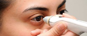
Hyperemia of the conjunctiva of the eye is not considered as an independent pathology. This is just a symptom that can manifest itself with various abnormalities. Therefore, when it is detected, it is necessary to diagnose and prescribe targeted treatment. First of all, the doctor conducts a survey, visual examination of the patient and palpation of the lacrimal sac (the latter to exclude or confirm dacryocystitis). In some cases, this is enough to make a diagnosis. Sometimes additional examinations of the affected eye are required:
- Biomicroscopy is an examination of optical media and organ tissues using a microscope and directed light.
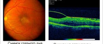
- Ophthalmoscopy is a non-invasive technique for examining the fundus of the eye. It is carried out using an ophthalmoscope. Allows you to identify venous congestion of the fundus, anomalies in the retina, choroid, and optic nerve head.
- Visometry is a standard test of visual acuity using a special table.
- Perimetry - testing the visual field and detecting blind spots (scotoma).
- Gonioscopy is a contact method for studying the structures of the anterior chamber of the eye.
Review of Treatment Methods
At the early stage of the disease, you can use various cosmetic products, creams with an anti-varicose effect, and special tonics. They help improve blood microcirculation in the facial area and prevent the development of pathologies of the circulatory system.
Special products help eliminate bags under the eyes, increase cellular turgor, and reduce swelling.
For varicose veins, you can use creams with collagen, products with hyaluronic acid and elastin. They give the skin a well-groomed appearance and at the same time improve local blood circulation. For varicose veins, venotonic gels made from natural ingredients are also used.
In advanced forms of the disease, to eliminate problems with the veins, they resort to:
- laser therapy;
- ozone therapy;
- chemical peeling;
- photocoagulation;
- lipofilling;
- miniphlebectomy;
- sclerotherapy;
- microfigrafting.
It is recommended to use medications that eliminate the main symptoms of varicose veins under the supervision of a doctor. Self-use of such drugs can be harmful to health.
| Belonging to a pharmacological group | Name of drugs |
| Drugs from the group of venotonics | Venarus Phlebodia 600 Troxevasin |
| Anticoagulants | Lyoton Heparin Fraxiparine Venolife |
| Medicines from the group of angioprotectors | Askorutin Venoruton Ginkor fort Corvitin |
| Vitamin and mineral preparations | Duovit Vita-Supradin Active |