Eye diseases are widespread in the modern world. This is facilitated by many factors: the rapid development of computer technology, the poor environmental situation... Often the disease is asymptomatic, so it is periodically recommended to undergo preventive examinations with an ophthalmologist.
There are many different diseases that affect the visual organs. Let's look at the most common diseases that anyone can encounter and their symptoms.
“Veil of fatigue” - what is it?
Fog, haze before the eyes, blurred images, unclear pictures seem to be unusual symptoms, but office employees often complain about such visual impairments. At the same time, problems with focusing disappear as soon as your eyes take a little break from the computer. However, sometimes veil and fatigue occur when:
- vascular diseases;
- cataracts;
- glaucoma;
- pathologies of the cornea.
Also, the appearance of a veil before the eyes can occur with diabetes. In some cases, patients with myopia complain of this symptom. In other words, the cause of the “veil of fatigue” can be a variety of diseases, and they are not always associated with the organs of vision.
Common eye diseases
Among the ophthalmic disorders, the most common are the following:
- conjunctivitis;
- keratitis;
- retinal dystrophy;
- iridocyclitis;
- destruction of the vitreous body;
- retinal detachment;
- tumors of the lacrimal glands;
- myopia and farsightedness;
- nystagmus;
- blepharitis;
- barley;
- pinguecula of the eye, cataracts, glaucoma and other age-related pathologies.
These are the main pathologies treated by an ophthalmologist. To make an accurate diagnosis, a specialist can conduct instrumental and laboratory tests. Ophthalmology closely interacts with traumatology, surgery, endocrinology, and allergology.
Cholesterol slides
What are cholesterol “slides”? This is also sometimes called xanthelasma - white or yellowish formations on the eyelids or in the corners of the eyes. They may not go away for years without causing any discomfort to the person. In some patients they begin to grow. Xanthelasmas are benign formations. The exact reasons for their appearance are unknown. It is assumed that such plaques arise due to lipid metabolism disorders in the body. Most often they are observed in people with diabetes, obesity, liver cirrhosis, pancreatitis, and high cholesterol levels in the blood.
Xanthelasma is detected during a routine ophthalmological examination. There is no pain when palpating the cholesterol “mountain.” The plaques do not cause itching or burning. Xanthelasmas can be single or multiple. In the second case, as a rule, they merge into one continuous bumpy growth. There is no specific treatment for this disease. It is necessary to find out against what pathology xanthelasma occurs. If this is due to a violation of fat metabolism, the patient is advised to adhere to a diet. The growths themselves are removed surgically. They look unaesthetic and it is better to get rid of them. Moreover, the operation is performed on an outpatient basis. The wound after skin excision heals within 1-1.5 weeks.
Distichiasis
An anomaly in the development of eyelashes, when another one grows behind the usual row. Outwardly it even looks beautiful - the eyelashes are thick and lush. For example, Elizabeth Taylor had distichiasis - this explains her unusually alluring and expressive gaze. However, when additional eyelashes grow through the conjunctiva, they cause discomfort: lacrimation, irritation, and blepharospasm occur. In this case, either surgical treatment is prescribed - removal of part of the mucous membrane of the eyelids with hair follicles from which eyelashes grow, or cosmetic treatment - laser hair removal.
“Allergy to white” - does this happen?
In ophthalmological practice, there are cases when patients experience painful sensations from the sight of everything white. This “allergy to white” manifests itself in the following symptoms:
- pain in the eyes, temples and forehead;
- red vascular network on the sclera;
- “flies” before the eyes;
- discomfort when moving the eyeballs, trying to look up.
Another concern is increased photosensitivity. White color is very bright, which is why the listed symptoms most often occur when a person looks at objects of this color. This may be due to infectious and inflammatory eye diseases. Such signs may also indicate problems with the nervous system. If such an “allergy” occurs periodically when working at a computer, you should take breaks and do eye exercises. However, you yourself most likely will not be able to determine the exact cause of the painful perception of white color. If the symptom bothers you frequently, consult your doctor.
Types of iridocyclitis
According to the etiological criterion, i.e. Depending on the causes of iridocyclitis, the following variants are distinguished:
- infectious;
- allergic;
- mixed infectious-allergic;
- systemic or syndromic-related;
- post-traumatic (including surgical injuries);
- unknown etiology;
- others (due to various pathological conditions).
Like any inflammatory process, iridocyclitis can take on various types of course, incl. acute or subacute, chronic, recurrent. Depending on the nature of the symptoms and the development of the clinical picture, serous and exudative (purulent, accompanied by mucous-liquid discharge), hemorrhagic (bleeding), fibrinous-plastic (scarring) variants are distinguished.
Are “shots in the head” a reason to get examined?
Sharp shooting pains that begin in the eye area and radiate to the head may indicate the development of glaucoma. When intraocular pressure increases, the eyes and head hurt. A person may even experience discomfort, like a patient who has had a heart attack. Sometimes attacks of glaucoma are accompanied by severe lacrimation, nausea and vomiting. Usually, according to this scenario, the angle-closure form of this pathology develops. It is very dangerous, since visual functions deteriorate quickly and cannot always be restored later.
Glaucoma is treated in various ways. The choice of treatment method depends on the type of disease and stage. At the initial stage, special eye drops are used that reduce intraocular pressure. Surgery may also be required.
Glaucoma occurs most often in people over 40-45 years of age. At the same time, a variety of reasons can lead to its development: injuries, systemic pathologies, congenital abnormalities of the visual organs, eye diseases. It is worth saying that glaucoma is a very dangerous pathology. About 15% of all blind people have lost their vision because of it.
Shooting pain in the head can also be a consequence of damage to the central nervous system. In addition, this symptom occurs with diseases of the ears, nose and throat.
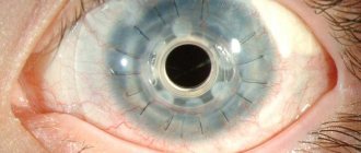
Aniridia
Refers to orphan (rare) genetic diseases - this is the complete absence of the iris. Often combined with other eye pathologies: photophobia, nystagmus, optic nerve hypoplasia, cataracts. The cause of congenital aniridia is a mutation in the PAX6 gene, which is located on chromosome 11. For treatment, an artificial iris implant is used, made of a special hydrogel, with a hole corresponding to the pupil. The patient may also choose to wear colored contact lenses. To prevent the development of keratopathy (drying) of the cornea, you need to use moisturizing drops or gels. You must wear sunglasses when going outside
"Eternal lack of sleep"
Closing eyelids, a feeling of sand in the eyes, a desire to sleep even after sleeping for 8-9 hours can signal a person about the disease. Such symptoms develop with dry eye syndrome. This ophthalmological disease can occur due to prolonged work at the computer without breaks. However, dry cornea also occurs with diseases of the genitourinary system. Of course, in most cases, when the above symptoms occur, it is enough to rest or drop moisturizing and relaxing drops into your eyes. Constant malaise and lack of sleep should be a reason for examination. It is not a fact that the cause of fatigue is an eye disease.
So, “veil of fatigue,” “shots in the head,” “allergy to white” are controversial, ambiguous symptoms. They can hardly be called specific, that is, characteristic of a specific disease. They are not always associated with ophthalmic pathologies. This means that you cannot self-medicate. You may make the situation worse. However, there is no need to panic ahead of time. Perhaps you just need to rest a little, play sports, and balance your diet. If you are bothered by several symptoms at the same time, there is no need to delay a visit to the ophthalmologist. We list the most typical signs of eye diseases:
- decreased visual acuity;
- dry eyes or, on the contrary, increased lacrimation;
- photophobia, or painful photosensitivity;
- redness of the tunica albuginea;
- blurred image, veil, “spots”, black dots before the eyes;
- pain, burning, itching, feeling of sand under the eyelids;
- rapid fatigue of the visual organs;
- headaches and dizziness.
The International Classification of Diseases devotes quite a lot of space to ophthalmological diseases. Some pathologies are not so rare, but still dangerous, for example, cataracts and glaucoma. Get tested often. This way you can detect the disease in time and cure it faster.
What laser correction methods are the newest?
The most popular laser procedure for correcting refractive errors is laser keratomileusis (LASIK/LASEK). All other techniques (FemtoLASIK, SuperLASIK, FemtoSuperLASIK, EpiLASIK, PresbyLASIK) are modifications of this basic procedure. The choice in favor of one or another type of operation is made based on medical indications, the patient’s lifestyle and his financial capabilities. Moreover, any LASIK laser operation is carried out in three stages.
First, anesthetic drops are instilled into the patient's eyes, after which the surgeon uses a microkeratome to make a small incision in the cornea, forming a flap from its upper epithelial layer. He moves aside until the procedure is completed. The second stage is laser correction itself. The doctor directs the laser at the cornea and vaporizes its tissue until it acquires the correct shape. The final stage of the operation is the return of the corneal flap to its original place. In some cases, a bandage lens is placed on the eyeball. The entire procedure lasts no more than 30 minutes. During this time, the doctor corrects vision in both eyes. After 2-3 hours the patient leaves the clinic. Vision is restored within a day. Complete healing occurs within a few weeks.
Laser surgery is not the cheapest treatment option, but today it is available to many people.
Pathologies of the optic nerves
- 8.1 Video: Eye diseases: which ones are curable?
- 9.1 Video: Primary angle-closure glaucoma
Ischemic neuropathy is a violation of blood flow in the intrabulbar or intraorbital region. Symptoms: decreased visual acuity and viewing angle, “blind” zones appear in certain areas.
Ischemic neuropathy
Neuritis is an infectious disease in which an inflammatory process occurs in the optic nerve. Symptoms: pain, loss of sensitivity in the area around the eye, weakening of the muscles that are located near the affected nerve.
Nerve atrophy is a disease in which conduction in nerve fibers is disrupted. Symptoms: decreased visual acuity, up to complete blindness, impaired color perception, decreased viewing angle.
Stages of optic nerve atrophy
Ophthalmoplegia is a condition in which the motor nerves of the eye stop functioning normally, often resulting in muscle paralysis and the inability to rotate the eyes. Symptoms: the eyes are shifted and fixed in one position.
Ophthalmoplegia
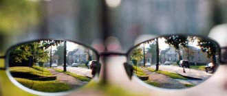
Diplopia – with this disease, a person constantly has double vision, which causes a lot of unpleasant sensations.
This is how a person with diplopia sees