Causes of the disease
The causes of xanthelasma are currently not fully understood. It is believed that these yellow plaques are a type of xanthoma and occur when cholesterol metabolism is disrupted. It has been noted that eyelid xanthelasmas most often occur in people suffering from obesity, pancreatitis, diabetes mellitus, liver disease and in cases of high cholesterol in the blood. In some cases, xanthelasmas are a manifestation of xanthomatosis, which may be hereditary.
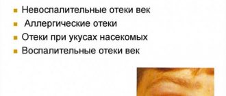
Symptoms and course of the disease
The first signs of xanthelasma of the eyelids are the appearance of a convex plaque in the area of the inner corner of the upper eyelid (sometimes the lower eyelid, but rarely). The size of this plaque usually varies from 0.5 to 1.5 cm. Most often, xanthelasmas have single elements of manifestation that tend to merge, but sometimes there are multiple formations that look like a solid convex line with uneven edges. The presence of xanthelasma brings psychological discomfort rather than physical: patients do not experience any unpleasant sensations or reasons interfering with the full functioning of the eyelid. Therefore, removal of xanthelasma is a correction of a cosmetic defect.
The course of xanthelasma is usually complemented by abnormal fat metabolism with the presence of humoral changes. The disease occurs suddenly, without any previous pathologies on the skin of the eyelid. Developed xanthelasma remains in a person forever; over time, its size increases, and the number of lesions also increases.
Reasons for the development of the disease
The exact origin of xanthelasma has not yet been established. It is assumed that neoplasms develop against the background of increased accumulation of fat molecules in the body.
The problem may arise due to metabolic disorders. The following pathologies lead to changes in lipid metabolism:
- diabetes;
- inflammatory process in the pancreas;
- diffuse toxic goiter;
- cirrhotic liver;
- lipoid nephrosis – fatty degeneration of the kidney tissue.
The patient’s poor diet also contributes to the onset of the disease. An abundance of fatty foods in the diet leads to excess cholesterol entering the body.
It is deposited in organs and tissues, including the eyelid area, where xanthelasmas are formed.
The cause of neoplasms may be a hereditary predisposition. The child has a genetic defect that contributes to the rapid accumulation of fats in the body and disruption of their metabolism. Usually the disease appears very early - in the first year of life.
Treatment prognosis and possible complications, consequences
Xanthelasma is more of a cosmetic defect than a disease. Cholesterol stains do not cause discomfort to the patient and, when small in size, do not interfere with vision. What brings a patient to the doctor’s office is only the desire to get rid of an aesthetic defect in appearance.
Inflammation or infection of xanthelasma occurs exclusively when the skin in the area of the plaque is injured. Suppuration and swelling, as complications of the postoperative period, can appear if the wound is not treated thoroughly or if an infection is introduced into it with dirty hands.
The operated eyelid heals in no longer than a month; no visible marks or scars remain on it. However, if the disorder of lipid metabolism in the body is not eliminated, xanthelasma may appear again (recur) after removal.
To date, medicine does not know of a single case of degeneration of these neoplasms into a malignant form.
After a competent operation to remove xanthelasmas, almost no traces remain on the eyelids
Appearance of formation (xanthelasma)
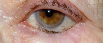
The photo of xanthelasma of the eyelids shows that it can be of small size. It resembles a plaque that forms in the upper eyelid area. Most often, the formation is located closer to the inner corner of the patient's eye.
The plaque protrudes slightly above the surface of the skin, making it visible to the naked eye.
The tumor differs in color from the surrounding skin - it has a yellowish tint. The formation has a soft consistency; when pressed, the patient does not feel pain.
Xanthelasmas can be single or multiple. The tumors usually affect the eyelids of both eyes.
Multiple xanthelasmas can merge into single plaques that have fuzzy edges and a bumpy surface. Often the neoplasms form a continuous thin line that runs along the entire surface of the eyelid.
The formations appear suddenly, without any redness or other changes in the skin of the eyelids. They grow very slowly and gradually spread over the entire surface of the eyelid.
Xanthelasmas usually grow to the size of a small pea; in rare cases, they grow to the size of a bean (approximately 4-5 cm). Tumors do not cause any unpleasant sensations to the patient, with the exception of psychological discomfort from their appearance.
However, tumors significantly affect the patient’s appearance and prevent them from feeling healthy, so they are considered a rather large cosmetic problem.
In addition to tumors on the eyelids, patients often develop tumors on other parts of the body - face, neck, limbs, joint surfaces.
Such formations are called xanthomas. Their appearance indicates that the patient has a specific disease - xanthomatosis.
What is xanthelasma of the eyelids (photo)?
Xanthelasma, or, in other words, flat xanthoma, is a benign fatty growth on the eyelid that resembles a yellow plaque. Outwardly, it looks like a blister or wen, slightly convex, and has gentle edges. It may be the only one on the eyelid, but in especially severe cases it happens that one or both eyelids are completely covered with xanthelasmas.
Usually the disease begins with a single plaque in the corner of the upper eyelid
The disease occurs with equal frequency in all parts of the globe, but it is noted that women suffer from it more often than men. Xanthelasma primarily affects the eyelids of people over 40 years of age. Children and adolescents rarely get sick. At an early age, this disease is usually a sign of genetically determined hypercholesterolemic xanthomatosis, in which fatty plaques are located not only on the eyelids, but throughout the body. As an adult, such a child usually suffers from diseases of the cardiovascular system, musculoskeletal system and liver.
Like xanthoma, it is also a sign of a lipid metabolism disorder. Xanthelasma of the eyelids is a frequent and faithful companion to diabetes mellitus, hypertension, pancreatitis, liver disease and excess body weight.
Once occurring, xanthelasmas do not spontaneously disappear and persist for life. They slowly increase in size; Gradually more and more new plaques appear.
Gradually, more and more new plaques appear around the eye
TOP 3 disease risk factors
Risk factors for the development of xanthelasma are:
| Factor | Explanation |
| Female | The disease occurs in men, but much less frequently. |
| Patients over 45 years of age | Against the background of age-related changes, the lipid balance in the body is often disturbed |
| People with chronic diseases | Diabetes mellitus, obesity and other endocrine pathologies |
Patients at risk are recommended to undergo regular lipid profile testing to prevent the occurrence of neoplasms.
Prevention
To prevent relapses after surgical removal of xanthelasma, the doctor recommends that the patient follow a plant-dairy diet, exclude animal fats from his diet and limit carbohydrates. You can eat no more than 30 g of butter and no more than 70 g of vegetable oil per day.
After surgery, it is necessary to regularly (at least once every six months) undergo a preventive examination by an ophthalmologist and dermatologist. The risk of early development of atherosclerosis and myocardial ischemia should be taken into account, so such patients need to especially closely monitor their blood pressure. If it persistently increases, you must register with a therapist and follow all his prescriptions.
The occurrence of xanthelasma is difficult to predict, so all people who are not yet familiar with this disease are advised to:
- promptly and correctly treat diseases of the liver and endocrine system;
- carefully observe eye and facial hygiene;
- use only high-quality, hypoallergenic eyelid cosmetics;
- Protect the delicate skin around the eyes from injury, and if it is damaged, immediately contact an ophthalmologist.
Diagnostics
If a tumor occurs in the eyelid area, the patient is advised to make an appointment with a dermatologist. A specialist determines the presence of xanthelasma based on its appearance - a flat yellowish formation in a characteristic location.
Additionally, during the examination, the dermatologist uses the diascopy technique. The formation is compressed with a transparent glass slide. This can achieve bleeding of xanthelasma. Due to the outflow of blood, the yellow color of the tumor is more clearly visible.
It is necessary to differentiate xanthelasma with the following diseases:
- malignant neoplasms of the skin;
- elastic pseudoxanthoma;
- syringoma.
Based on the clinical manifestations of the disease, the specialist diagnoses the patient and sends him for a full examination.
After determining xanthelasma, the patient must undergo a blood lipid test. For this purpose, the level of cholesterol, triglycerides and lipoproteins is determined.
Diagnostics allows you to assess the level of risk for the development of such dangerous diseases as atherosclerosis, hypertension and myocardial infarction.
Eliminating the cause of the pathology
Treatment of xanthelasma can be divided into several main stages. Therapy of the disease is a complex treatment that affects not only the tumor itself, but also the factors contributing to its appearance.
A course of specific treatment is necessary to prevent the recurrence of tumors.
An important component of therapy is the influence on the cause of the development of xanthelasma. Thus, in case of diabetes mellitus, the patient’s insulin therapy is adjusted. If the thyroid gland is malfunctioning, hormone replacement therapy, for example, thyroidin, is indicated.
To reduce cholesterol levels, the patient is prescribed a special therapeutic diet. It involves strict limitation of foods containing large amounts of animal fats. They are replaced with vegetable oils.
Preference is given to olive and flaxseed oil, as they are more beneficial than sunflower oil.
More plant foods are introduced into the diet - vegetables, fruits and berries. It is recommended to stop drinking alcohol during the treatment period.
Nicotine consumption prevents the body from restoring normal metabolism, so the patient should try to minimize the number of cigarettes smoked.
To reduce blood cholesterol levels, special lipotropic drugs are used that normalize the fat balance in the body.
The most effective means include:
- Pyricarbate.
- Lipoic acid.
- Clofibrate and its analogs.
- Thioctic acid.
- Cetamifene.
- Esters of polyunsaturated acids.
To reduce cholesterol, herbal preparations that have fewer side effects can also be used.
Drugs with this effect include:
- dandelion root;
- corn silk;
- Birch buds;
- dog-rose fruit;
- immortelle flowers.
Lipotropic drugs have a pronounced choleretic effect - they stimulate the secretion of bile from the liver. Therefore, their use is prohibited if there are any disorders of the biliary tract.
The use of medications must be agreed with your doctor. The patient may have contraindications to the prescription of drugs, which can be diagnosed by a specialist.
Additionally, to normalize metabolism in the body, the administration of vitamins and microelements is required. The use of multivitamin preparations has a restorative effect on the patient’s body, helping to restore normal metabolism.
TOP 5 methods of tumor removal
After treating the underlying pathology, the patient can remove the tumor. Typically, treatment takes place on an outpatient basis, meaning the patient does not need to go to the hospital to have surgery.
The tumor removal procedure can be performed using several methods. Each of them has its own advantages and disadvantages.
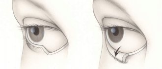
Surgical intervention
The operation involves excision of the xanthelasma using a scalpel. The area of its localization is treated with iron sesquichloride, after which sutures are applied to the edges of the surgical wound.
Surgical intervention injures the human skin and leaves a noticeable scar on the eyelid, so it is usually used for large tumors that cannot be removed by other methods.
In other cases, more minimally invasive treatment methods are used. Surgery is often performed when patients have xanthelasmas and drooping upper eyelids.
In this case, along with excision of the tumor, excess skin tissue above the eye is removed.
The average price for removing eyelid xanthelasma ranges from 5,000 to 12,000 depending on the stage of the disease.
Laser removal
The use of a carbon dioxide laser is an effective and safe method of tumor removal. The device allows you to excise only the tumor tissue without damaging the healthy skin around it.
The main advantages of the technique include:
- Complete sterility.
- No mechanical or thermal injury to the skin.
- High precision of tumor removal.
- Rapid bleeding stop.
- Contactless procedure.
Due to its advantages, laser excision produces a minimal cosmetic defect, which allows it to be used for excision of small xanthelasmas.
The price of the procedure ranges from 6-20 thousand rubles.
Electrocoagulation
Electrocoagulation is the destruction of tumor cells using electric current.
At the first stage of exposure, the protruding parts of the neoplasm are removed using tweezers or scissors. After this, electrodes are applied to the base of the tumor, with the help of which the skin is cauterized.
After the procedure, the patient is left with a small crust on the eyelid that has a reddish color. With proper care, the postoperative wound heals in about 10 days.
In the first few months, a reddish spot remains in its place, which then completely disappears.
The main advantage of electrocoagulation is the elimination of possible relapses.
Treatment of the tumor with electric current leads to the fact that all cells of the tumor die. Therefore, xanthelasma cannot re-develop at the surgical site. The price of electrocoagulation is 1200-1400 rubles.
Cryodestruction
Cryodestruction involves the destruction of neoplasm tissue using targeted exposure to ultra-low temperatures. The device supplies liquid nitrogen, which kills tumor cells. Further growth of the tumor stops completely.
At the site of the procedure, a dry scab is formed - a small fragment of necrotic tissue, which disappears after about 10-15 days. In its place, pink skin is formed, indistinguishable in color from healthy tissue.
The cost of the procedure is approximately 4-6 thousand.
Radio wave tumor removal
The use of radio waves to excise xanthelasmas ensures high accuracy of tumor removal. During the action of waves on the skin, heat is generated by a thin electrode.
Thermal exposure leads to the destruction and evaporation of tumor cells. The use of radio wave radiation provides the finest cut, much more accurate than with a scalpel.
Thanks to this, the skin around the tumor is practically not injured, and the cosmetic defect disappears. The average price of the procedure is 600-1500 rubles.
Question answer
An important quality of xanthelasma is that the neoplasm never degenerates into cancer.
Therefore, the risk to the patient’s health is practically minimal. Very rarely, patients experience bleeding at the extraction site, and an unsightly scar may also form. In addition, such a plaque can form again. For everything to go well, you need to go to a good specialist.
There are certain rules of prevention. To do this, doctors recommend adhering to proper nutrition, reducing the consumption of carbohydrates and animal fats. It is worth treating diseases that provoke tumor formation in a timely manner, not injuring the eyelids, removing makeup, using good cosmetics, and regularly seeing a dermatologist.
TOP 5 medicinal mixtures (folk methods)
To treat xanthelasma of the eyelid with folk remedies, compresses are often used:
- honey - a mixture of an egg, a spoonful of flour and a spoonful of honey;
- onion – soft onion baked in the oven, mixed with laundry soap;
- sour cream – sour cream in equal proportions with honey;
- chestnut – 5 crushed baked chestnuts with the addition of crushed aloe leaf;
- wheat – ground wheat grains diluted with olive oil.
The prepared mixture must be regularly applied to the surface of the tumor, left for 15-20 minutes, and then washed off with water.
Treatment of xanthelasma of the eyelids with folk remedies using compresses has excellent reviews among patients.
To normalize cholesterol levels in the body, you can use propolis tincture. It must be diluted in purified water, 7 drops per 30 ml. The resulting solution must be taken once every day during lunch.
To treat xanthelasma of the eyelids with folk remedies, you can use herbs. Patients need to brew yarrow, mint or chamomile. They need to be drunk twice a day, 100-150 ml. The effect of the procedure is achieved in approximately 1-2 months.
Patient reviews
The cost of eyelid xanthelasma removal can vary significantly, so it is recommended to read patient reviews before scheduling the procedure.
LARISSA, MOSCOW:
“I had a small plaque in the upper eyelid area, so I decided to remove it with a laser. The procedure was done very quickly, it didn’t hurt me, of course, there were some unpleasant sensations. Everything healed quickly, there were no traces left.”
ALEXEY, ST. PETERSBURG:
“I took my mother for treatment - some kind of yellow tumor appeared on her eyelid.
They said that it was already too big and could only be removed surgically. The operation was done quickly, under local anesthesia. We are pleased with the result, although there is a small scar left on the upper eyelid.”
Prices
| Disease | Approximate price, $ |
| Prices for breast reconstruction after cancer treatment | 41 130 — 59 740 |
| Disease | Approximate price, $ |
| Prices for diagnosing atopic dermatitis | 2 950 |
| Prices for diagnosing systemic lupus erythematosus | 6 350 |
| Prices for melanoma treatment | 32 620 — 57 620 |
| Prices for treatment of basal cell carcinoma | 7 700 — 8 880 |
| Prices for the treatment of malignant skin tumors | 4 420 — 5 420 |
| Prices for diagnosing psoriasis | 2 640 |
Expert opinion
- Cosmetologist
- Surgeon
Anna Avaliani
practicing cosmetologist
I think that it is better not to self-medicate using traditional methods, but to go to the doctor. To be sure to get rid of the plaque, it is better to resort to radical excision by any method (laser, liquid nitrogen, etc.). These are minimally invasive techniques, as they do not require the use of general anesthesia.
Aisha Baron
plastic surgeon
You can get confused in the variety of xanthelasma removal methods.
But the most effective way can be considered coagulation using a laser. The procedure has many advantages. After it, rehabilitation does not last long, the plaque will not appear again, the session itself is painless, and the risk of complications is reduced to zero. The procedure will take about 15-30 minutes. Xanthelasmas are neoplasms that are an important prognostic sign. Their occurrence indicates that the patient’s body has a disturbance in the metabolism of fat molecules.
Therefore, you should not delay treatment of the disease. You need to consult a specialist and undergo an examination to discover the cause of the formation of tumors.
Removal methods
Surgical treatment is performed on an outpatient basis using local anesthesia. The following methods are used to remove xanthelasma:
- Mechanical excision.
- Laser destruction.
- Electrocoagulation.
- Radio wave method.
Electrocoagulation is usually used to treat small lesions. Extensive plaques are removed using surgical scissors and tweezers. The choice of the exact method for removing xanthelasma depends on the technical equipment of the clinic.